S-44
The Three-Dimensional
Structure of Proteins
chapter 4
1. Properties of the Peptide Bond In x-ray studies of crystalline peptides, Linus Pauling and Robert
Corey found that the CON bond in the peptide link is intermediate in length (1.32 Å) between a typical
CON single bond (1.49 Å) and a CPN double bond (1.27 Å). They also found that the peptide bond is pla-
nar (all four atoms attached to the C—N group are located in the same plane) and that the two a-carbon
atoms attached to the CON are always trans to each other (on opposite sides of the peptide bond).
(a) What does the length of the CON bond in the peptide linkage indicate about its strength and its
bond order (i.e., whether it is single, double, or triple)?
(b) What do the observations of Pauling and Corey tell us about the ease of rotation about the CON
peptide bond?
Answer
2. Structural and Functional Relationships in Fibrous Proteins William Astbury discovered that
the x-ray diffraction pattern of wool shows a repeating structural unit spaced about 5.2 Å along the
length of the wool fiber. When he steamed and stretched the wool, the x-ray pattern showed a new
repeating structural unit at a spacing of 7.0 Å. Steaming and stretching the wool and then letting it
shrink gave an x-ray pattern consistent with the original spacing of about 5.2 Å. Although these
observations provided important clues to the molecular structure of wool, Astbury was unable to
interpret them at the time.
(a) Given our current understanding of the structure of wool, interpret Astbury’s observations.
(b) When wool sweaters or socks are washed in hot water or heated in a dryer, they shrink. Silk, on
the other hand, does not shrink under the same conditions. Explain.
Answer
(a) The principal structural units in the wool fiber polypeptide, a-keratin, are successive
c04TheThree-DimensionalStructureofProteins.qxd 12/6/12 4:15 PM Page S-44
Chapter 4 The Three-Dimensional Structure of Proteins S-45
(b) Freshly sheared wool is primarily in its a-keratin (a-helical coiled coil) form (see
3. Rate of Synthesis of Hair a-Keratin Hair grows at a rate of 15 to 20 cm/yr. All this growth is con-
centrated at the base of the hair fiber, where a-keratin filaments are synthesized inside living epider-
mal cells and assembled into ropelike structures (see Fig. 4–11). The fundamental structural element
of a-keratin is the ahelix, which has 3.6 amino acid residues per turn and a rise of 5.4 Å per turn
(see Fig. 4–4a). Assuming that the biosynthesis of a-helical keratin chains is the rate-limiting factor
in the growth of hair, calculate the rate at which peptide bonds of a-keratin chains must be synthe-
sized (peptide bonds per second) to account for the observed yearly growth of hair.
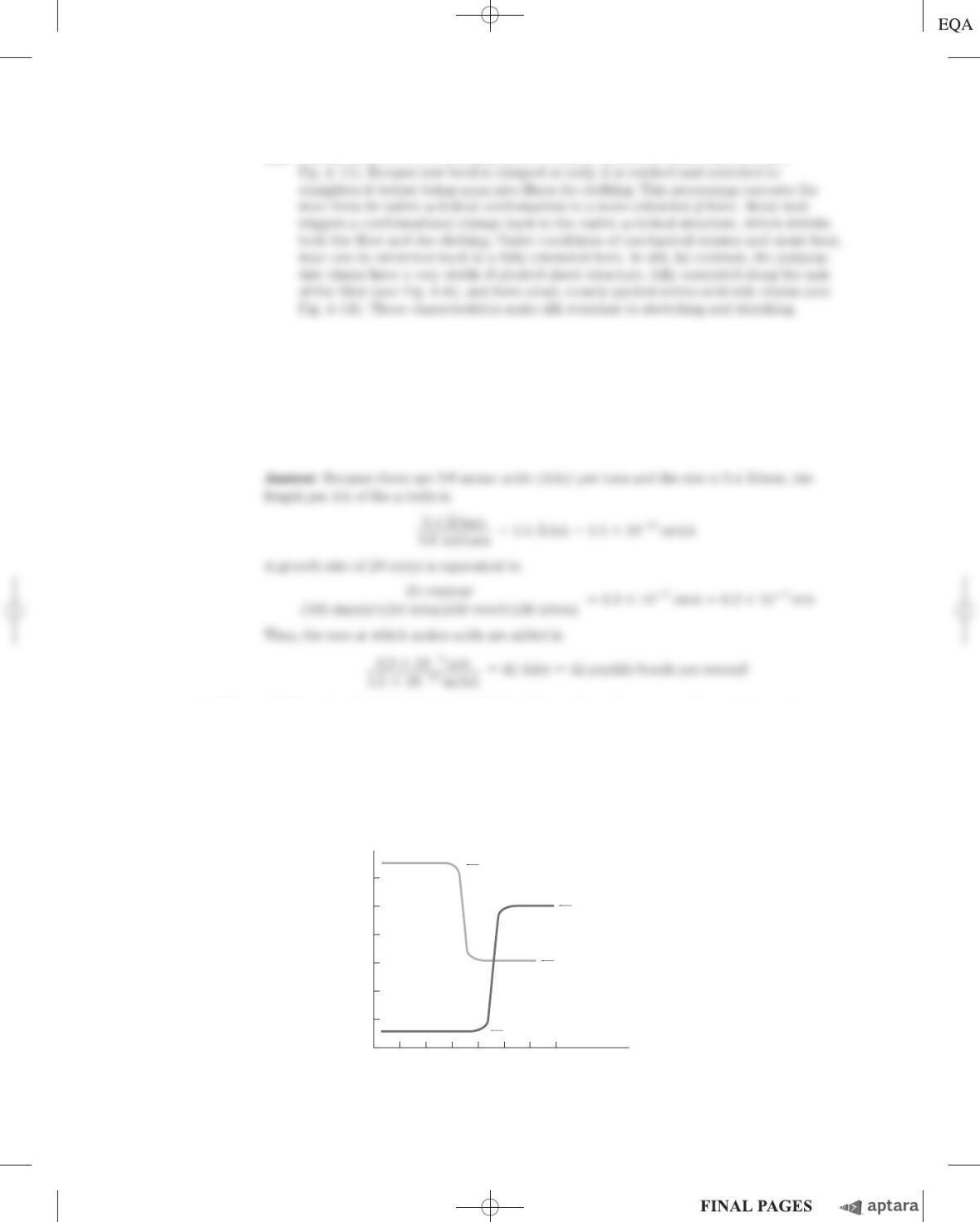
4. Effect of pH on the Conformation of a-Helical Secondary Structures The unfolding of the
ahelix of a polypeptide to a randomly coiled conformation is accompanied by a large decrease in a
property called specific rotation, a measure of a solution’s capacity to rotate circularly polarized light.
Polyglutamate, a polypeptide made up of only
L
-Glu residues, has the a-helical conformation at pH 3.
When the pH is raised to 7, there is a large decrease in the specific rotation of the solution. Similarly,
polylysine (
L
-Lys residues) is an ahelix at pH 10, but when the pH is lowered to 7 the specific rotation
also decreases, as shown by the following graph.
0
Poly(Glu)
Random conformation
Poly(Lys)
pH
Specific rotation
2 4 6 8 10 12 14
␣ Helix
Random
conformation
␣ Helix
c04TheThree-DimensionalStructureofProteins.qxd 12/6/12 4:15 PM Page S-45
S-46 Chapter 4 The Three-Dimensional Structure of Proteins
What is the explanation for the effect of the pH changes on the conformations of poly(Glu) and
poly(Lys)? Why does the transition occur over such a narrow range of pH?
5. Disulfide Bonds Determine the Properties of Many Proteins Some natural proteins are rich in
disulfide bonds, and their mechanical properties (tensile strength, viscosity, hardness, etc.) are corre-
lated with the degree of disulfide bonding.
(a) Glutenin, a wheat protein rich in disulfide bonds, is responsible for the cohesive and elastic char-
acter of dough made from wheat flour. Similarly, the hard, tough nature of tortoise shell is due to
the extensive disulfide bonding in its a-keratin. What is the molecular basis for the correlation
between disulfide-bond content and mechanical properties of the protein?
(b) Most globular proteins are denatured and lose their activity when briefly heated to 65 C.
However, globular proteins that contain multiple disulfide bonds often must be heated longer at
higher temperatures to denature them. One such protein is bovine pancreatic trypsin inhibitor
(BPTI), which has 58 amino acid residues in a single chain and contains three disulfide bonds.
On cooling a solution of denatured BPTI, the activity of the protein is restored. What is the
molecular basis for this property?
Answer
(b) As the temperature is raised, the increased thermal motion of the polypeptide chains
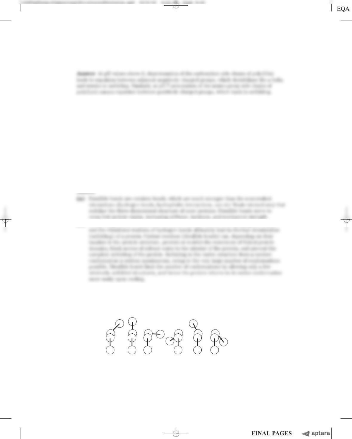
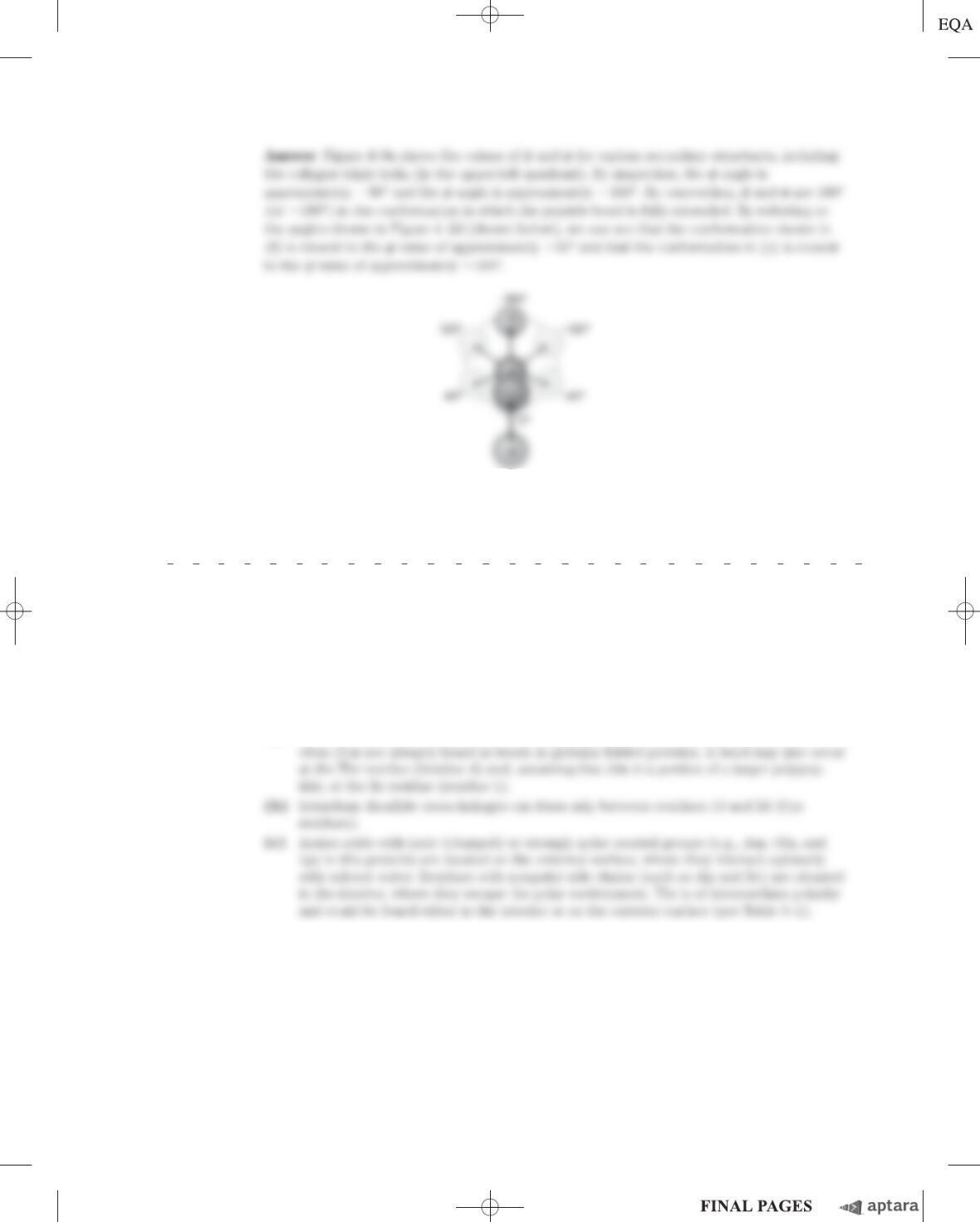
6. Dihedral Angles A series of torsion angles, and , that might be taken up by the peptide backbone
is shown below. Which of these closely correspond to and for an idealized collagen triple helix?
Refer to Figure 4
–
9 as a guide.
(a) (b) (c) (d) (e) (f)
Chapter 4 The Three-Dimensional Structure of Proteins S-47
12 3 4 5 6 7 8 910
Ile Ala His Thr Tyr Gly Pro Phe Glu Ala
11 12 13 14 15 16 17 18 19 20
Ala Met Cys Lys Trp Glu Ala Gln Pro Asp
21 22 23 24 25 26 27 28
Gly Met Glu Cys Ala Phe His Arg
7. A
mino Acid Sequence and Protein Structure Our growing understanding of how proteins fold allows
researchers to make predictions about protein structure based on primary amino acid sequence data.
Consider the following amino acid sequence.
(a) Where might bends or bturns occur?
(b) Where might intrachain disulfide cross-linkages be formed?
(c) Assuming that this sequence is part of a larger globular protein, indicate the probable location
(the external surface or interior of the protein) of the following amino acid residues: Asp, Ile,
Thr, Ala, Gln, Lys. Explain your reasoning. (Hint: See the hydropathy index in Table 3–1.)
Answer
(a) Bends or turns are most likely to occur at residues 7 and 19 because Pro residues are
8. Bacteriorhodopsin in Purple Membrane Proteins Under the proper environmental conditions,
the salt-loving archaeon Halobacterium halobium synthesizes a membrane protein (M
r
26,000)
known as bacteriorhodopsin, which is purple because it contains retinal (see Fig. 10–21). Molecules of
this protein aggregate into “purple patches” in the cell membrane. Bacteriorhodopsin acts as a light-
activated proton pump that provides energy for cell functions. X-ray analysis of this protein reveals
that it consists of seven parallel a-helical segments, each of which traverses the bacterial cell mem-
brane (thickness 45 Å). Calculate the minimum number of amino acids necessary for one segment of
ahelix to traverse the membrane completely. Estimate the fraction of the bacteriorhodopsin protein
that is involved in membrane-spanning helices. (Use an average amino acid residue weight of 110.)
c04TheThree-DimensionalStructureofProteins.qxd 12/6/12 4:15 PM Page S-47
S-48 Chapter 4 The Three-Dimensional Structure of Proteins
9. Protein Structure Terminology Is myoglobin a motif, a domain, or a complete three-dimensional
structure?
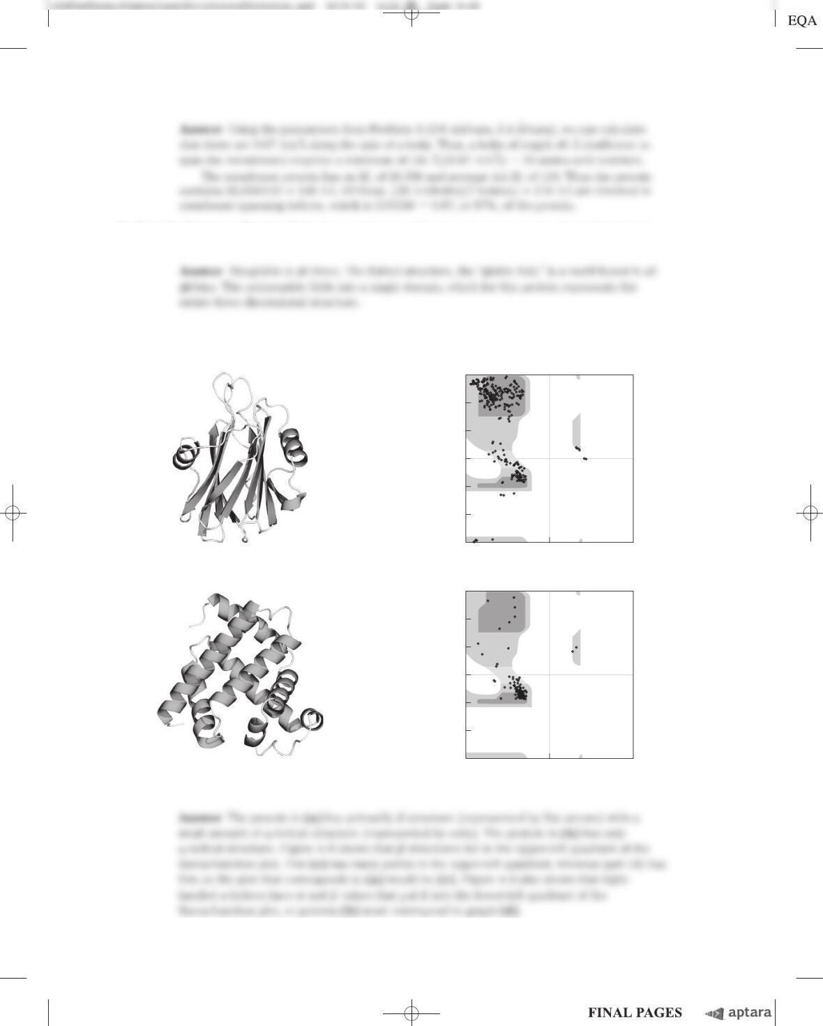
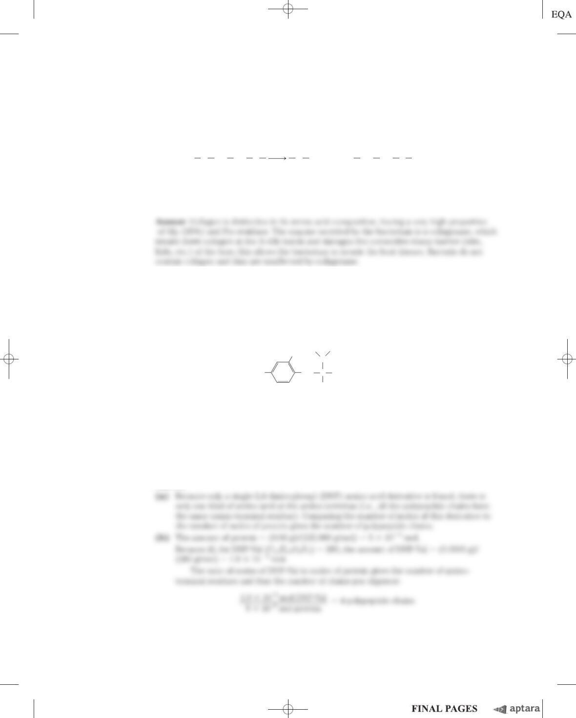
10. Interpreting Ramachandran Plots Examine the two proteins labeled (a) and (b) below. Which of the
two Ramachandran plots, labeled (c) and (d), is more likely to be derived from which protein? Why?
(b) (d) ⫹180
120
60
0
⫺60
⫺120
⫺180
⫹1800
⫺180
w (degrees)
f (degrees)
(c) ⫹180
120
60
0
⫺60
⫺120
⫺180
⫹1800
⫺180
w (degrees)
f (degrees)
(a)
2,4-Dinitrophenyl derivatives of the a-amino groups of other amino acids could not be found.
(a) Explain how this information can be used to determine the number of polypeptide chains in an
oligomeric protein.
(b) Calculate the number of polypeptide chains in this protein.
(c) What other protein analysis technique could you employ to determine whether the polypeptide
chains in this protein are similar or different?
Answer
Chapter 4 The Three-Dimensional Structure of Proteins S-49
XGly Pro Y H2OX COO H
3
N Gly Pro Y
⫹
*⫹
⫺
O
2
N
NO
2
NH C
C
CH
3
CH
3
H
H
COOH
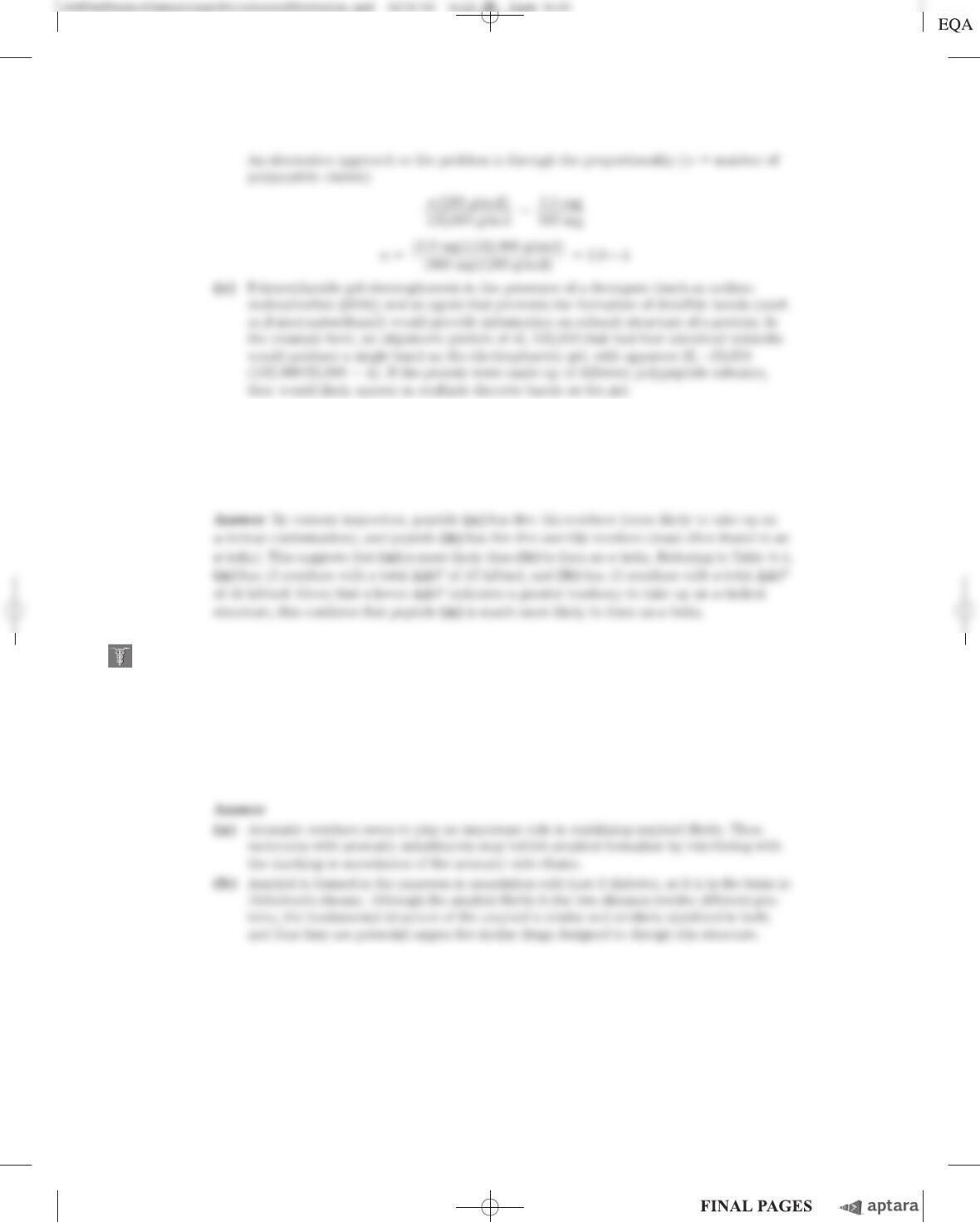
11. P
athogenic Action of Bacteria That Cause Gas Gangrene The highly pathogenic anaerobic bac-
terium Clostridium perfringens is responsible for gas gangrene, a condition in which animal tissue
structure is destroyed. This bacterium secretes an enzyme that efficiently catalyzes the hydrolysis of
the peptide bond indicated by an asterisk:
where X and Y are any of the 20 common amino acids. How does the secretion of this enzyme
contribute to the invasiveness of this bacterium in human tissues? Why does this enzyme not affect
the bacterium itself?
12. Number of Polypeptide Chains in a Multisubunit Protein A sample (660 mg) of an oligomeric
protein of M
r
132,000 was treated with an excess of 1-fluoro-2,4-dinitrobenzene (Sanger’s reagent)
under slightly alkaline conditions until the chemical reaction was complete. The peptide bonds of the
protein were then completely hydrolyzed by heating it with concentrated HCl. The hydrolysate was
found to contain 5.5 mg of the following compound:
c04TheThree-DimensionalStructureofProteins.qxd 12/6/12 4:15 PM Page S-49
S-50 Chapter 4 The Three-Dimensional Structure of Proteins
13. Predicting Secondary Structure Which of the following peptides is more likely to take up an
␣-helical structure, and why?
(a) LKAENDEAARAMSEA
(b) CRAGGFPWDQPGTSN
14. Amyloid Fibers in Disease Several small aromatic molecules, such as phenol red (used as a non-
toxic drug model), have been shown to inhibit the formation of amyloid in laboratory model systems.
A goal of the research on these small aromatic compounds is to find a drug that would efficiently
inhibit the formation of amyloid in the brain in people with incipient Alzheimer disease.
(a) Suggest why molecules with aromatic substituents would disrupt the formation of amyloid.
(b) Some researchers have suggested that a drug used to treat Alzheimer disease may also be
effective in treating type 2 (non-insulin-dependent) diabetes mellitus. Why might a single drug
be effective in treating these two different conditions?
Using the Web
15. Protein Modeling on the Internet A group of patients with Crohn disease (an inflammatory bowel
disease) underwent biopsies of their intestinal mucosa in an attempt to identify the causative agent.
Researchers identified a protein that was present at higher levels in patients with Crohn disease than
in patients with an unrelated inflammatory bowel disease or in unaffected controls. The protein was
isolated, and the following partial amino acid sequence was obtained (reads left to right):
EAELCPDRCI HSFQNLGIQC VKKRDLEQAI SQRIQTNNNP FQVPIEEQRG
DYDLNAVRLC FQVTVRDPSG RPLRLPPVLP HPIFDNRAPN TAELKICRVN
RNSGSCLGGD EIFLLCDKVQ KEDIEVYFTG PGWEARGSFS QADVHRQVAI
VFRTPPYADP SLQAPVRVSM QLRRPSDREL SEPMEFQYLP DTDDRHRIEE
KRKRTYETFK SIMKKSPFSG PTDPRPPPRR IAVPSRSSAS VPKPAPQPYP
(a) You can identify this protein using a protein database on the Internet. Some good places to start
include Protein Information Resource (PIR; http://pir.georgetown.edu), Structural Classification
of Proteins (SCOP; http://scop.mrc-lmb.cam.ac.uk/scop), and Prosite (http://prosite.expasy.org).
At your selected database site, follow links to the sequence comparison engine. Enter about
30 residues from the protein sequence in the appropriate search field and submit it for analysis.
What does this analysis tell you about the identity of the protein?
(b) Try using different portions of the amino acid sequence. Do you always get the same result?
(c) A variety of websites provide information about the three-dimensional structure of proteins. Find
information about the protein’s secondary, tertiary, and quaternary structures using database
sites, such as the Protein Data Bank (PDB; www.pdb.org) or SCOP.
(d) In the course of your Web searches, what did you learn about the cellular function of the
protein?
Chapter 4 The Three-Dimensional Structure of Proteins S-51
c04TheThree-DimensionalStructureofProteins.qxd 12/6/12 4:15 PM Page S-51
S-52 Chapter 4 The Three-Dimensional Structure of Proteins
Data Analysis Problem
16. Mirror-Image Proteins As noted in Chapter 3, “The amino acid residues in protein molecules are
exclusively
L
stereoisomers.” It is not clear whether this selectivity is necessary for proper protein
function or is an accident of evolution. To explore this question, Milton and colleagues (1992) pub-
lished a study of an enzyme made entirely of
D
stereoisomers. The enzyme they chose was HIV pro-
tease, a proteolytic enzyme made by HIV that converts inactive viral preproteins to their active forms.
Previously, Wlodawer and coworkers (1989) had reported the complete chemical synthesis of HIV
protease from
L
-amino acids (the
L
-enzyme), using the process shown in Figure 3–32. Normal HIV
protease contains two Cys residues at positions 67 and 95. Because chemical synthesis of proteins
containing Cys is technically difficult, Wlodawer and colleagues substituted the synthetic amino acid
L
–␣-amino-n-butyric acid (Aba) for the two Cys residues in the protein. In the authors’ words, this was
done to “reduce synthetic difficulties associated with Cys deprotection and ease product handling.”
(a) The structure of Aba is shown below. Why was this a suitable substitution for a Cys residue?
Under what circumstances would it not be suitable?
L–␣-Amino-n-butyric acid
C
O
CH3
CH CH2
⫺O
⫹NH3
Wlodawer and coworkers denatured the newly synthesized protein by dissolving it in 6
M
guani-
dine HCl, and then allowed it to fold slowly by dialyzing away the guanidine against a neutral buffer
(10% glycerol, 25 m
M
NaPO
4
, pH 7).
(b) There are many reasons to predict that a protein synthesized, denatured, and folded in this man-
ner would not be active. Give three such reasons.
(c) Interestingly, the resulting
L
-protease was active. What does this finding tell you about the role of
disulfide bonds in the native HIV protease molecule?
In their new study, Milton and coworkers synthesized HIV protease from
D
-amino acids, using the
same protocol as the earlier study (Wlodawer et al.). Formally, there are three possibilities for the
folding of the
D
-protease: it would give (1) the same shape as the
L
-protease, (2) the mirror image of
the
L
-protease, or (3) something else, possibly inactive.
(d) For each possibility, decide whether or not it is a likely outcome and defend your position.
In fact, the
D
-protease was active: it cleaved a particular synthetic substrate and was inhibited by
specific inhibitors. To examine the structure of the
D
– and
L
-enzymes, Milton and coworkers tested
both forms for activity with
D
and
L
forms of a chiral peptide substrate and for inhibition by
D
and
L
forms of a chiral peptide-analog inhibitor. Both forms were also tested for inhibition by the achiral in-
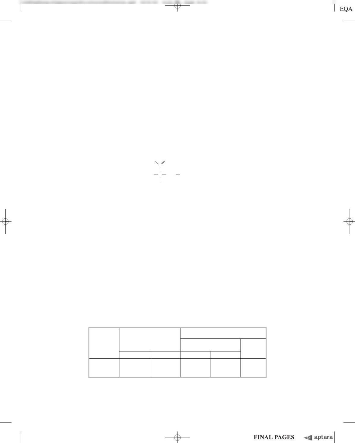
hibitor Evans blue. The findings are given in the table.
Inhibition
Substrate Peptide
hydrolysis inhibitor Evans
HIV blue
protease
D
-substrate
L
-protease
D
-inhibitor
L
-inhibitor (achiral)
L
-protease
D
-protease +
(e) Which of the three models proposed above is supported by these data? Explain your reasoning.
(f) Why does Evans blue inhibit both forms of the protease?
(g) Would you expect chymotrypsin to digest the
D
-protease? Explain your reasoning.
(h) Would you expect total synthesis from
D
-amino acids followed by renaturation to yield active
enzyme for any enzyme? Explain your reasoning.
Answer
References
Chapter 4 The Three-Dimensional Structure of Proteins S-53
c04TheThree-DimensionalStructureofProteins.qxd 12/6/12 4:15 PM Page S-53