S-29
1. Absolute Configuration of Citrulline The citrulline isolated from watermelons has the structure
shown below. Is it a
D
– or
L
-amino acid? Explain.
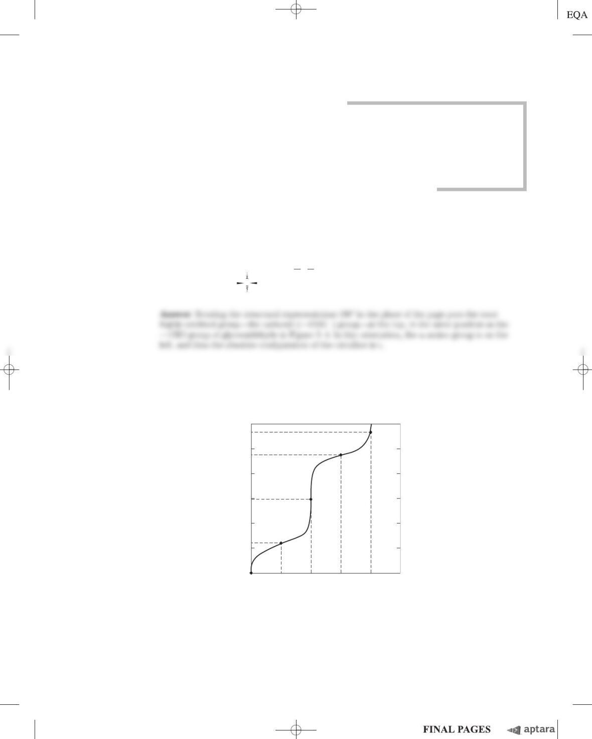
2. Relationship between the Titration Curve and the Acid-Base Properties of Glycine A 100 mL
solution of 0.1
M
glycine at pH 1.72 was titrated with 2
M
NaOH solution. The pH was monitored and the
results were plotted as shown in the following graph. The key points in the titration are designated I to V.
For each of the statements (a) to (o), identify the appropriate key point in the titration and justify your
choice.
12
2
4
6
8
0
11.30
0.5
OH
⫺
(equivalents)
pH
1.0 1.5 2.0
(V)
9.60
(IV)
(III)
2.34
(I)
(II)
5.97
10
CC
O
)H (CH NH
2
NH
222
P
HCN
⫹H
3
COO
⫺
(a) Glycine is present predominantly as the species
⫹
H
3
NOCH
2
OCOOH.
(b) The average net charge of glycine is ⫹.
(c) Half of the amino groups are ionized.
(d) The pH is equal to the pK
a
of the carboxyl group.
1
ᎏ
2
Amino Acids, Peptides,
and Proteins
chapter 3
c03AminoAcidsPeptidesandProteins.qxd 12/6/12 4:14 PM Page S-29
S-30 Chapter 3 Amino Acids, Peptides, and Proteins
(e) The pH is equal to the pK
a
of the protonated amino group.
(f) Glycine has its maximum buffering capacity.
(g) The average net charge of glycine is zero.
(h) The carboxyl group has been completely titrated (first equivalence point).
(i) Glycine is completely titrated (second equivalence point).
(j) The predominant species is
⫹
H
3
NOCH
2
OCOO
⫺
.
(k) The average net charge of glycine is ⫺1.
(l) Glycine is present predominantly as a 50:50 mixture of
⫹
H
3
NOCH
2
OCOOH and
⫹
H
3
NOCH
2
OCOO
⫺
.
(m) This is the isoelectric point.
(n) This is the end of the titration.
(o) These are the worst pH regions for buffering power.
Answer
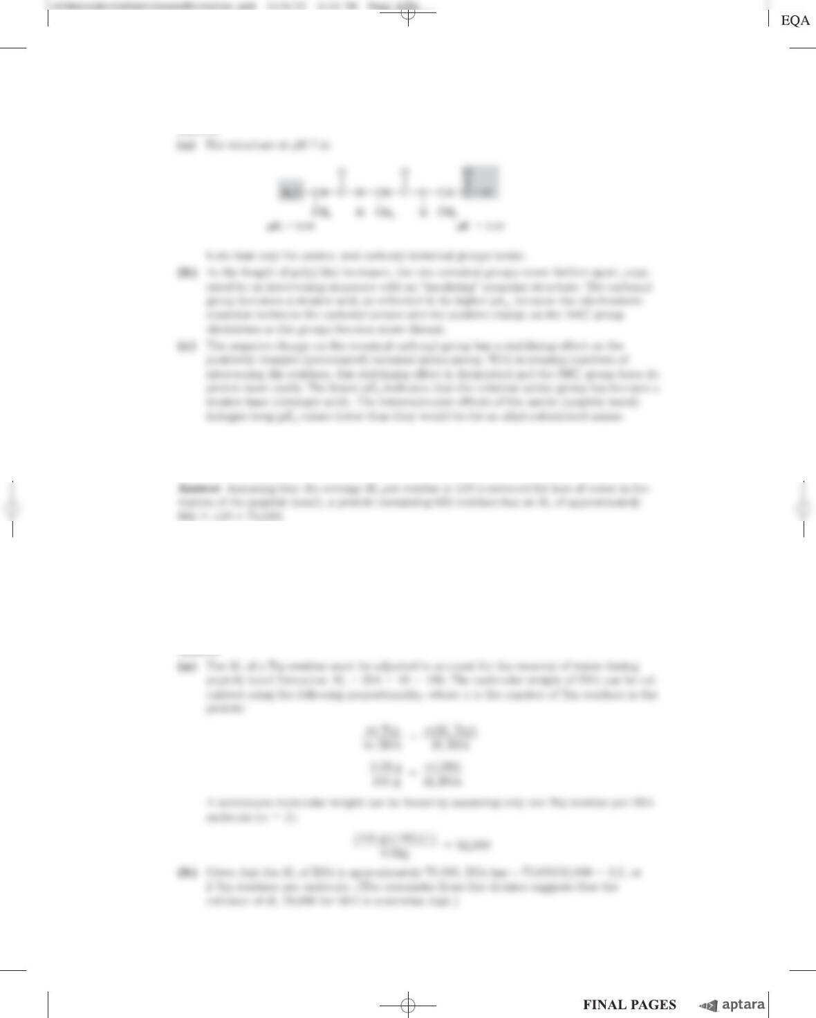
3. How Much Alanine Is Present as the Completely Uncharged Species? At a pH equal to the iso-
electric point of alanine, the net charge on alanine is zero. Two structures can be drawn that have a
net charge of zero, but the predominant form of alanine at its pI is zwitterionic.
⫹C
CH3
H3N
H
C
O
O⫺
Zwitterionic Uncharged
C
CH3
H2N
H
C
O
OH
(a) Why is alanine predominantly zwitterionic rather than completely uncharged at its pI?
(b) What fraction of alanine is in the completely uncharged form at its pI? Justify your assumptions.
c03AminoAcidsPeptidesandProteins.qxd 12/6/12 4:14 PM Page S-30
Chapter 3 Amino Acids, Peptides, and Proteins S-31
Answer
4. Ionization State of Histidine Each ionizable group of an amino acid can exist in one of two states,
charged or neutral. The electric charge on the functional group is determined by the relationship be-
tween its pK
a
and the pH of the solution. This relationship is described by the Henderson-Hasselbalch
equation.
(a) Histidine has three ionizable functional groups. Write the equilibrium equations for its three ion-
izations and assign the proper pK
a
for each ionization. Draw the structure of histidine in each
ionization state. What is the net charge on the histidine molecule in each ionization state?
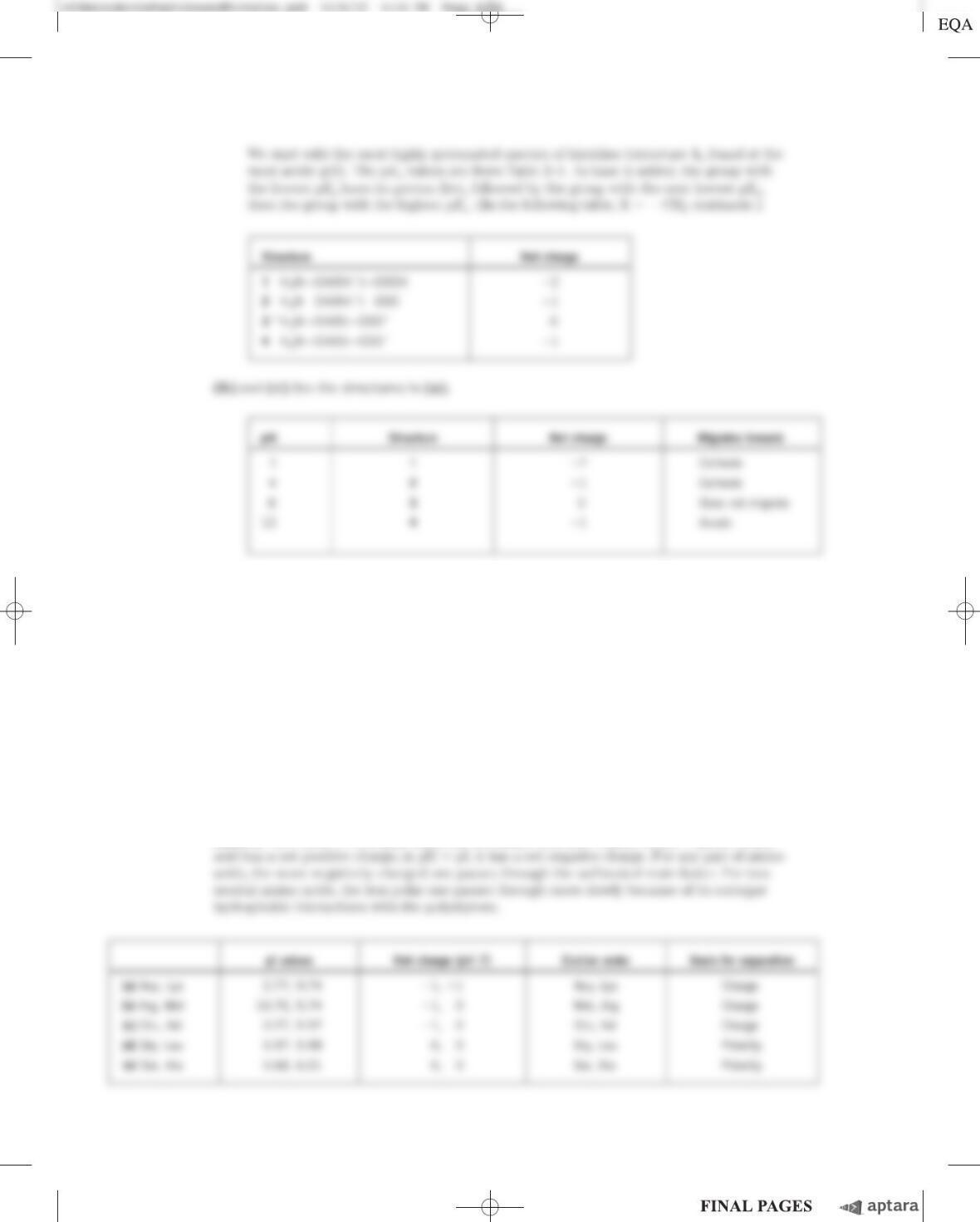
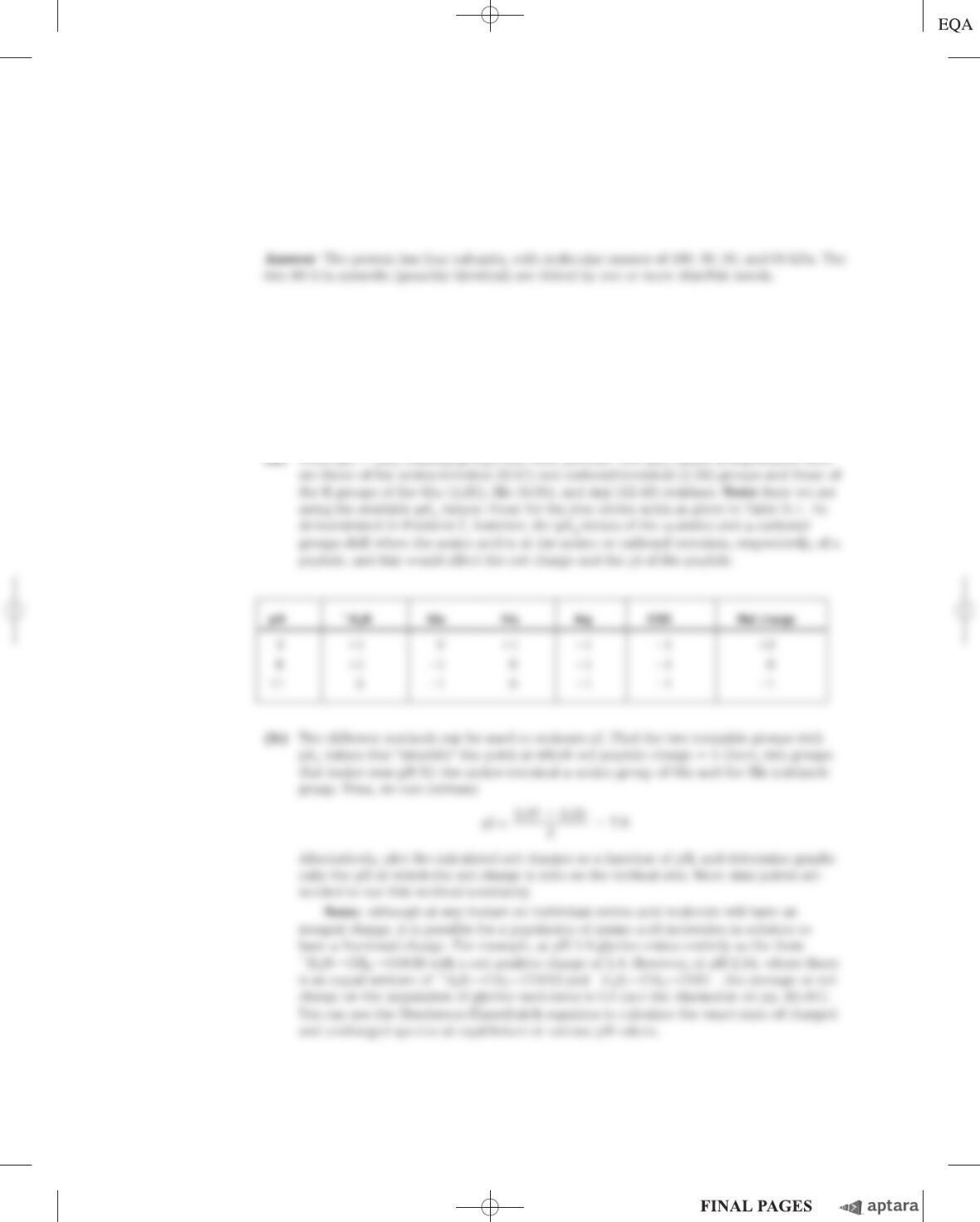
(b) Draw the structures of the predominant ionization state of histidine at pH 1, 4, 8, and 12. Note
that the ionization state can be approximated by treating each ionizable group independently.
(c) What is the net charge of histidine at pH 1, 4, 8, and 12? For each pH, will histidine migrate to-
ward the anode (⫹) or cathode (⫺) when placed in an electric field?
Answer
(a)
c03AminoAcidsPeptidesandProteins.qxd 12/6/12 4:14 PM Page S-31
S-32 Chapter 3 Amino Acids, Peptides, and Proteins
5. Separation of Amino Acids by Ion-Exchange Chromatography Mixtures of amino acids can be
analyzed by first separating the mixture into its components through ion-exchange chromatography.
Amino acids placed on a cation-exchange resin (see Fig. 3–17a) containing sulfonate (OSO
3
⫺
) groups
flow down the column at different rates because of two factors that influence their movement: (1)
ionic attraction between the sulfonate residues on the column and positively charged functional
groups on the amino acids, and (2) hydrophobic interactions between amino acid side chains and the
strongly hydrophobic backbone of the polystyrene resin. For each pair of amino acids listed, determine
which will be eluted first from an ion-exchange column by a pH 7.0 buffer.
(a) Asp and Lys
(b) Arg and Met
(c) Glu and Val
(d) Gly and Leu
(e) Ser and Ala
Answer See Table 3–1 for pK
a
values for the amino acid side chains. At pH ⬍pI, an amino
Chapter 3 Amino Acids, Peptides, and Proteins S-33
6. Naming the Stereoisomers of Isoleucine The structure of the amino acid isoleucine is
HC
H3N
H
C
COO⫺
H
CH2
CH3
CH3
(a) How many chiral centers does it have?
(b) How many optical isomers?
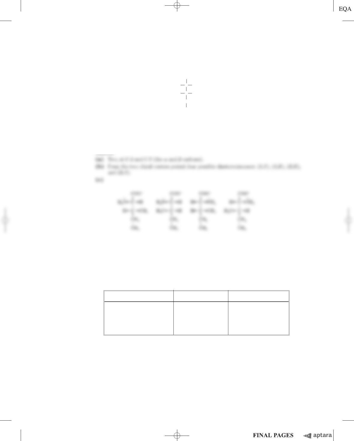
(c) Draw perspective formulas for all the optical isomers of isoleucine.
Answer
7. Comparing the pK
a
Values of Alanine and Polyalanine The titration curve of alanine shows the
ionization of two functional groups with pK
a
values of 2.34 and 9.69, corresponding to the ionization
of the carboxyl and the protonated amino groups, respectively. The titration of di-, tri-, and larger
oligopeptides of alanine also shows the ionization of only two functional groups, although the experi-
mental pK
a
values are different. The trend in pK
a
values is summarized in the table.
(a) Draw the structure of Ala–Ala–Ala. Identify the functional groups associated with pK
1
and pK
2
.
(b) Why does the value of pK
1
increase with each additional Ala residue in the Ala
oligopeptide?
(c) Why does the value of pK
2
decrease with each additional Ala residue in the Ala
oligopeptide?
Amino acid or peptide p
K
1
p
K
2
Ala 2.34 9.69
Ala–Ala 3.12 8.30
Ala–Ala–Ala 3.39 8.03
Ala–(Ala)
n
–Ala,
n
ⱖ4 3.42 7.94
c03AminoAcidsPeptidesandProteins.qxd 12/6/12 4:14 PM Page S-33
S-34 Chapter 3 Amino Acids, Peptides, and Proteins
Answer
8. The Size of Proteins What is the approximate molecular weight of a protein with 682 amino acid
residues in a single polypeptide chain?
9. The Number of Tryptophan Residues in Bovine Serum Albumin A quantitative amino acid
analysis reveals that bovine serum albumin (BSA) contains 0.58% tryptophan (M
r
204) by weight.
(a) Calculate the minimum molecular weight of BSA (i.e., assuming there is only one tryptophan
residue per protein molecule).
(b) Gel filtration of BSA gives a molecular weight estimate of 70,000. How many tryptophan residues
are present in a molecule of serum albumin?
Answer
Chapter 3 Amino Acids, Peptides, and Proteins S-35
10. Subunit Composition of a Protein A protein has a molecular mass of 400 kDa when measured by gel
filtration. When subjected to gel electrophoresis in the presence of sodium dodecyl sulfate (SDS), the pro-
tein gives three bands with molecular masses of 180, 160, and 60 kDa. When electrophoresis is carried out
in the presence of SDS and dithiothreitol, three bands are again formed, this time with molecular masses
of 160, 90, and 60 kDa. Determine the subunit composition of the protein.
11. Net Electric Charge of Peptides A peptide has the sequence
Glu–His–Trp–Ser–Gly–Leu–Arg–Pro–Gly
(a) What is the net charge of the molecule at pH 3, 8, and 11? (Use pK
a
values for side chains and
terminal amino and carboxyl groups as given in Table 3–1.)
(b) Estimate the pI for this peptide.
Answer
12. Isoelectric Point of Pepsin Pepsin is the name given to a mix of several digestive enzymes secreted
(as larger precursor proteins) by glands that line the stomach. These glands also secrete hydrochloric
acid, which dissolves the particulate matter in food, allowing pepsin to enzymatically cleave individual
c03AminoAcidsPeptidesandProteins.qxd 12/6/12 4:14 PM Page S-35
S-36 Chapter 3 Amino Acids, Peptides, and Proteins
protein molecules. The resulting mixture of food, HCl, and digestive enzymes is known as chyme and
has a pH near 1.5. What pI would you predict for the pepsin proteins? What functional groups must be
present to confer this pI on pepsin? Which amino acids in the proteins would contribute such groups?
Answer Pepsin proteins have a relatively low pI (near the pH of gastric juice) in order to
13. The Isoelectric Point of Histones Histones are proteins found in eukaryotic cell nuclei, tightly
bound to DNA, which has many phosphate groups. The pI of histones is very high, about 10.8. What
amino acid residues must be present in relatively large numbers in histones? In what way do these
residues contribute to the strong binding of histones to DNA?
14. Solubility of Polypeptides One method for separating polypeptides makes use of their different
solubilities. The solubility of large polypeptides in water depends upon the relative polarity of their R
groups, particularly on the number of ionized groups: the more ionized groups there are, the more soluble
the polypeptide. Which of each pair of the polypeptides that follow is more soluble at the indicated pH?
(a) (Gly)
20
or (Glu)
20
at pH 7.0
(b) (Lys–Ala)
3
or (Phe–Met)
3
at pH 7.0
(c) (Ala–Ser–Gly)
5
or (Asn–Ser–His)
5
at pH 6.0
(d) (Ala–Asp–Gly)
5
or (Asn–Ser–His)
5
at pH 3.0
Answer
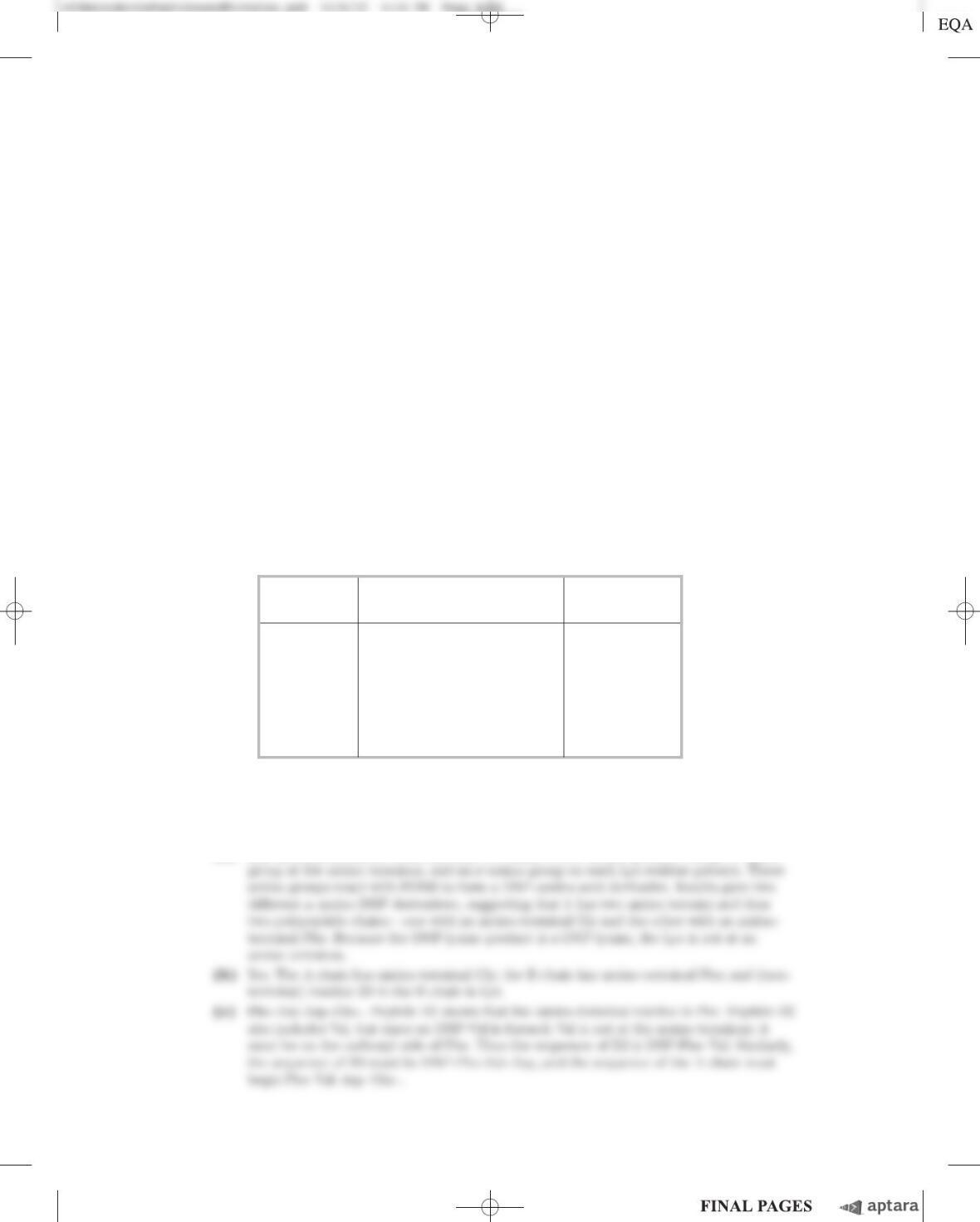
15. Purification of an Enzyme A biochemist discovers and purifies a new enzyme, generating the purifi-
cation table below.
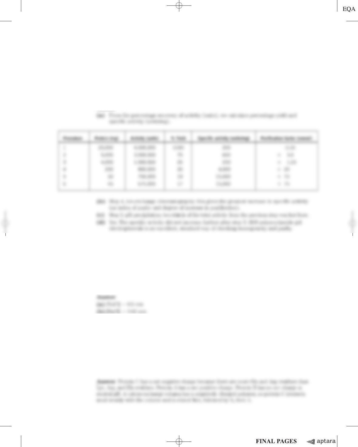
Procedure Total protein (mg) Activity (units)
1. Crude extract 20,000 4,000,000
2. Precipitation (salt) 5,000 3,000,000
3. Precipitation (pH) 4,000 1,000,000
4. Ion-exchange chromatography 200 800,000
5. Affinity chromatography 50 750,000
6. Size-exclusion chromatography 45 675,000
Chapter 3 Amino Acids, Peptides, and Proteins S-37
(a) From the information given in the table, calculate the specific activity of the enzyme after each
purification procedure.
(b) Which of the purification procedures used for this enzyme is most effective (i.e., gives the greatest
relative increase in purity)?
(c) Which of the purification procedures is least effective?
(d) Is there any indication based on the results shown in the table that the enzyme after step 6 is
now pure? What else could be done to estimate the purity of the enzyme preparation?
Answer
16. Dialysis A purified protein is in a Hepes (N-(2-hydroxyethyl)piperazine-N⬘-(2-ethanesulfonic acid)) buffer
at pH 7 with 500 m
M
NaCl. A sample (1 mL) of the protein solution is placed in a tube made of dialysis mem-
brane and dialyzed against 1 L of the same Hepes buffer with 0 m
M
NaCl. Small molecules and ions (such as
Na
⫹
, Cl
⫺
, and Hepes) can diffuse across the dialysis membrane, but the protein cannot.
(a) Once the dialysis has come to equilibrium, what is the concentration of NaCl in the protein sam-
ple? Assume no volume changes occur in the sample during the dialysis.
(b) If the original 1 mL sample were dialyzed twice, successively, against 100 mL of the same Hepes
buffer with 0 m
M
NaCl, what would be the final NaCl concentration in the sample?
17. Peptide Purification At pH 7.0, in what order would the following three peptides be eluted from a col-
umn filled with a cation-exchange polymer? Their amino acid compositions are:
Peptide A: Ala 10%, Glu 5%, Ser 5%, Leu 10%, Arg 10%, His 5%, Ile 10%, Phe 5%, Tyr 5%, Lys 10%,
Gly 10%, Pro 5%, and Trp 10%.
Peptide B: Ala 5%, Val 5%, Gly 10%, Asp 5%, Leu 5%, Arg 5%, Ile 5%, Phe 5%, Tyr 5%, Lys 5%,
Trp 5%, Ser 5%, Thr 5%, Glu 5%, Asn 5%, Pro 10%, Met 5%, and Cys 5%.
Peptide C: Ala 10%, Glu 10%, Gly 5%, Leu 5%, Asp 10%, Arg 5%, Met 5%, Cys 5%, Tyr 5%, Phe 5%,
His 5%, Val 5%, Pro 5%, Thr 5%, Ser 5%, Asn 5%, and Gln 5%.
c03AminoAcidsPeptidesandProteins.qxd 12/6/12 4:14 PM Page S-37
S-38 Chapter 3 Amino Acids, Peptides, and Proteins
18. Sequence Determination of the Brain Peptide Leucine Enkephalin A group of peptides that in-
fluence nerve transmission in certain parts of the brain has been isolated from normal brain tissue.
These peptides are known as opioids because they bind to specific receptors that also bind opiate
drugs, such as morphine and naloxone. Opioids thus mimic some of the properties of opiates. Some re-
searchers consider these peptides to be the brain’s own painkillers. Using the information below, deter-
mine the amino acid sequence of the opioid leucine enkephalin. Explain how your structure is consis-
tent with each piece of information.
(a) Complete hydrolysis by 6
M
HCl at 110 ⬚C followed by amino acid analysis indicated the presence
of Gly, Leu, Phe, and Tyr in a 2:1:1:1 molar ratio.
(b) Treatment of the peptide with 1-fluoro-2,4-dinitrobenzene followed by complete hydrolysis and
chromatography indicated the presence of the 2,4-dinitrophenyl derivative of tyrosine. No free
tyrosine could be found.
(c) Complete digestion of the peptide with chymotrypsin followed by chromatography yielded free
tyrosine and leucine, plus a tripeptide containing Phe and Gly in a 1:2 ratio.
Answer
19. Structure of a Peptide Antibiotic from Bacillus brevis Extracts from the bacterium Bacillus
brevis contain a peptide with antibiotic properties. This peptide forms complexes with metal ions and
seems to disrupt ion transport across the cell membranes of other bacterial species, killing them. The
structure of the peptide has been determined from the following observations.
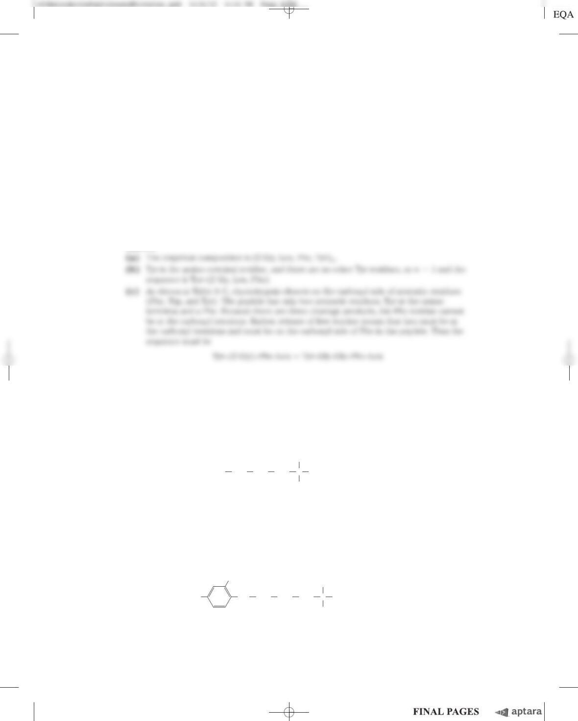
(a) Complete acid hydrolysis of the peptide followed by amino acid analysis yielded equimolar
amounts of Leu, Orn, Phe, Pro, and Val. Orn is ornithine, an amino acid not present in proteins
but present in some peptides. It has the structure
(b) The molecular weight of the peptide was estimated as about 1,200.
(c) The peptide failed to undergo hydrolysis when treated with the enzyme carboxypeptidase. This en-
zyme catalyzes the hydrolysis of the carboxyl-terminal residue of a polypeptide unless the
residue is Pro or, for some reason, does not contain a free carboxyl group.
(d) Treatment of the intact peptide with 1-fluoro-2,4-dinitrobenzene, followed by complete hydrolysis
and chromatography, yielded only free amino acids and the following derivative:
NO
2
CH
2
CH
2
⫹
NH
3
O
2
N COO
⫺
CH
2
NH C
H
CH
2
CH
2
CH
2
C COO
⫺
H
3
N
H
⫹
NH
3
⫹
(Hint: The 2,4-dinitrophenyl derivative involves the amino group of a side chain rather than the
a-amino group.)
Chapter 3 Amino Acids, Peptides, and Proteins S-39
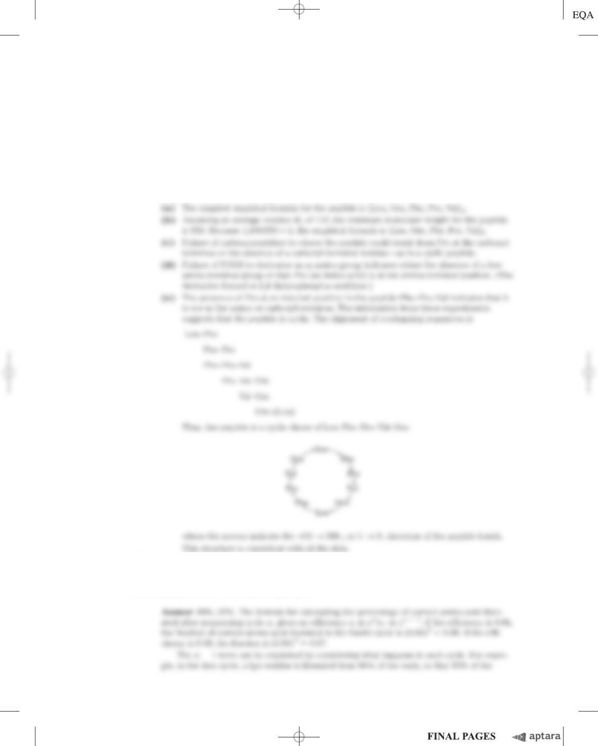
(e) Partial hydrolysis of the peptide followed by chromatographic separation and sequence analysis
yielded the following di- and tripeptides (the amino-terminal amino acid is always at the left):
Leu–Phe Phe–Pro Orn–Leu Val–Orn
Val–Orn–Leu Phe–Pro–Val Pro–Val–Orn
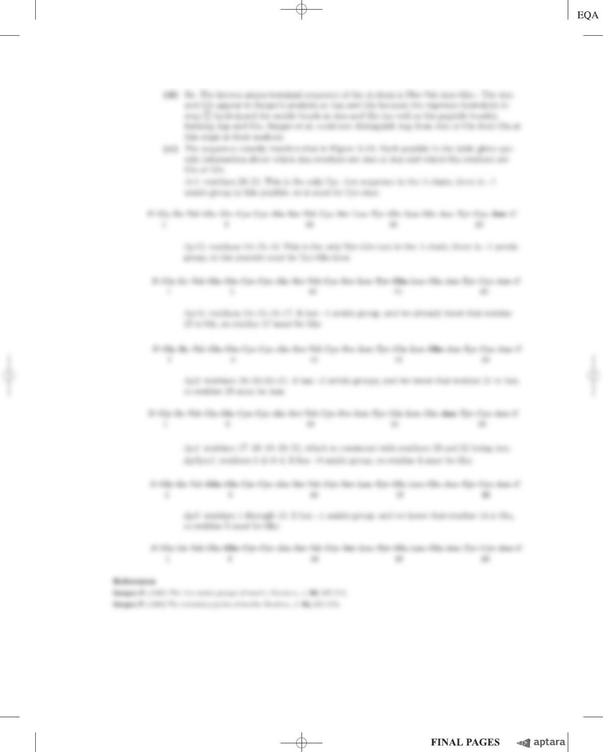
Given the above information, deduce the amino acid sequence of the peptide antibiotic. Show
your reasoning. When you have arrived at a structure, demonstrate that it is consistent with each
experimental observation.
Answer The information obtained from each experiment is as follows.
20. Efficiency in Peptide Sequencing A peptide with the primary structure Lys–Arg–Pro–Leu–
Ile–Asp–Gly–Ala is sequenced by the Edman procedure. If each Edman cycle is 96% efficient, what
percentage of the amino acids liberated in the fourth cycle will be leucine? Do the calculation a second
time, but assume a 99% efficiency for each cycle.
c03AminoAcidsPeptidesandProteins.qxd 12/6/12 4:14 PM Page S-39
S-40 Chapter 3 Amino Acids, Peptides, and Proteins
21. Sequence Comparisons Proteins called molecular chaperones (described in Chapter 4) assist in the
process of protein folding. One class of chaperone found in organisms from bacteria to mammals is
heat shock protein 90 (Hsp90). All Hsp90 chaperones contain a 10 amino acid “signature sequence,”
which allows for ready identification of these proteins in sequence databases. Two representations of
this signature sequence are shown below.
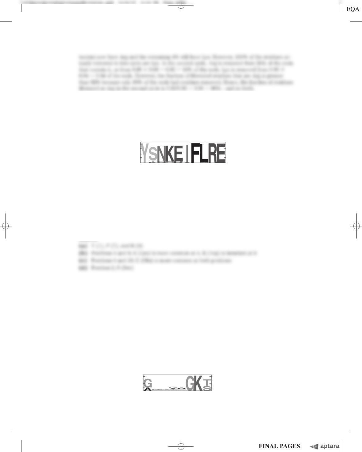
(a) In this sequence, which amino acid residues are invariant (conserved across all species)?
(b) At which position(s) are amino acids limited to those with positively charged side chains? For
each position, which amino acid is more commonly found?
(c) At which positions are substitutions restricted to amino acids with negatively charged side
chains? For each position, which amino acid predominates?
(d) There is one position that can be any amino acid, although one amino acid appears much more often
than any other. What position is this, and which amino acid appears most often?
Answer
22. Chromatographic Methods Three polypeptides, the sequences of which are represented below us-
ing the one-letter code for their amino acids, are present in a mixture:
1. ATKNRASCLVPKHGALMFWRHKQLVSDPILQKRQHILVCRNAAG
2. GPYFGDEPLDVHDEPEEG
3. PHLLSAWKGMEGVGKSQSFAALIVILA
Of the three, which one would migrate most slowly during chromatography through:
(a) an ion-exchange resin; beads coated with positively charged groups?
(b) an ion-exchange resin; beads coated with negatively charged groups?
(c) a size-exclusion (get-filtration) column designed to separate small peptides such as these?
(d) Which peptide contains the ATP-binding motif shown in the following sequence logo?
Y-x-[NQHD]-[KHR]-[DE]-[IVA]-F-[LM]-R-[ED].
45 6 7 8 91021
NC
3
4
3
1
0
2
Bits
456 7 8
C
21
N
3
4
0
2
Bits
Chapter 3 Amino Acids, Peptides, and Proteins S-41
Answer
Data Analysis Problem
23. Determining the Amino Acid Sequence of Insulin Figure 3–24 shows the amino acid sequence of
the hormone insulin. This structure was determined by Frederick Sanger and his coworkers. Most of this
work is described in a series of articles published in the Biochemical Journal from 1945 to 1955.
When Sanger and colleagues began their work in 1945, it was known that insulin was a small protein
consisting of two or four polypeptide chains linked by disulfide bonds. Sanger and his coworkers had
developed a few simple methods for studying protein sequences.
Treatment with FDNB. FDNB (1-fluoro-2,4-dinitrobenzene) reacted with free amino (but not
amido or guanidino) groups in proteins to produce dinitrophenyl (DNP) derivatives of amino acids:
Acid Hydrolysis. Boiling a protein with 10% HCl for several hours hydrolyzed all of its peptide and
amide bonds. Short treatments produced short polypeptides; the longer the treatment, the more com-
plete the breakdown of the protein into its amino acids.
Oxidation of Cysteines. Treatment of a protein with performic acid cleaved all the disulfide bonds
and converted all Cys residues to cysteic acid residues (Fig. 3–28).
Paper Chromatography. This more primitive version of thin-layer chromatography (see Fig. 10–25)
separated compounds based on their chemical properties, allowing identification of single amino acids
and, in some cases, dipeptides. Thin-layer chromatography also separates larger peptides.
As reported in his first paper (1945), Sanger reacted insulin with FDNB and hydrolyzed the result-
ing protein. He found many free amino acids, but only three DNP–amino acids: ␣-DNP-glycine (DNP
group attached to the ␣-amino group); ␣-DNP-phenylalanine; and -DNP-lysine (DNP attached to the
-amino group). Sanger interpreted these results as showing that insulin had two protein chains: one
with Gly at its amino terminus and one with Phe at its amino terminus. One of the two chains also
contained a Lys residue, not at the amino terminus. He named the chain beginning with a Gly residue
“A” and the chain beginning with Phe “B.”
(a) Explain how Sanger’s results support his conclusions.
(b) Are the results consistent with the known structure of insulin (Fig. 3–24)?
In a later paper (1949), Sanger described how he used these techniques to determine the first few
amino acids (amino-terminal end) of each insulin chain. To analyze the B chain, for example, he carried
out the following steps:
1. Oxidized insulin to separate the A and B chains.
2. Prepared a sample of pure B chain with paper chromatography.
3. Reacted the B chain with FDNB.
4. Gently acid-hydrolyzed the protein so that some small peptides would be produced.
5. Separated the DNP-peptides from the peptides that did not contain DNP groups.
6. Isolated four of the DNP-peptides, which were named B1 through B4.
7. Strongly hydrolyzed each DNP-peptide to give free amino acids.
8. Identified the amino acids in each peptide with paper chromatography.
⫹HF
⫹
O
2
N
NO
2
N
H
RNH
2
R
O
2
N
NO
2
F
Amine FDNB DNP-amine
c03AminoAcidsPeptidesandProteins.qxd 12/7/12 10:03 PM Page S-41
S-42 Chapter 3 Amino Acids, Peptides, and Proteins
The results were as follows:
B1: ␣-DNP-phenylalanine only
B2: ␣-DNP-phenylalanine; valine
B3: aspartic acid; ␣-DNP-phenylalanine; valine
B4: aspartic acid; glutamic acid; ␣-DNP-phenylalanine; valine
(c) Based on these data, what are the first four (amino-terminal) amino acids of the B chain?
Explain your reasoning.
(d) Does this result match the known sequence of insulin (Fig. 3–24)? Explain any discrepancies.
Sanger and colleagues used these and related methods to determine the entire sequence of the A
and B chains. Their sequence for the A chain was as follows (amino terminus on left):
Because acid hydrolysis had converted all Asn to Asp and all Gln to Glu, these residues had to be desig-
nated Asx and Glx, respectively (exact identity in the peptide unknown). Sanger solved this problem by
using protease enzymes that cleave peptide bonds, but not the amide bonds in Asn and Gln residues, to
prepare short peptides. He then determined the number of amide groups present in each peptide by
measuring the NH
4
⫹
released when the peptide was acid-hydrolyzed. Some of the results for the A chain
are shown below. The peptides may not have been completely pure, so the numbers were approximate—
but good enough for Sanger’s purposes.
(e) Based on these data, determine the amino acid sequence of the A chain. Explain how you
reached your answer. Compare it with Figure 3–24.
Answer
(a) Any linear polypeptide chain has only two kinds of free amino groups: a single ␣-amino
Gly–Ile–Val–Glx–Glx–Cys–Cys–Ala–Ser–Val–
1051
2015
Cys–Ser–Leu–Tyr–Glx–Leu–Glx–Asx–Tyr–Cys–Asx
Peptide name Peptide sequence Number of amide
groups in peptide
Ac1 Cys–Asx 0.7
Ap15 Tyr–Glx–Leu 0.98
Ap14 Tyr–Glx–Leu–Glx 1.06
Ap3 Asx–Tyr–Cys–Asx 2.10
Ap1 Glx–Asx–Tyr–Cys–Asx 1.94
Ap5pa1 Gly–Ile–Val–Glx 0.15
Ap5 Gly–Ile–Val–Glx–Glx–Cys–Cys–
Ala–Ser–Val–Cys–Ser–Leu 1.16
Chapter 3 Amino Acids, Peptides, and Proteins S-43
c03AminoAcidsPeptidesandProteins.qxd 12/6/12 4:14 PM Page S-43