S-120
Biological Membranes
and Transport
chapter
11
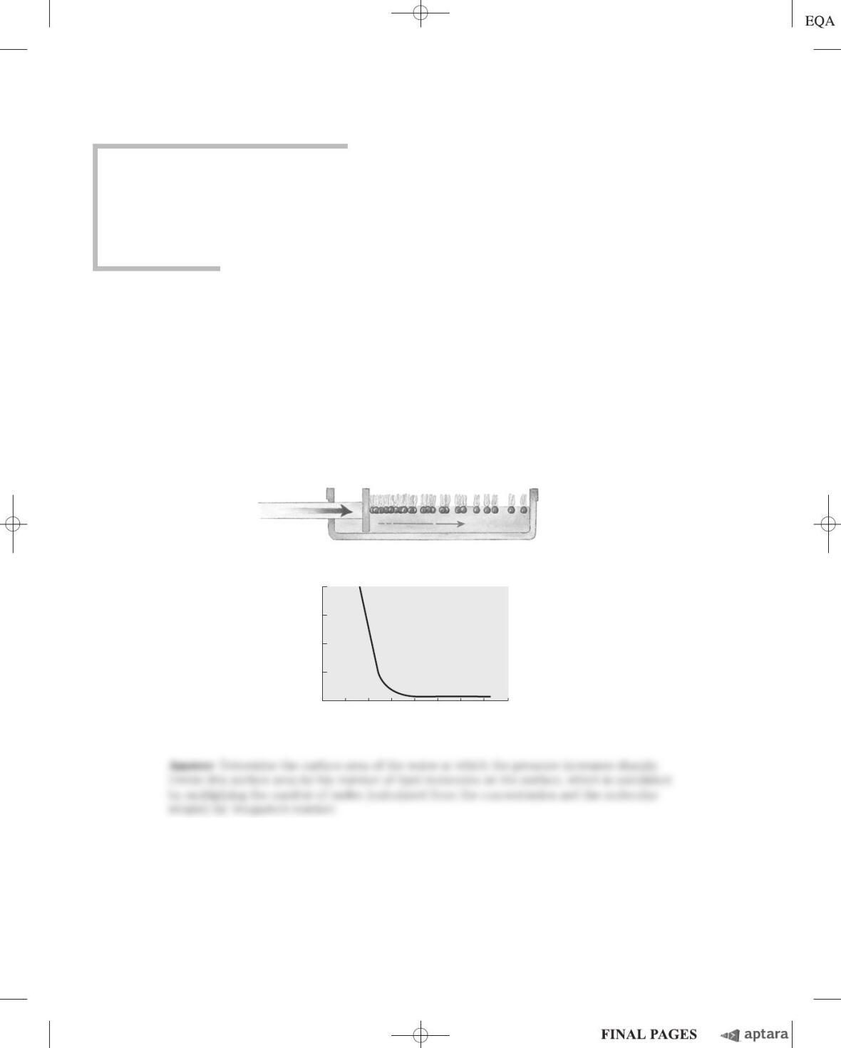
1. Determining the Cross-Sectional Area of a Lipid Molecule When phospholipids are layered gen-
tly onto the surface of water, they orient at the air-water interface with their head groups in the water
and their hydrophobic tails in the air. An experimental apparatus (a) has been devised that reduces the
surface area available to a layer of lipids. By measuring the force necessary to push the lipids together, it
is possible to determine when the molecules are packed tightly in a continuous monolayer; as that area is
approached, the force needed to further reduce the surface area increases sharply (b). How would you
use this apparatus to determine the average area occupied by a single lipid molecule in the monolayer?
Force applied here
to compress
monolayer
(a)
Force (dyne/cm)
Area (nm2/molecule)
(b)
40
30
1.41.00.60.2
20
10
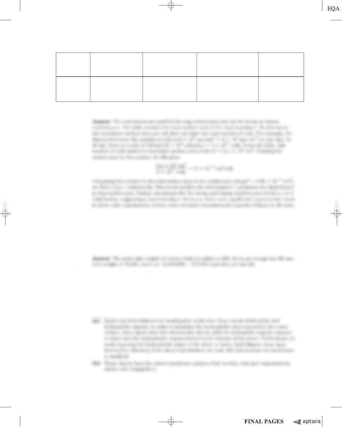
2. Evidence for a Lipid Bilayer In 1925, E. Gorter and F. Grendel used an apparatus like that de-
scribed in Problem 1 to determine the surface area of a lipid monolayer formed by lipids extracted
from erythrocytes of several animal species. They used a microscope to measure the dimensions of
individual cells, from which they calculated the average surface area of one erythrocyte. They obtained
the data shown in the following table. Were these investigators justified in concluding that “chromocytes
[erythrocytes] are covered by a layer of fatty substances that is two molecules thick” (i.e., a lipid bilayer)?
c11BiologicalMembranesandTransport.qxd 12/6/12 4:29 PM Page S-120
Chapter 11 Biological Membranes and Transport S-121
3. Number of Detergent Molecules per Micelle When a small amount of the detergent sodium dode-
cyl sulfate (SDS; Na
⫹
CH
3
(CH
2
)
11
OSO
3
⫺
) is dissolved in water, the detergent ions enter the solution as
monomeric species. As more detergent is added, a concentration is reached (the critical micelle con-
centration) at which the monomers associate to form micelles. The critical micelle concentration of
SDS is 8.2 m
M
. The micelles have an average particle weight (the sum of the molecular weights of the
constituent monomers) of 18,000. Calculate the number of detergent molecules in the average micelle.
4. Properties of Lipids and Lipid Bilayers Lipid bilayers formed between two aqueous phases have
this important property: they form two-dimensional sheets, the edges of which close upon each other
and undergo self-sealing to form liposomes.
(a) What properties of lipids are responsible for this property of bilayers? Explain.
(b) What are the consequences of this property for the structure of biological membranes?
Answer
Total surface
Volume of Number area of lipid Total surface
packed of cells monolayer area of one
Animal cells (mL) (per mm
3
) from cells (m
2
) cell (mm
2
)
Dog 40 8,000,000 62 98
Sheep 10 9,900,000 6.0 29.8
Human 1 4,740,000 0.92 99.4
Source: Data from Gorter, E. & Grendel, F. (1925) On bimolecular layers of lipoids on the chromocytes of the blood.
J. Exp. Med.
41, 439–443.
c11BiologicalMembranesandTransport.qxd 12/6/12 4:29 PM Page S-121
S-122 Chapter 11 Biological Membranes and Transport
5. Length of a Fatty Acid Molecule The carbon–carbon bond distance for single-bonded carbons
such as those in a saturated fatty acyl chain is about 1.5 Å. Estimate the length of a single molecule of
palmitate in its fully extended form. If two molecules of palmitate were placed end to end, how would
their total length compare with the thickness of the lipid bilayer in a biological membrane?
6. Temperature Dependence of Lateral Diffusion The experiment described in Figure 11–18 was
performed at 37 ⬚C. If the experiment were carried out at 10 ⬚C, what effect would you expect on the
rate of diffusion? Why?
7. Synthesis of Gastric Juice: Energetics Gastric juice (pH 1.5) is produced by pumping HCl from
blood plasma (pH 7.4) into the stomach. Calculate the amount of free energy required to concentrate
the H
⫹
in 1 L of gastric juice at 37 ⬚C. Under cellular conditions, how many moles of ATP must be
hydrolyzed to provide this amount of free energy? The free-energy change for ATP hydrolysis under
cellular conditions is about ⫺58 kJ/mol (as explained in Chapter 13). Ignore the effects of the trans-
membrane electrical potential.
8. Energetics of the Na
ⴙ
K
ⴙ
ATPase For a typical vertebrate cell with a membrane potential of
⫺0.070 V (inside negative), what is the free-energy change for transporting 1 mol of Na
⫹
out of the
cell and into the blood at 37 ⬚C? Assume the concentration of Na
⫹
inside the cell is 12 m
M
, and that
in blood plasma is 145 m
M
.
Answer
9. Action of Ouabain on Kidney Tissue Ouabain specifically inhibits the Na
⫹
K
⫹
ATPase activity of
animal tissues but is not known to inhibit any other enzyme. When ouabain is added to thin slices
of living kidney tissue, it inhibits oxygen consumption by 66%. Why? What does this observation tell
us about the use of respiratory energy by kidney tissue?
10. Energetics of Symport Suppose that you determined experimentally that a cellular transport system
for glucose, driven by symport of Na
⫹
, could accumulate glucose to concentrations 25 times greater
than in the external medium, while the external [Na
⫹
] was only 10 times greater than the intracellular
[Na
⫹
]. Would this violate the laws of thermodynamics? If not, how could you explain this observation?
11. Location of a Membrane Protein The following observations are made on an unknown membrane
protein, X. It can be extracted from disrupted erythrocyte membranes into a concentrated salt solution,
and it can be cleaved into fragments by proteolytic enzymes. Treatment of erythrocytes with proteolytic
enzymes followed by disruption and extraction of membrane components yields intact X. However, treat-
ment of erythrocyte “ghosts” (which consist of just plasma membranes, produced by disrupting the cells
and washing out the hemoglobin) with proteolytic enzymes followed by disruption and extraction yields
extensively fragmented X. What do these observations indicate about the location of X in the plasma
membrane? Do the properties of X resemble those of an integral or peripheral membrane protein?
12. Membrane Self-sealing Cellular membranes are self-sealing—if they are punctured or disrupted
mechanically, they quickly and automatically reseal. What properties of membranes are responsible for
this important feature?
13. Lipid Melting Temperatures Membrane lipids in tissue samples obtained from different parts of the
leg of a reindeer have different fatty acid compositions. Membrane lipids from tissue near the hooves
contain a larger proportion of unsaturated fatty acids than those from tissue in the upper leg. What is
the significance of this observation?
14. Flip-Flop Diffusion The inner leaflet (monolayer) of the human erythrocyte membrane consists
predominantly of phosphatidylethanolamine and phosphatidylserine. The outer leaflet consists predomi-
nantly of phosphatidylcholine and sphingomyelin. Although the phospholipid components of the
membrane can diffuse in the fluid bilayer, this sidedness is preserved at all times. How?
Chapter 11 Biological Membranes and Transport S-123
c11BiologicalMembranesandTransport.qxd 12/6/12 4:29 PM Page S-123

S-124 Chapter 11 Biological Membranes and Transport
15. Membrane Permeability At pH 7, tryptophan crosses a lipid bilayer at about one-thousandth the
rate of indole, a closely related compound:
Suggest an explanation for this observation.
16. Water Flow through an Aquaporin A human erythrocyte has about 2 ⫻10
5
AQP-1 monomers.
If water molecules flow through the plasma membrane at a rate of 5 ⫻10
8
per AQP-1 tetramer per
second, and the volume of an erythrocyte is 5 ⫻10
⫺11
mL, how rapidly could an erythrocyte halve
its volume as it encountered the high osmolarity (1
M
) in the interstitial fluid of the renal medulla?
Assume that the erythrocyte consists entirely of water.
17. Labeling the Lactose Transporter A bacterial lactose transporter, which is highly specific for lactose,
contains a Cys residue that is essential to its transport activity. Covalent reaction of N-ethylmaleimide
(NEM) with this Cys residue irreversibly inactivates the transporter. A high concentration of lactose
in the medium prevents inactivation by NEM, presumably by sterically protecting the Cys residue,
which is in or near the lactose-binding site. You know nothing else about the transporter protein.
Suggest an experiment that might allow you to determine the M
r
of the Cys-containing transporter
polypeptide.
18. Predicting Membrane Protein Topology from Sequence You have cloned the gene for a human ery-
throcyte protein, which you suspect is a membrane protein. From the nucleotide sequence of the gene,
you know the amino acid sequence. From this sequence alone, how would you evaluate the possibility that
N
H
Uptake in presence of Na
ⴙ
Uptake in absence of Na
ⴙ
Substrate
V
max
K
t
(m
M
)
V
max
K
t
(m
M
)
L
-Leucine 420 0.24 23 0.2
D
-Leucine 310 4.7 5 4.7
L
-Valine 225 0.31 19 0.31
the protein is an integral protein? Suppose the protein proves to be an integral protein, either type I or
type II. Suggest biochemical or chemical experiments that might allow you to determine which type it is.
19. Intestinal Uptake of Leucine You are studying the uptake of
L
-leucine by epithelial cells of the mouse
intestine. Measurements of the rate of uptake of
L
-leucine and several of its analogs, with and without Na
⫹
in the assay buffer, yield the results given in the table. What can you conclude about the properties and
mechanism of the leucine transporter? Would you expect
L
-leucine uptake to be inhibited by ouabain?
20. Effect of an Ionophore on Active Transport Consider the leucine transporter described in
Problem 19. Would V
max
and/or K
t
change if you added a Na
⫹
ionophore to the assay solution
containing Na
⫹
? Explain.
21. Surface Density of a Membrane Protein E. coli can be induced to make about 10,000 copies of
the lactose transporter (M
r
31,000) per cell. Assume that E. coli is a cylinder 1 mm in diameter and
2 mm long. What fraction of the plasma membrane surface is occupied by the lactose transporter mole-
cules? Explain how you arrived at this conclusion.
Chapter 11 Biological Membranes and Transport S-125
c11BiologicalMembranesandTransport.qxd 12/6/12 4:29 PM Page S-125
S-126 Chapter 11 Biological Membranes and Transport
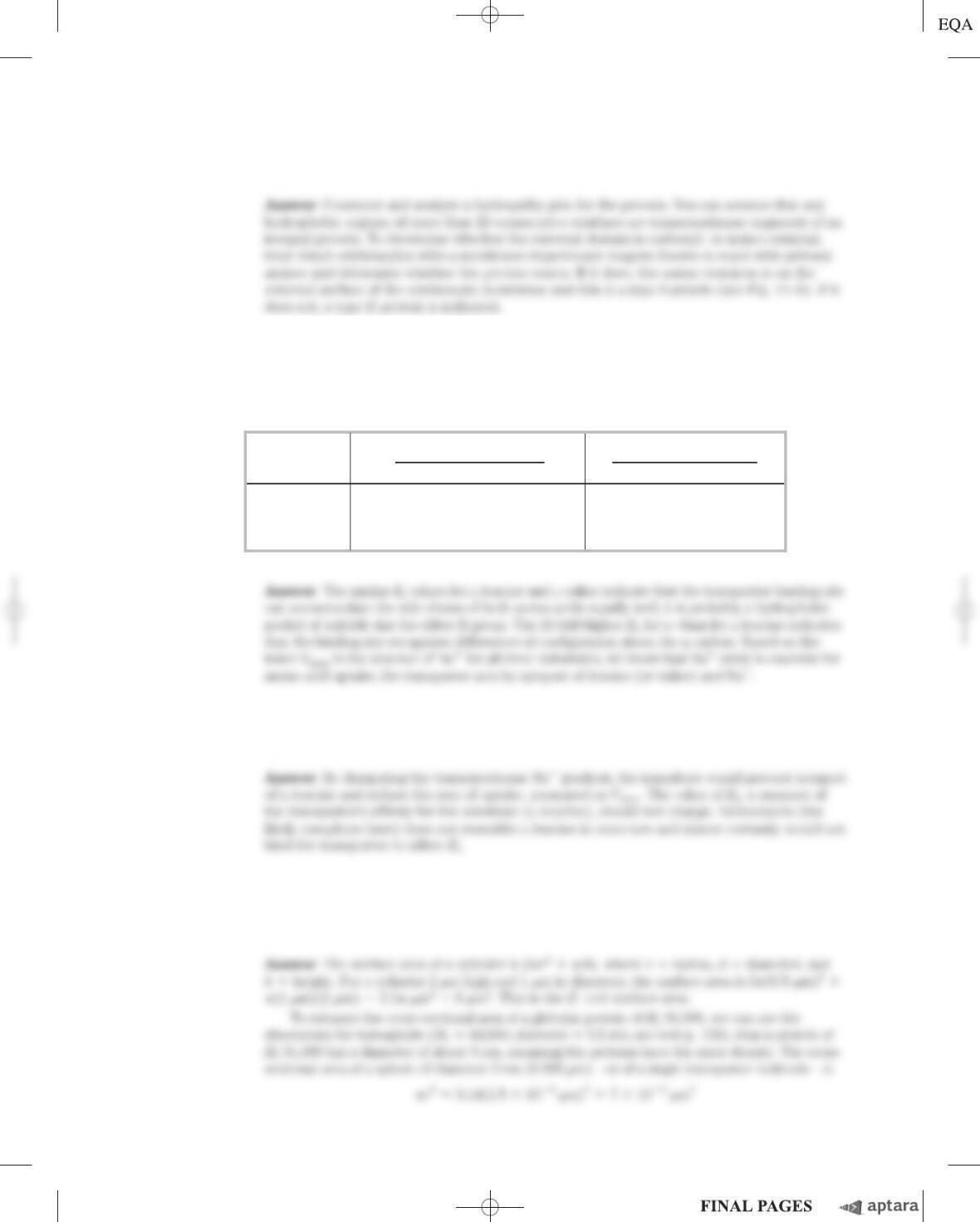
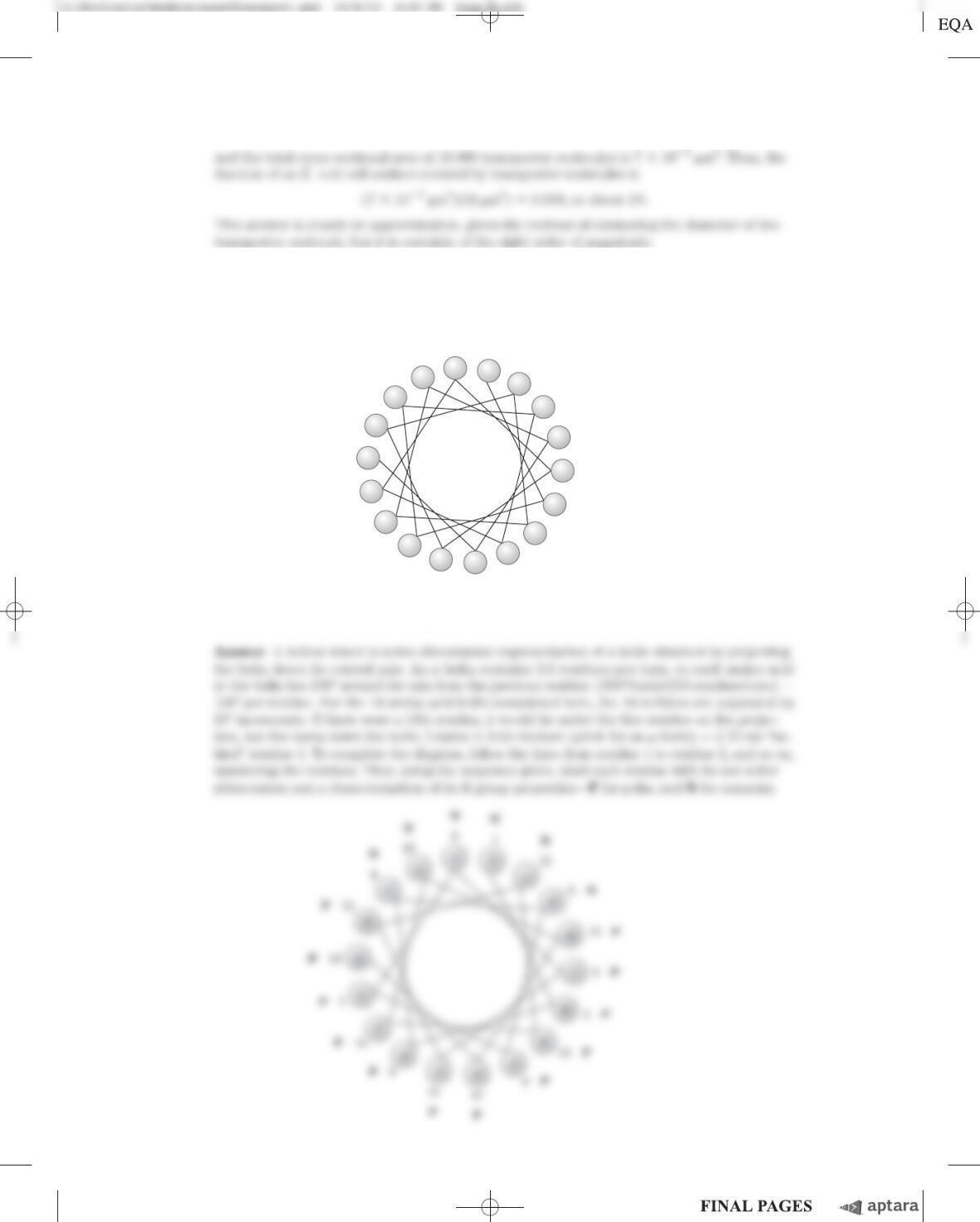
22. Use of the Helical Wheel Diagram A helical wheel is a two-dimensional representation of a helix, a
view along its central axis (see Fig. 11–30b; see also Fig. 4–4d). Use the helical wheel diagram below
to determine the distribution of amino acid residues in a helical segment with the sequence –Val–
Asp–Arg–Val–Phe–Ser–Asn–Val–Cys–Thr–His–Leu–Lys–Thr–Leu–Gln–Asp–Lys–
What can you say about the surface properties of this helix? How would you expect the helix to be
oriented in the tertiary structure of an integral membrane protein?
1

23. Molecular Species in the E. coli Membrane The plasma membrane of E. coli is about 75% pro-
tein and 25% phospholipid by weight. How many molecules of membrane lipid are present for each
molecule of membrane protein? Assume an average protein M
r
of 50,000 and an average phospho-
lipid M
r
of 750. What more would you need to know to estimate the fraction of the membrane sur-
face that is covered by lipids?
Using the Web
24. Membrane Protein Topology The receptor for the hormone epinephrine in animal cells is an inte-
gral membrane protein (M
r
64,000) that is believed to have seven membrane-spanning regions.
(a) Show that a protein of this size is capable of spanning the membrane seven times.
(b) Given the amino acid sequence of this protein, how would you predict which regions of the protein
form the membrane-spanning helices?
(c) Go to the Protein Data Bank (www.pdb.org). Use the PDB identifier 1DEP to retrieve the data
page for a portion of the b-adrenergic receptor (one type of epinephrine receptor) from a turkey.
Using Jmol to explore the structure, predict where this portion of the receptor is located: within
the membrane or at the membrane surface. Explain.
(d) Retrieve the data for a portion of another receptor, the acetylcholine receptor of neurons and
myocytes, using the PDB identifier 1A11. As in (c), predict where this portion of the receptor is
located and explain your answer.
If you have not used the PDB, see Box 4–4 (p. 132) for more information.
Answer
Chapter 11 Biological Membranes and Transport S-127
c11BiologicalMembranesandTransport.qxd 12/6/12 4:29 PM Page S-127
S-128 Chapter 11 Biological Membranes and Transport
(c) This portion of the epinephrine receptor is an intracellular loop that connects adjacent
membrane-spanning regions of the protein. You can predict that this ␣helix is not lo-
Data Analysis Problem
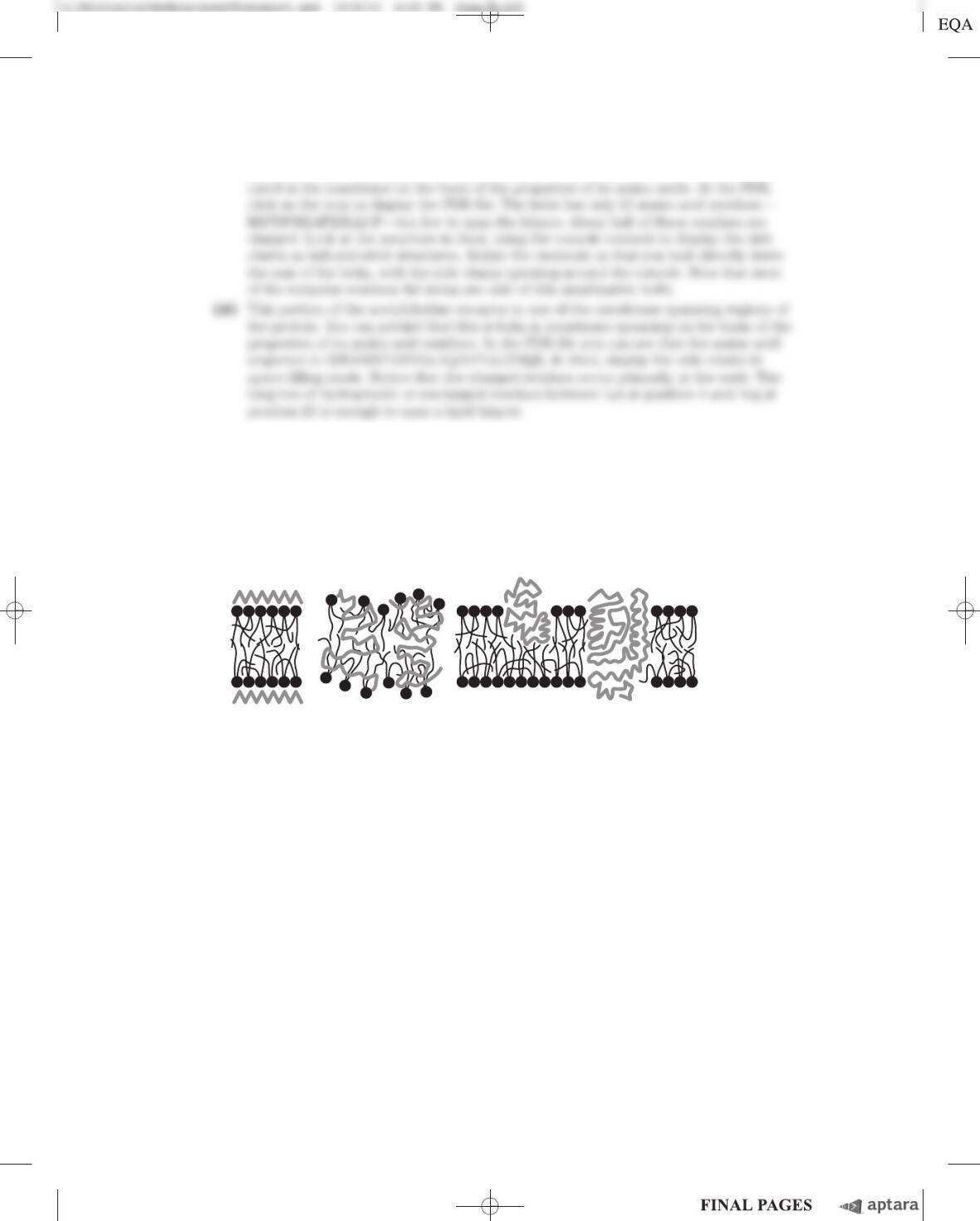
25. The Fluid Mosaic Model of Biological Membrane Structure Figure 11–3 shows the currently
accepted fluid mosaic model of biological membrane structure. This model was presented in detail in
a review article by S. J. Singer in 1971. In the article, Singer presented the three models of membrane
structure that had been proposed by that time:
A
++++
+
+
+
−
+
++
−
−
−−
−
−
−
−−
−
−−
BC
A. The Davson-Danielli-Robertson Model. This was the most widely accepted model in 1971, when
Singer’s review was published. In this model, the phospholipids are arranged as a bilayer. Proteins are
found on both surfaces of the bilayer, attached to it by ionic interactions between the charged head
groups of the phospholipids and charged groups in the proteins. Crucially, there is no protein in the
interior of the bilayer.
B. The Benson Lipoprotein Subunit Model. Here, the proteins are globular and the membrane is a
protein-lipid mixture. The hydrophobic tails of the lipids are embedded in the hydrophobic parts of
the proteins. The lipid head groups are exposed to the solvent. There is no lipid bilayer.
C. The Lipid-Globular Protein Mosaic Model. This is the model shown in Figure 11–3. The lipids
form a bilayer and proteins are embedded in it, some extending through the bilayer and others not.
Proteins are anchored in the bilayer by hydrophobic interactions between the hydrophobic tails of the
lipids and hydrophobic portions of the protein.
For the data given below, consider how each piece of information aligns with each of the three
models of membrane structure. Which model(s) are supported, which are not supported, and what
reservations do you have about the data or their interpretation? Explain your reasoning.
Chapter 11 Biological Membranes and Transport S-129
(a) When cells were fixed, stained with osmium tetroxide, and examined in the electron mi-
croscope, the membranes showed a “railroad track” appearance, with two dark-staining
lines separated by a light space.
(b) The thickness of membranes in cells fixed and stained in the same way was found to be 5 to
9 nm. The thickness of a “naked” phospholipid bilayer, without proteins, was 4 to 4.5 nm.
The thickness of a single monolayer of proteins was about 1 nm.
(c) Singer wrote in his article: “The average amino acid composition of membrane proteins is
not distinguishable from that of soluble proteins. In particular, a substantial fraction of the
residues is hydrophobic” (p. 165).
(d) As described in Problems 1 and 2 of this chapter, researchers had extracted membranes
from cells, extracted the lipids, and compared the area of the lipid monolayer with the
area of the original cell membrane. The interpretation of the results was complicated by
the issue illustrated in the graph of Problem 1: the area of the monolayer depended on
how hard it was pushed. With very light pressures, the ratio of monolayer area to cell
membrane area was about 2.0. At higher pressures—thought to be more like those found
in cells—the ratio was substantially lower.
(e) Circular dichroism spectroscopy uses changes in polarization of UV light to make infer-
ences about protein secondary structure (see Fig. 4–10). On average, this technique showed
that membrane proteins have a large amount of ␣helix and little or no sheet. This finding
was consistent with most membrane proteins having a globular structure.
(f) Phospholipase C is an enzyme that removes the polar head group (including the phos-
phate) from phospholipids. In several studies, treatment of intact membranes with phos-
pholipase C removed about 70% of the head groups without disrupting the “railroad track”
structure of the membrane.
(g) Singer described a study in which “a glycoprotein of molecular weight about 31,000 in
human red blood cell membranes is cleaved by tryptic treatment of the membranes into
soluble glycopeptides of about 10,000 molecular weight, while the remaining portions are
quite hydrophobic” (p. 199). Trypsin treatment did not cause gross changes in the mem-
branes, which remained intact.
Singer’s review also included many more studies in this area. In the end, though, the data
available in 1971 did not conclusively prove Model C was correct. As more data have accumu-
lated, this model of membrane structure has been accepted by the scientific community.
Answer
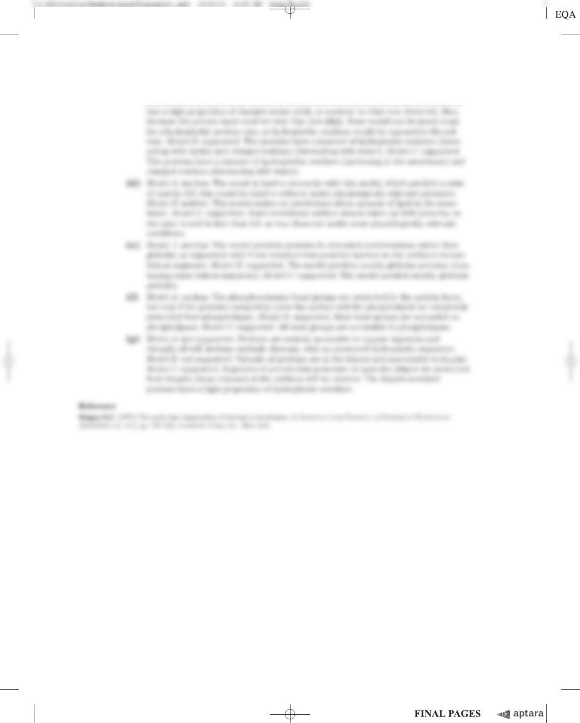
(a) Model A: supported. The two dark lines are either the protein layers or the phospholipid
heads, and the clear space is either the bilayer or the hydrophobic core, respectively.
c11BiologicalMembranesandTransport.qxd 12/6/12 4:29 PM Page S-129
S-130 Chapter 11 Biological Membranes and Transport
(c) Model A: unclear. The result is hard to reconcile with this model. If the proteins are
bound to the membrane by ionic interactions, the model predicts that the proteins con-