CHAPTER
19
The Cardiovascular System:
Blood Vessels
Objectives
Part 1: Blood Vessel Structure and Function
Structure of Blood Vessel Walls
Arterial System
3. Compare and contrast the structure and function of the three types of arteries.
Capillaries
4. Describe the structure and function of a capillary bed.
Part 2: Physiology of Circulation
Introduction to Blood Flow, Blood Pressure, and Resistance
6. Define blood flow, blood pressure, and resistance, and explain the relationships between
these factors.
Maintaining Blood Pressure
8. List and explain the factors that influence blood pressure, and describe how blood
pressure is regulated.
9. Define hypertension. Describe its manifestations and consequences.
Part 3: Circulatory Pathways: Blood Vessels of the Body
The Two Main Circulations of the Body
13. Trace the pathway of blood through the pulmonary circuit, and state the importance of
this special circulation.
14. Describe the general functions of the systemic circuit.
Suggested Lecture Outline
Part 1: Blood Vessel Structure and Function (pp. 693–701; Figs. 19.1–
19.5; Table 19.1)
I. Structure of Blood Vessel Walls (p. 693; Figs. 19.1–19.2; Table 19.1)
A. The walls of all blood vessels except the smallest consist of three layers: the tunica
intima, tunica media, and tunica externa (p. 693; Fig. 19.1).
II. Arterial System (pp. 693–696; Fig. 19.2; Tables 19.1–19.2)
A. Elastic, or conducting, arteries contain large amounts of elastin, which enables these
vessels to withstand and smooth out pressure fluctuations due to heart action (pp. 693–
695; Fig. 19.2; Table 19.1).
III. Capillaries (pp. 696–698; Figs. 19.3–19.4; Table 19.1)
A. Capillaries are the smallest vessels and allow for exchange of substances between the
blood and interstitial fluid (pp. 696–698; Fig. 19.3; Table 19.1).
B. Capillary beds are microcirculatory networks consisting of a vascular shunt and true
capillaries, which function as the exchange vessels (p. 698; Fig. 19.4).
IV. Venous System (pp. 698–699; Fig. 19.5; Table 19.1)
A. Venules are formed where capillaries converge and allow fluid and white blood cells to
move easily between the blood and tissues (p. 698; Table 19.1).
Part 2: Physiology of Circulation (pp. 701–720; Figs. 19.6–19.18;
Table 19.2)
VI. Introduction to Blood Flow, Blood Pressure, and Resistance (pp. 701–702)
A. Blood flow is the volume of blood flowing through a vessel, organ, or the entire circula-
tion in a given period and may be expressed as ml/min (p. 701).
D. Relationship Between Flow, Pressure, and Resistance (p. 702)
1. If blood pressure increases, blood flow increases; if peripheral resistance increases,
blood flow decreases.
2. Peripheral resistance is the most important factor influencing local blood flow,
because vasoconstriction or vasodilation can dramatically alter local blood flow, while
systemic blood pressure remains unchanged.
VII. Systemic Blood Pressure (pp. 702–704; Figs. 19.6–19.7)
A. The pumping action of the heart generates blood flow; pressure results when blood flow
is opposed by resistance (p. 702).
1. When the left ventricle contracts, blood is forced into the aorta, producing a peak in
pressure called systolic pressure (120 mm Hg).
2. Diastolic pressure occurs when blood is prevented from flowing back into the ventri-
cles by the closed semilunar valve and the aorta recoils (70–80 mm Hg).
D. Capillary blood pressure is low, ranging from 15–40 mm Hg, which protects the capillar-
ies from rupture but is still adequate to ensure exchange between blood and tissues
(p. 703; Fig. 19.6).
VIII. Maintaining Blood Pressure (pp. 704–711; Figs. 19.8–19.12; Table 19.2)
A. Blood pressure varies directly with changes in blood volume and cardiac output, which
are determined primarily by venous return and neural and hormonal controls (p. 704;
Fig. 19.8).
B. Short-term neural controls of peripheral resistance alter blood distribution to meet
specific tissue demands and maintain adequate MAP by altering blood vessel diameter
(pp. 704–707; Fig. 19.9).
1. Clusters of neurons in the medulla oblongata, the cardioacceleratory, cardioinhibitory,
and vasomotor centers, form the cardiovascular center that regulates blood pressure by
altering cardiac output and blood vessel diameter.
centers.
C. Chemical controls influence blood pressure by acting on vascular smooth muscle or the
vasomotor center (p. 707; Table 19.2).
1. Norepinephrine and epinephrine promote an increase in cardiac output and generalized
vasoconstriction.
2. Atrial natriuretic peptide acts as a vasodilator and an antagonist to aldosterone,
resulting in a drop in blood volume.
3. Antidiuretic hormone promotes vasoconstriction and water conservation by the
kidneys, resulting in an increase in blood volume.
vasodilation.
D. Long-Term Regulation: Renal Mechanisms (pp. 708–710; Figs. 19.10–19.11)
1. The direct renal mechanism counteracts an increase in blood pressure by altering
blood volume, which increases the rate of kidney filtration.
2. The indirect renal mechanism is the renin-angiotensin-aldosterone mechanism, which
counteracts a decline in arterial blood pressure by causing systemic vasoconstriction.
E. Monitoring circulatory efficiency is accomplished by measuring pulse and blood
pressure; these values together with respiratory rate and body temperature are called vital
signs (p. 710; Fig. 19.12).
1. A pulse is generated by the alternating stretch and recoil of elastic arteries during each
cardiac cycle.
IX. Blood Flow Through Body Tissues: Tissue Perfusion (pp. 711–720;
Figs. 19.13–19.18)
A. Tissue perfusion is involved in delivery of oxygen and nutrients to, and removal of
wastes from, tissue cells; gas exchange in the lungs; absorption of nutrients from the
digestive tract; and urine formation in the kidneys (pp. 711–712; Fig. 19.13).
C. Autoregulation: Local Regulation of Blood Flow (pp. 712–713; Fig. 19.15)
1. Autoregulation is the automatic adjustment of blood flow to each tissue in proportion to
its needs and is controlled intrinsically by modifying the diameter of local arterioles.
2. Metabolic controls of autoregulation are most strongly stimulated by a shortage of
oxygen at the tissues.
D. Blood Flow in Special Areas (pp. 713–716)
1. Blood flow to skeletal muscles varies with level of activity and fiber type.
2. Muscular autoregulation occurs almost entirely in response to decreased oxygen
concentrations.
3. Cerebral blood flow is tightly regulated to meet neuronal needs, because neurons
cannot tolerate periods of ischemia, and increased blood carbon dioxide causes
marked vasodilation.
E. Blood Flow Through Capillaries and Capillary Dynamics (pp. 716–717; Figs. 19.16–19.17)
1. Vasomotion, the slow, intermittent flow of blood through the capillaries, reflects the
action of the precapillary sphincters in response to local autoregulatory controls.
5. Fluids will leave the capillaries if net HP exceeds net OP, but fluids will enter the
capillaries if net OP exceeds net HP.
F. Circulatory shock is any condition in which blood volume is inadequate and cannot
circulate normally, resulting in blood flow that cannot meet the needs of a tissue
(p. 717; Fig. 19.18).
1. Hypovolemic shock results from a large-scale loss of blood, and may be characterized
by an elevated heart rate and intense vasoconstriction.
2. Vascular shock is characterized by a normal blood volume accompanied by extreme
Part 3: Circulatory Pathways: Blood Vessels of the Body (pp. 721–745;
Figs. 19.19–19.30; Tables 19.3–19.13)
X. The Two Main Circulations of the Body (p. 721; Figs. 19.19–19.20; Table 19.3)
A. Two distinct pathways travel to and from the heart: pulmonary circulation runs from the
heart to the lungs and back to the heart; systemic circulation runs to all parts of the body
before returning to the heart.
XI. Systemic Arteries and Veins: Differences in Pathways and Courses (p. 721;
Fig. 19.29; Table 19.12)
A. There are some important differences between arteries and veins (p. 721; Table 19.12).
1. There is one terminal systemic artery, the aorta, but two terminal systemic veins: the
superior and inferior vena cava.
XII. Principal Vessels of the Systemic Circulation (pp. 721–745; Figs. 19.19–19.30;
Tables 19.3–19.13)
In the following sections (XII. A.–K.), vessels have been arranged in tables in order to
facilitate presentation by the instructor. More commonly taught vessels are presented, as
well as the branching pattern of these vessels, indicated as “levels.” Also, general areas
served by the listed vessels are presented.
A. The pulmonary circulation functions only to bring blood into close contact with the alveoli of
the lungs for gas exchange and then circulates it back to the heart to be pumped out to the rest
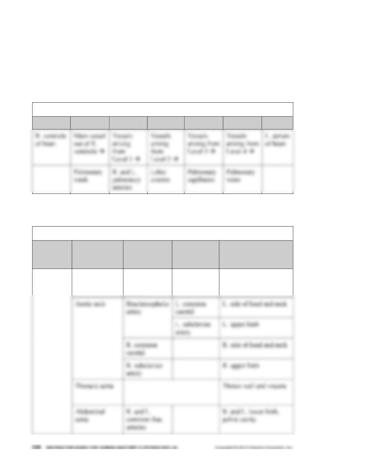
of the body (pp. 722–723; Fig. 19.19; Table 19.3).
Pulmonary Circulation
Start Level 1 Level 2 Level 3 Level 4 Level 5 End
B. The aorta is the largest artery in the body and has discretely named regions that extend from the
heart to the lower abdominal cavity (pp. 724–725; Fig. 19.21; Table 19.4).
Systemic Circulation: Aorta and Major Arteries
Level 1
Main
Vessel
Divisions of
Aorta
Level 2 Level 3 Area Served by
Vessel
Ascending aorta R. and
L. coronary
arteries
Myocardium of heart
Aorta
C. The common carotid arteries supply blood to the head and neck (pp. 726–727;
Fig. 19.22; Table 19.5).
Systemic Circulation: Arteries of the Head and Neck
Level 1
Main
Vessel
Level 2 Level 3 Level 4 Level 5 Area Served by
Vessel
Superficial
temporal
arteries
Parotid gland and
scalp
External
carotid
arteries
Facial
arteries
Skin and muscles
of face
Common
carotid
arteries
(R. and L.)
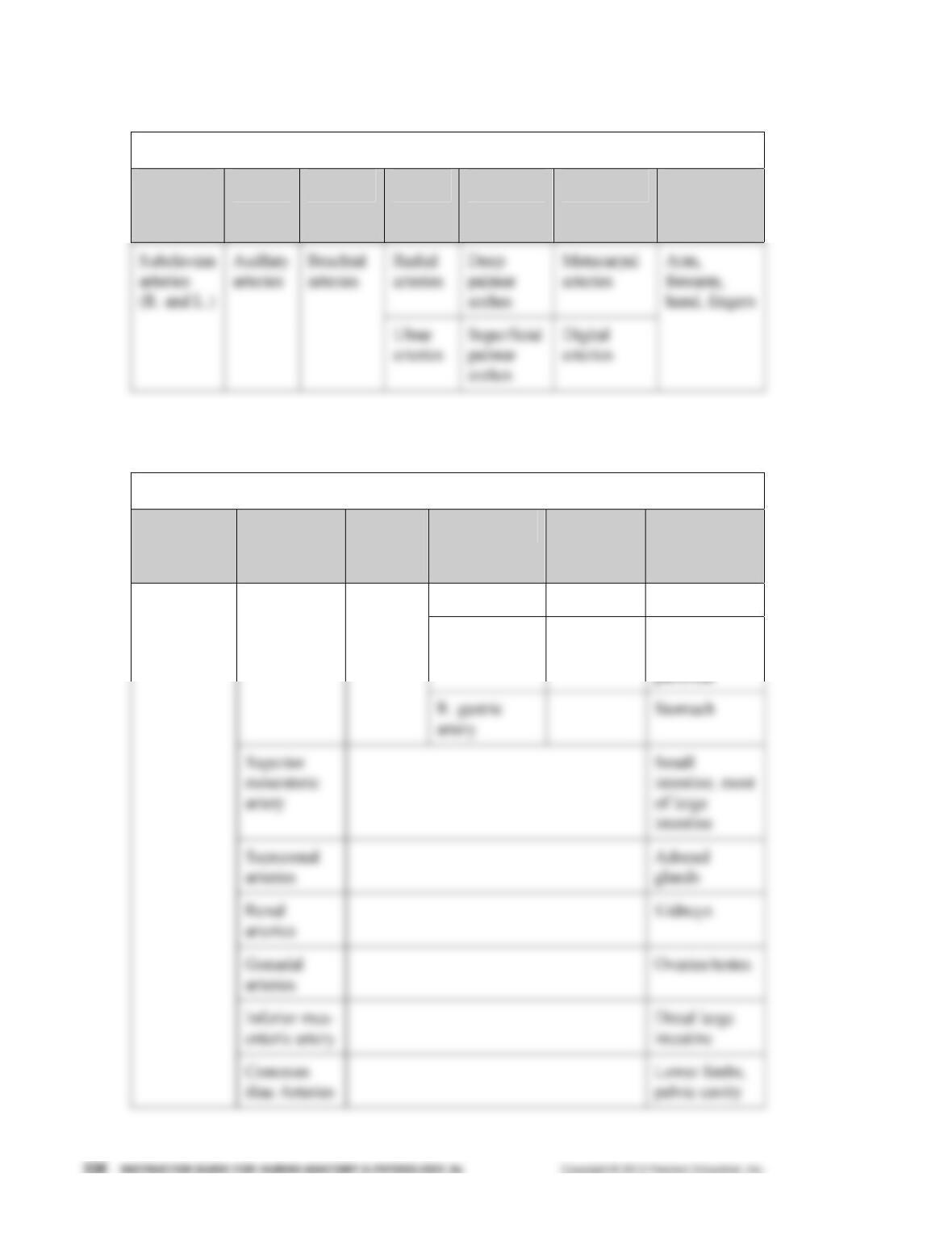
D. The subclavian arteries supply blood to the upper limbs (pp. 728–729; Fig. 19.23;
Table 19.6).
Systemic Circulation: Arteries of the Upper Limbs
Level 1
Main
Vessel
Level 2 Level 3 Level 4 Level 5 Level 6 Area
Served by
Vessel
E. The arteries supplying the abdomen arise from the abdominal aorta (pp. 730–733;
Fig. 19.24; Table 19.7).
Systemic Circulation: Arteries of the Abdomen
Level 1
Main
Vessel
Level 2 Level 3 Level 4 Level 5 Area Served
by Vessel
Hepatic artery Liver
Splenic artery Left gastric
artery
Stomach,
spleen,
pancreas
Celiac trunk Common
hepatic
artery
Abdominal
aorta
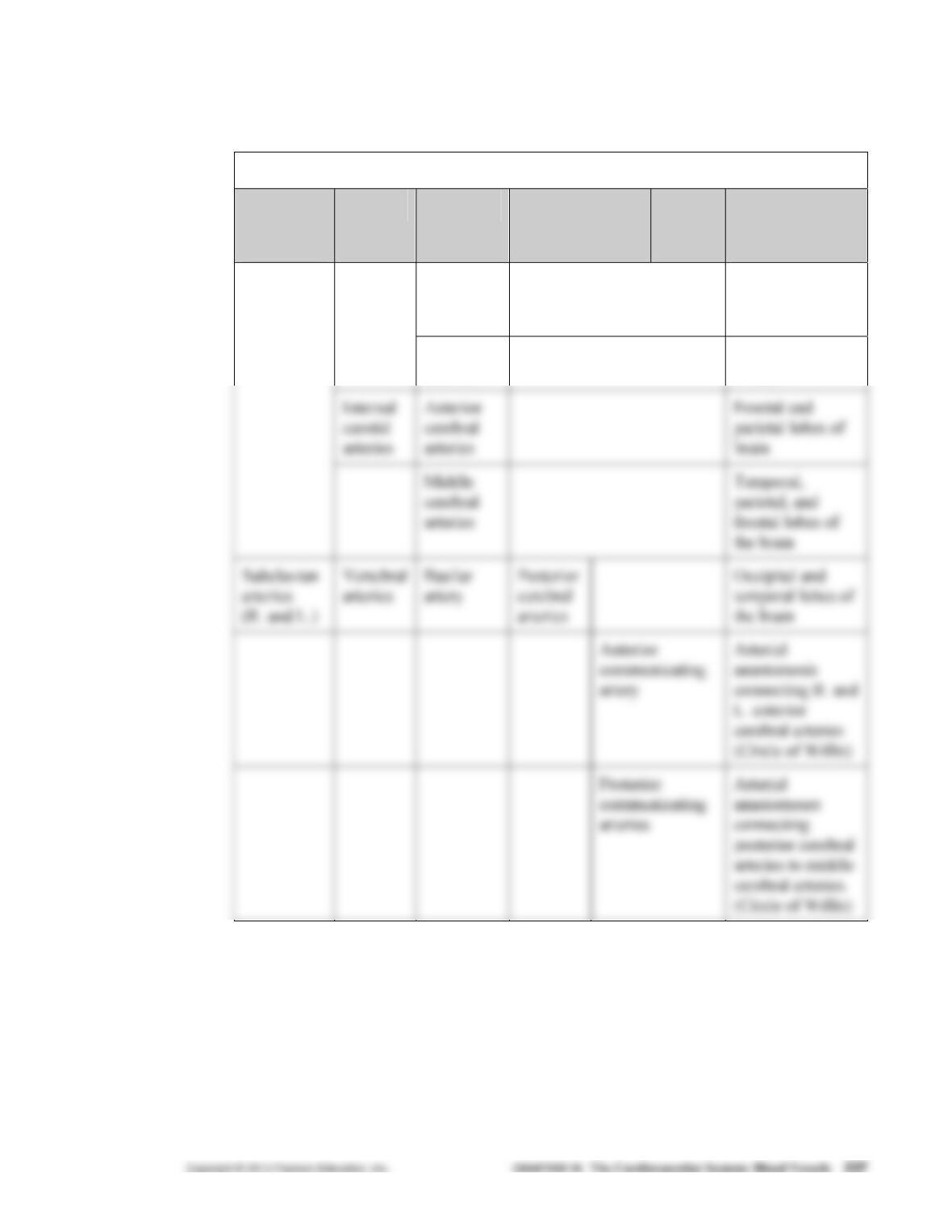
F. The arterial blood supply to the pelvis and lower limbs is provided by the common
iliac arteries, which branch from the distal end of the abdominal aorta (pp. 734–735;
Fig. 19.25; Table 19.8).
Systemic Circulation: Arteries of the Pelvis and Lower Limbs
Level 1
Main
Vessel
Level 2 Level 3 Level 4 Level 5 Level 6 Area
Served by
Vessel
Internal
iliac
arteries
Pelvic wall,
viscera,
gluteal
muscles
Common
iliac
arteries
(R. and L.)
arteries
arteries
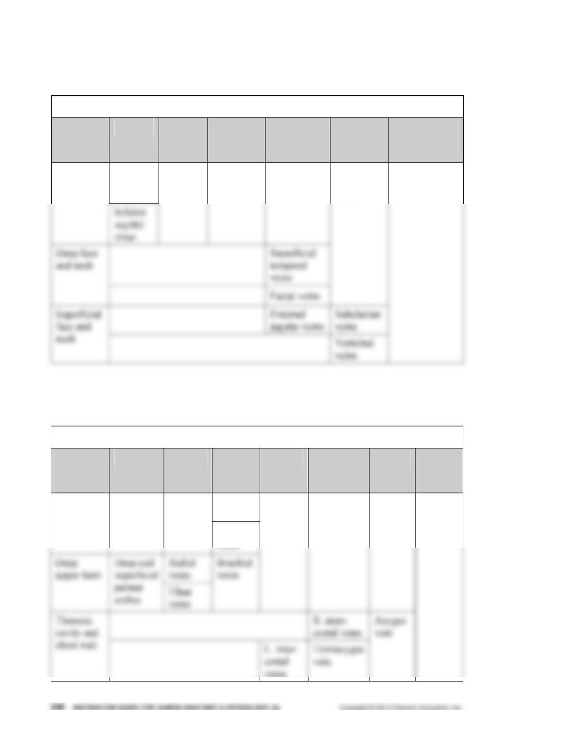
G. The vena cavae are the major veins that drain blood from the body back toward the heart
(pp. 736–737; Fig. 19.26; Table 19.9).
Systemic Circulation: The Vena Cavae and the Major Veins
Area Drained
by Vessel
Level 3 Level 2 Level 1 Main Vessel
H. Most of the blood drained from the head and neck is collected by three pairs of veins: the
external jugular veins, the internal jugular veins, and the vertebral veins (pp. 738–739;
Fig. 19.27; Table 19.10).
Systemic Circulation: Veins of the Head and Neck
Area
Drained
by Vessel
Level 6 Level 5 Level 4 Level 3 Level 2 Level 1 Main
Vessel
Superior
sagittal
sinus
Brain
Straight
sinus
Transverse
sinuses
Sigmoid
sinus
Internal
jugular
veins
Brachio-
cephalic veins
I. The deep veins of the upper limbs follow the same paths as the arteries of the same name; the
superficial veins are larger than the deep veins and easily seen beneath the skin (pp. 740–741;
Fig. 19.28; Table 19.11).
Systemic Circulation: Veins of the Upper Limbs and Thorax
Area
Drained
by Vessel
Level 7 Level 6 Level 5 Level 4 Level 3 Level 2 Level 1
Main
Vessel
Cephalic
veins
Superficial
forearm
Median
cubital
vein Basilic
Axillary
veins
Subclavian
veins
Brachio-
cephalic
veins
Superior
vena
cava