Unlock document.
This document is partially blurred.
Unlock all pages and 1 million more documents.
Get Access
CHAPTER
10
The Muscular System
Objectives
Actions and Interactions of Skeletal Muscles
Naming Skeletal Muscles
3. List the criteria used in naming muscles. Provide an example to illustrate the use of each
criterion.
Muscle Mechanics: Importance of Fascicle Arrangement and Leverage
4. Name the common patterns of muscle fascicle arrangement and relate them to power
generation.
Major Skeletal Muscles of the Body
7. Name and identify the muscles described in Tables 10.1 to 10.17. State the origin,
insertion, and action of each.
Suggested Lecture Outline
I. Actions and Interactions of Skeletal Muscles (pp. 319–320; Fig. 10.1)
A. Muscles only pull; they never push, and as a muscle shortens, the insertion is pulled
toward the origin (p. 319).
II. Naming Skeletal Muscles (pp. 320–322)
A. Criteria used to name skeletal muscles include location, shape, size, direction of muscle
fibers, number of origins, location of attachments, or action. A muscle name often incor-
porates more than one of these criteria. (p. 320)
1. An example of a muscle named for its location is the temporalis, which covers the
temporal bone.
2. An example of a muscle named for its shape is the deltoid, which has a triangular shape.
3. Terms such as maximus, minimus, longus, and brevis indicate relative muscle size.
III. Muscle Mechanics: Importance of Fascicle Arrangement and Leverage
(pp. 322–324; Figs. 10.2–10.4)
A. In skeletal muscles, the common arrangement of the fascicles varies, resulting in muscles
with different shapes and functional capabilities (pp. 322–323; Fig. 10.2).
1. The fascicular pattern is circular when the fascicles are arranged in concentric rings.
2. A convergent muscle has a broad origin and its fascicles converge toward a single
tendon of insertion.
B. The operation of most skeletal muscles involves the use of leverage and lever systems—
partnerships between the muscular and skeletal systems (pp. 323–324; Figs. 10.3–10.4).
1. A lever is a rigid bar that moves on a fixed point, or fulcrum, when a force is applied to it.
2. The applied force, or effort, is used to move a resistance, or load.
6. There are three types of levers: first-class, second-class, and third-class.
a. First-class levers have the effort applied at one end and the load at the other end,
with the fulcrum in between.
IV. Major Skeletal Muscles of the Body (pp. 324–382; Figs. 10.5–10.26; Tables
10.1–10.17)
The muscles listed in the following sections are outlined in tables that differ
from the tables in the textbook. Textbook tables are designed to be more ency-
clopedic, but not necessarily practical for a lecture outline. The following tables
condense the information found in tables in the main text in order to provide a
more concise, teachable outline of the more commonly taught muscles, along
with their actions, origins, and insertions.
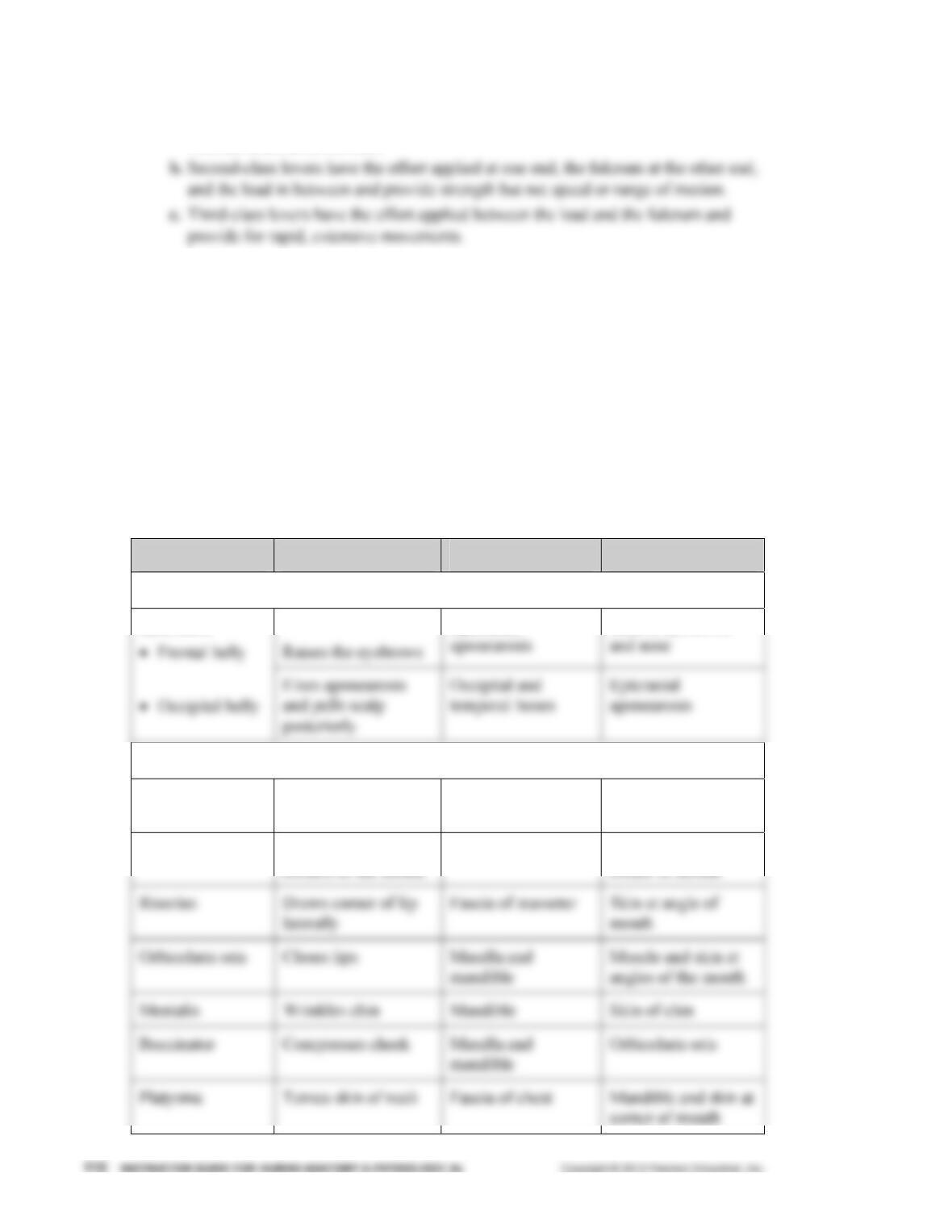
A. Muscles of the Head, Part I: Facial Expression (pp. 329–331; Fig. 10.7; Table 10.1)
1. Muscles of facial expression located in the scalp and face insert into skin or other
muscles, rather than bones, and are innervated by cranial nerve VII, the facial nerve.
Muscle Action Origin Insertion
Muscles of the Scalp
Epicranial
Skin of eyebrows
Epicranius
Muscles of the Face
Orbicularis oculi Closes eye Frontal and
maxillary bones
Eyelid
Zygomaticus Raises lateral
corners of the mouth
Zygomatic bone Skin and muscle at
corner of mouth
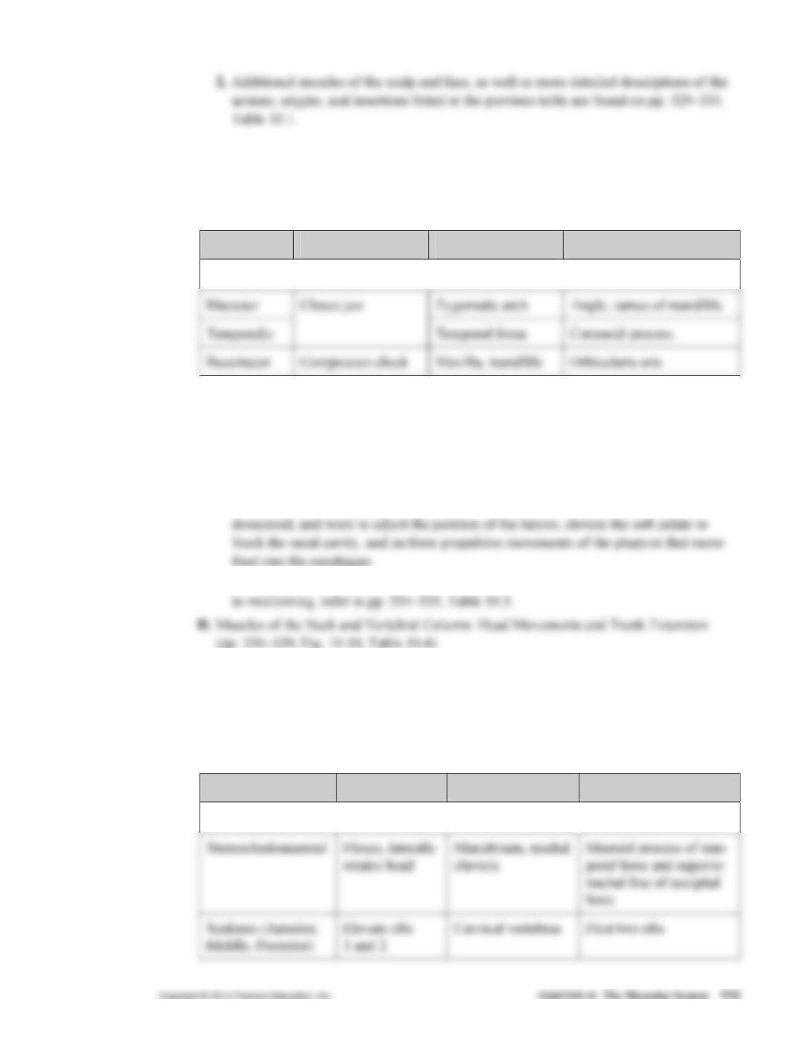
B. Muscles of the Head, Part II: Mastication and Tongue Movement (pp. 332–333;
Fig. 10.8; Table 10.2)
1. Muscles involved in mastication (chewing) move the mandible and anchor the tongue,
and are innervated by the mandibular branch of cranial nerve V, the trigeminal nerve.
Muscle Action Origin Insertion
Muscles of Mastication
2. Additional muscles controlling the mandible and tongue, as well as more detailed
descriptions of the actions, origins, and insertions listed above are found on pp. 332–
333, Table 10.2.
C. Muscles of the Anterior Neck and Throat: Swallowing (pp. 334–335; Fig. 10.9;
Table 10.3)
1. Muscles involved in swallowing are part of the anterior triangle next to the sternoclei-
2. For a detailed listing of the names, actions, origins, and insertions of muscles involved
1. Head movements are produced by muscles originating from the axial skeleton.
a. Movements of the head from side to side are accomplished by contraction of
muscles on only one side of the neck.
2. Extension of the back, and maintenance of normal spinal curvatures are produced by
deep back muscles originating from the sacrum to the skull.
Muscle Action Origin Insertion
Anterolateral Neck Muscles
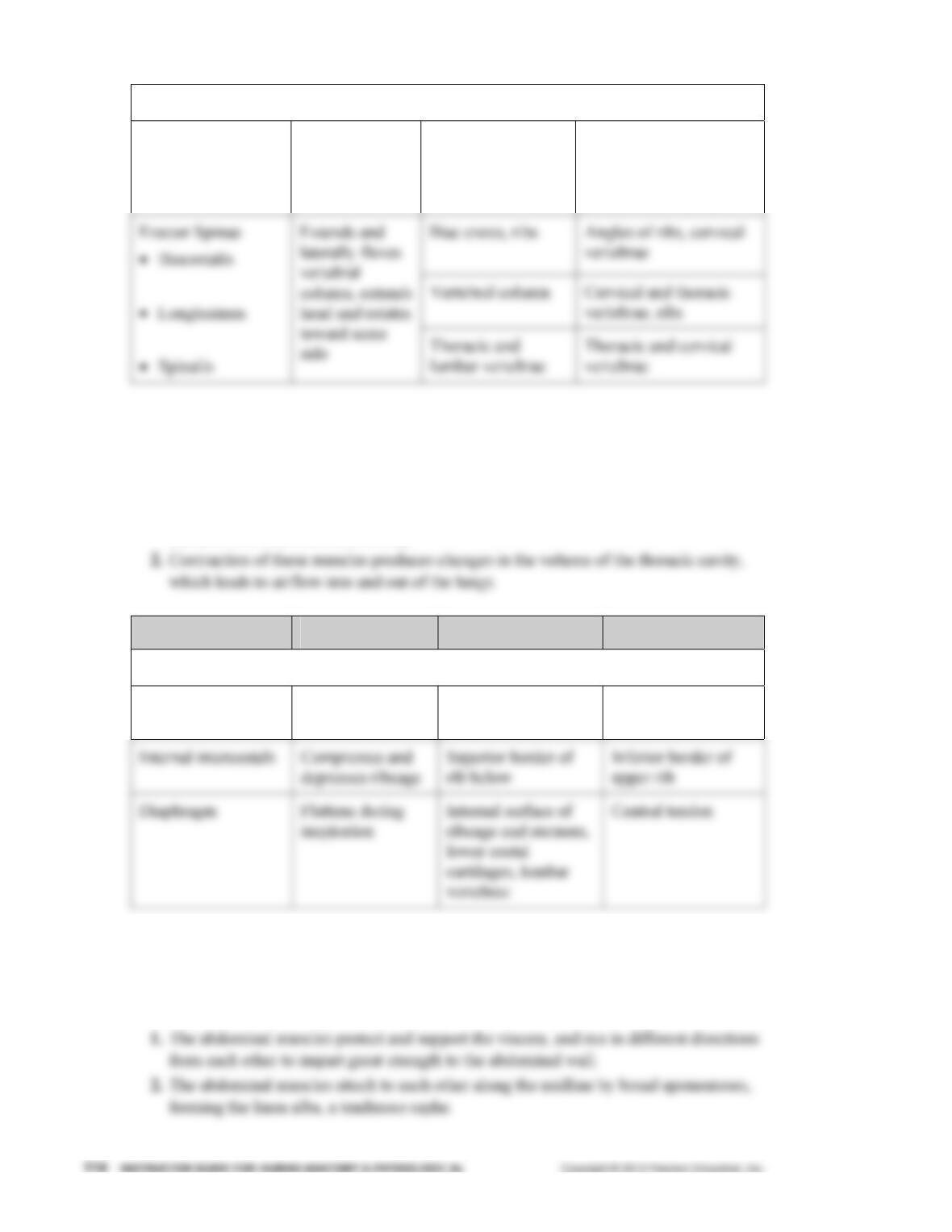
Intrinsic Muscles of the Back
Splenius capitis,
cervicis
Extends head Cervical and
thoracic vertebrae
Mastoid process of
temporal bone, occipital
bone, transverse
processes of C2–C4
3. Additional muscles of the neck and vertebral column, as well as more detailed descrip-
tions of the actions, origins, and insertions listed above are found on pp. 336–339,
Table 10.4.
E. Deep Muscles of the Thorax: Breathing (pp. 340–341; Fig. 10.11; Table 10.5)
1. Deep muscles of the thorax form the anterolateral wall of the thorax and partition the
thoracic from the abdominal cavity.
Muscle Action Origin Insertion
Muscles of the Thorax
External intercostals Elevates ribs Inferior border of
upper rib
Superior border of
rib below
3. More detailed descriptions of the actions, origins, and insertions listed above are found
on pp. 340–341, Table 10.5.
F. Muscles of the Abdominal Wall: Trunk Movements and Compression of Abdominal
Viscera (pp. 342–343; Fig. 10.12; Table 10.6)
Muscle Action Origin Insertion
Muscles of the Anterolateral Abdominal Wall
Rectus abdominis Flexes and rotates
vertebral column
Pubic crest and
symphysis
Xiphoid process,
lower costal
cartilages
3. More detailed descriptions of the actions, origins, and insertions of the abdominal
muscles listed above are found on pp. 342–343, Table 10.6.
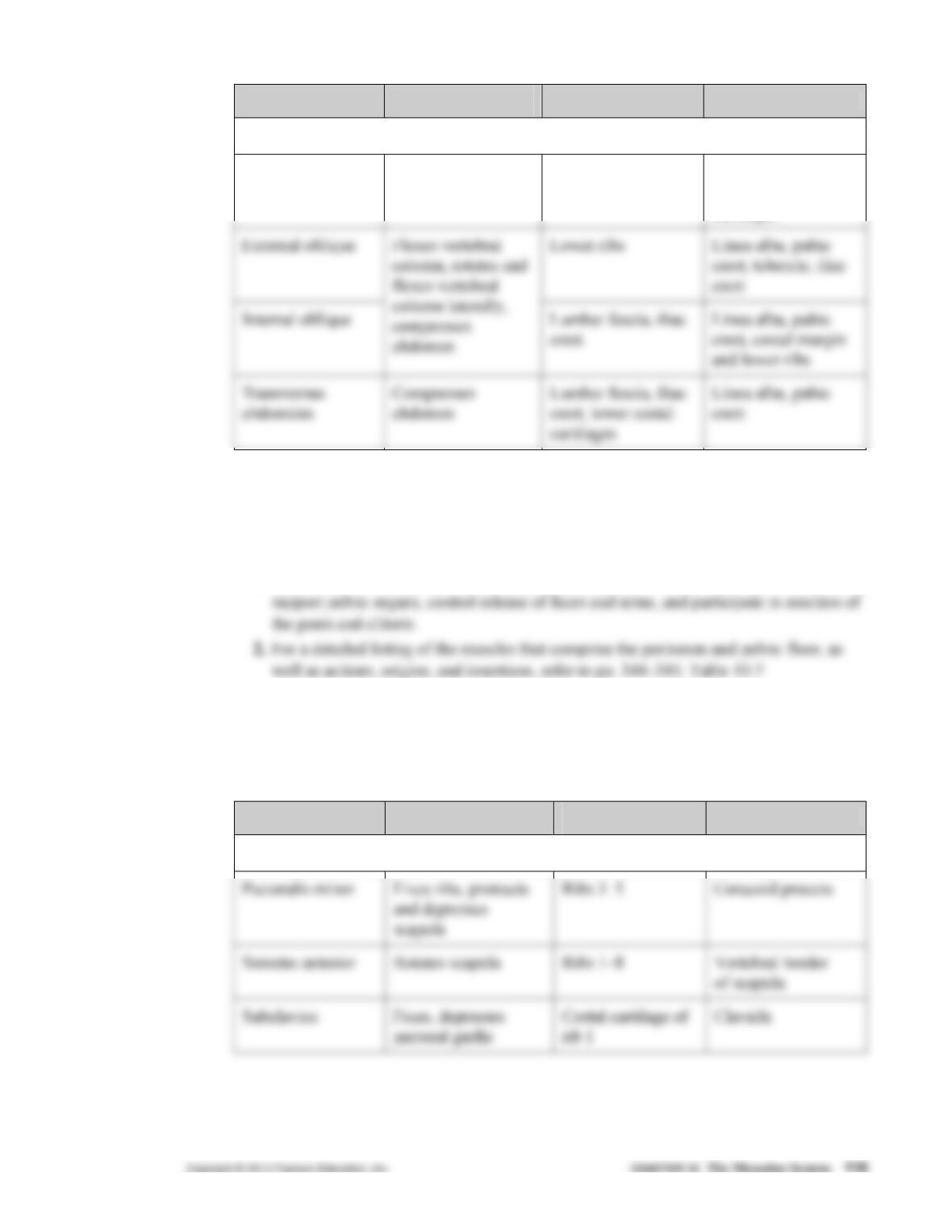
G. Muscles of the Pelvic Floor and Perineum: Support of Abdominopelvic Organs
(pp. 344–345; Fig. 10.13; Table 10.7)
1. The muscles of the pelvic floor and perineum close the inferior opening of the pelvis,
H. Superficial Muscles of the Anterior and Posterior Thorax: Movements of the Scapula and
Arm (pp. 346–349; Fig. 10.14; Table 10.8)
1. The superficial thorax muscles run from the ribs and vertebral column to the shoulder
girdle, and both fix the scapula and create greater range of motion of arm movements.
Muscle Action Origin Insertion
Muscles of the Anterior Thorax
Muscles of the Posterior Thorax
Trapezius Fixes, elevates,
retracts, and rotates
scapula
Occipital condyle,
vertebral column
Acromion, spine of
scapula, lateral third
of clavicle
2. Additional muscles moving the scapula and arm, as well as more detailed descriptions
of the actions, origins, and insertions listed above are found on pp. 346–349, Table
10.8.
I. Muscles Crossing the Shoulder Joint: Movements of the Arm (Humerus) (pp. 350–352;
Fig. 10.15; Table 10.9)
2. Muscles originating anterior to the shoulder flex the arm, while those originating pos-
terior to the shoulder extend the arm.
Muscle Action Origin Insertion
Muscles Moving the Arm
Pectoralis major Flexes, adducts,
medially rotates arm
Clavicle, sternum,
costal cartilages
Greater tubercle
3. More detailed descriptions of the actions, origins, and insertions of the muscles
moving the shoulder listed above are found on pp. 350–352, Table 10.9.
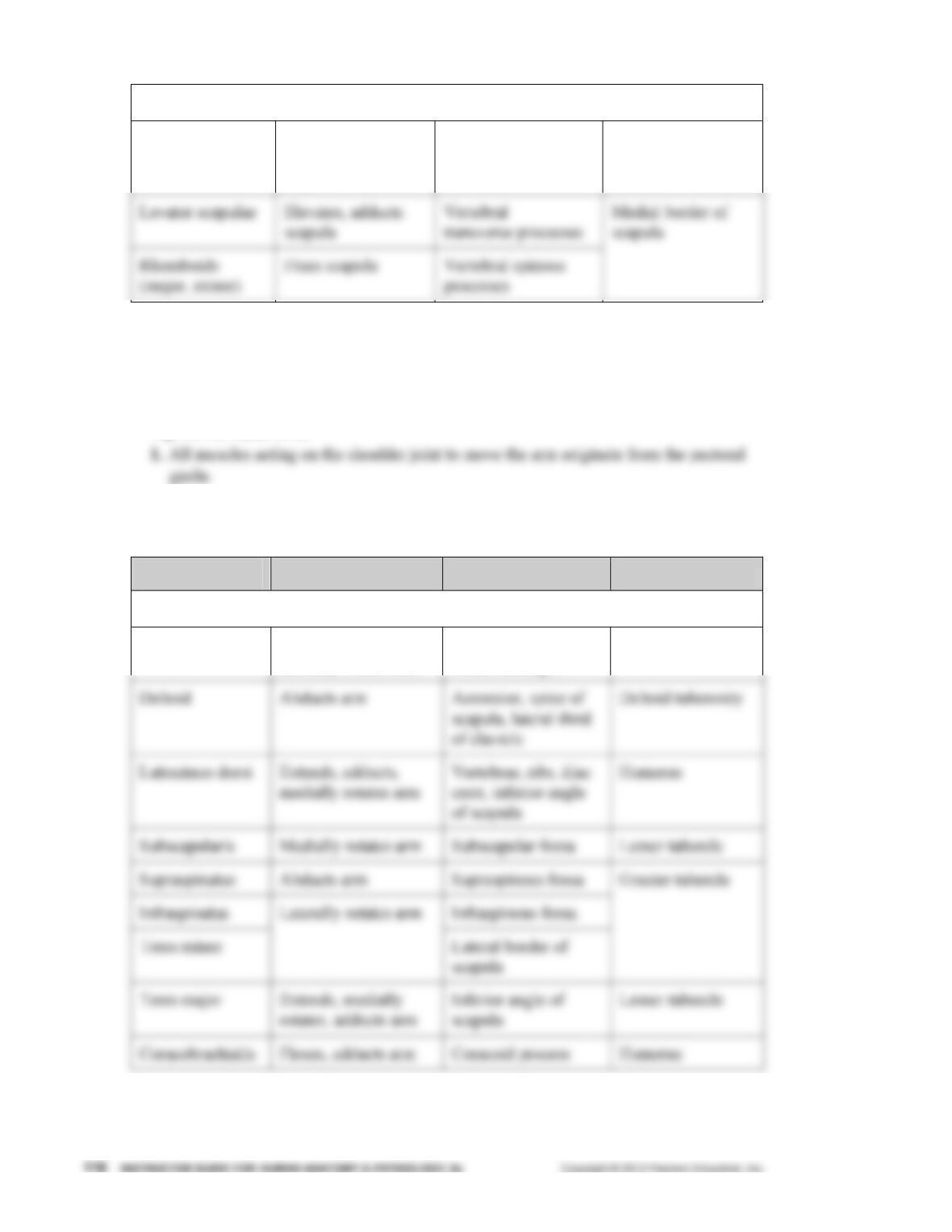
J. Muscles Crossing the Elbow Joint: Flexion and Extension of the Forearm (p. 353;
Fig. 10.15; Table 10.10)
1. There are two compartments in the arm: anterior flexors and posterior extensors, both
acting on the forearm.
Muscle Action Origin Insertion
Posterior Muscles
Triceps brachii Extends forearm Scapula, humerus Olecranon
2. Additional muscles crossing the elbow joint, as well as more detailed descriptions of
the actions, origins, and insertions listed above are found on p. 353, Table 10.10.
K. Muscles of the Forearm: Movements of the Wrist, Hand, and Fingers (pp. 354–357;
Figs. 10.16–10.17; Table 10.11)
1. Muscles of the forearm are divided by fascia into two compartments: anterior flexors
and posterior extensors.
Muscle Action Origin Insertion
Anterior Superficial Muscles
Pronator teres Pronates forearm Medial epicondyle
of humerus,
Radius
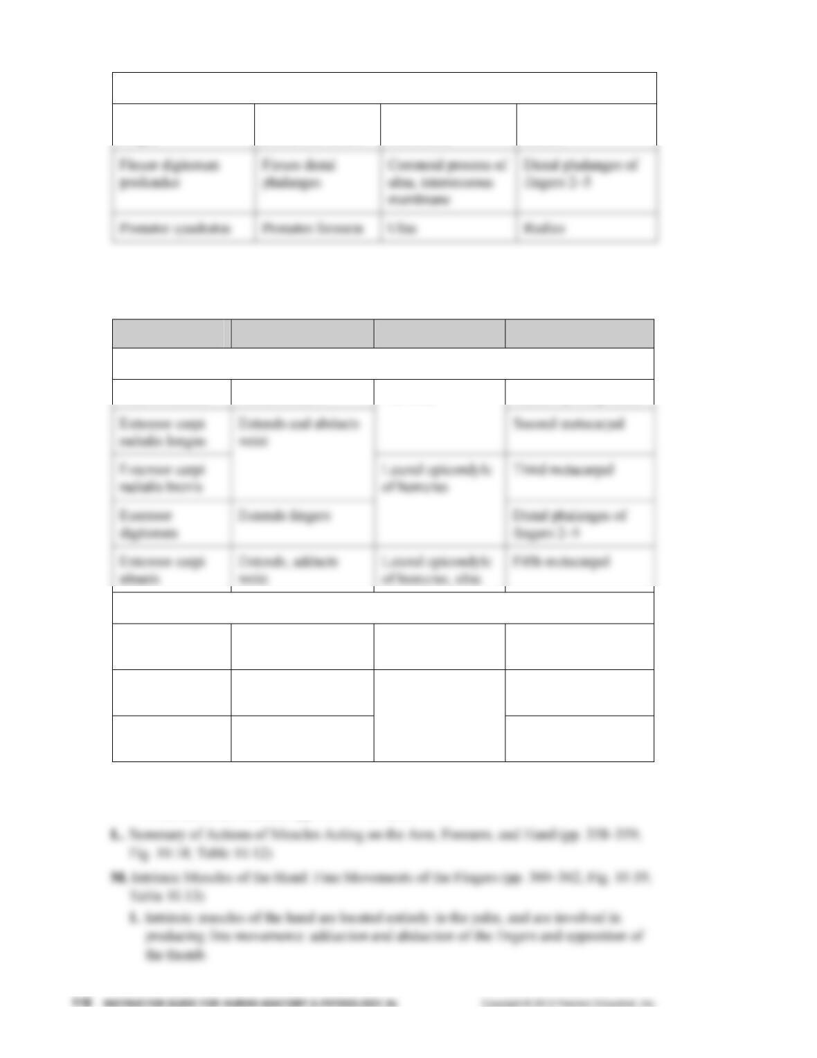
Anterior Deep Muscles
Flexor pollicis
longus
Flexes distal
phalanx of thumb
Radius, interosseous
membrane
Distal phalanx of
thumb
2. More detailed descriptions of the actions, origins, and insertions listed above are found
on pp. 354–355, Table 10.11.
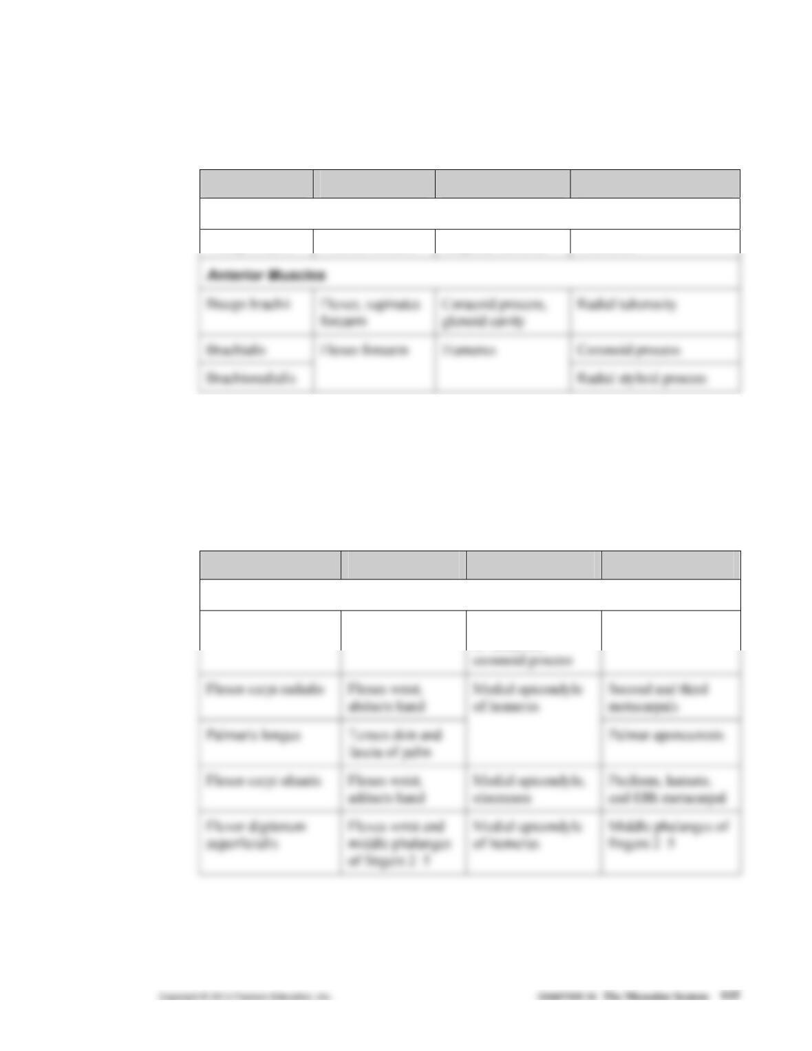
Muscle Action Origin Insertion
Posterior Superficial Muscles
Brachioradialis Flexes forearm Radial styloid process
Humerus
Posterior Deep Muscles
Supinator Supinates forearm Lateral epicondyle
of humerus, ulna
Radius
Abductor pollicis
longus
Abducts and extends
thumb
First metacarpal,
trapezium
Extensor pollicis
(brevis, longus)
Extends thumb
Radius, ulna,
interosseous
membrane Proximal, distal
phalanx of thumb
3. More detailed descriptions of the actions, origins, and insertions of forearm muscles
listed above are found on pp. 356–357, Table 10.11.
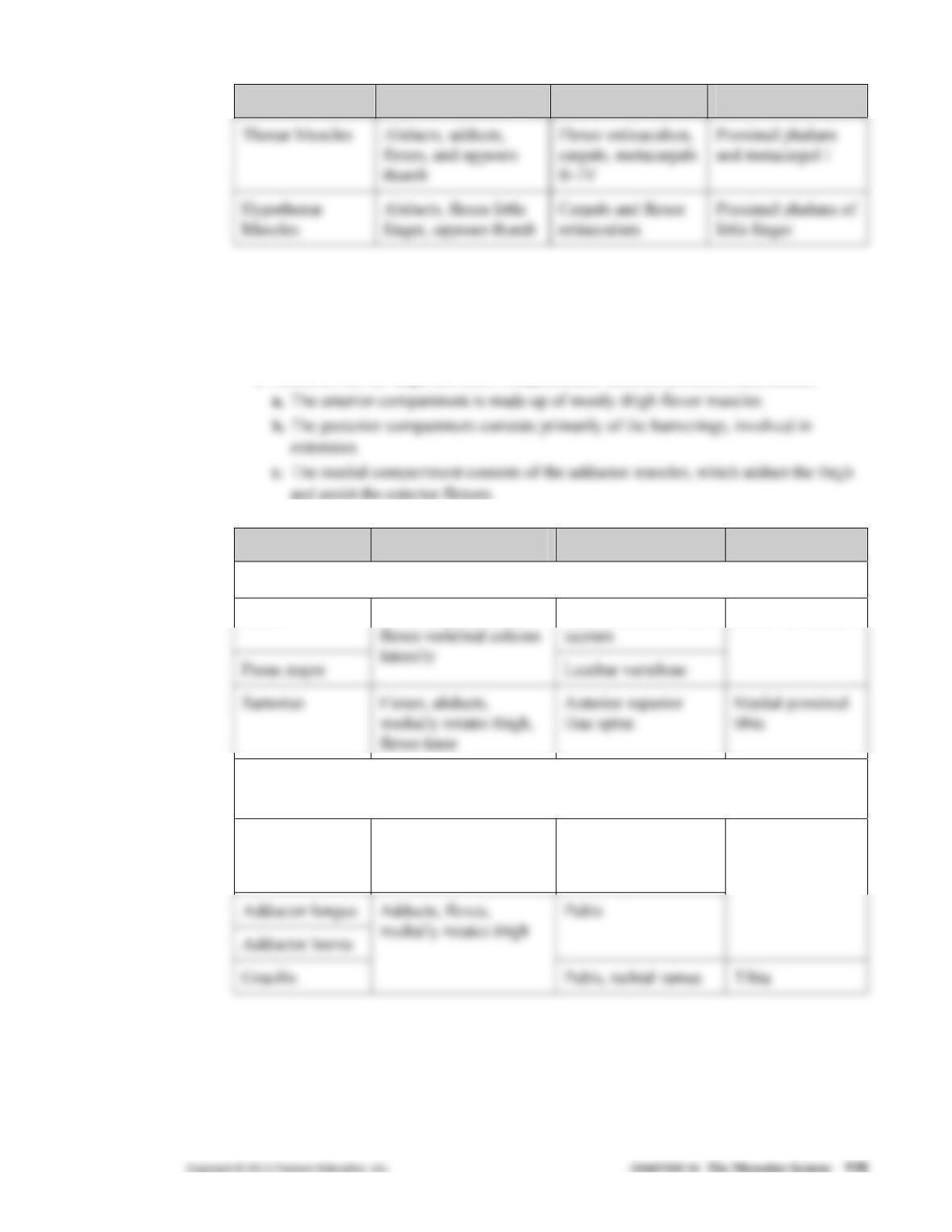
Muscle Action Origin Insertion
2. For a detailed listing of the names, actions, origins, and insertions of muscles of the
hand, refer to pp. 360–362, Table 10.13.
N. Muscles Crossing the Hip and Knee Joints: Movements of the Thigh and Leg
(pp. 363–369; Figs. 10.20–10.21; Table 10.14)
1. Fascia divide the thigh into three compartments: anterior, posterior, and medial.
Muscle Action Origin Insertion
Anterior and Medial Muscles: Iliopsoas and Sartorius
Iliacus Iliac fossa, crest, and
Flexes thigh at trunk,
Lesser trochanter
Muscles of the Medial Compartment: Adductor Group, Pectineus,
and Gracilis
Adductor
magnus
Adducts, flexes,
extends, medially
rotates thigh
Ischial and pubic
rami, ischial
tuberosity
Linea aspera
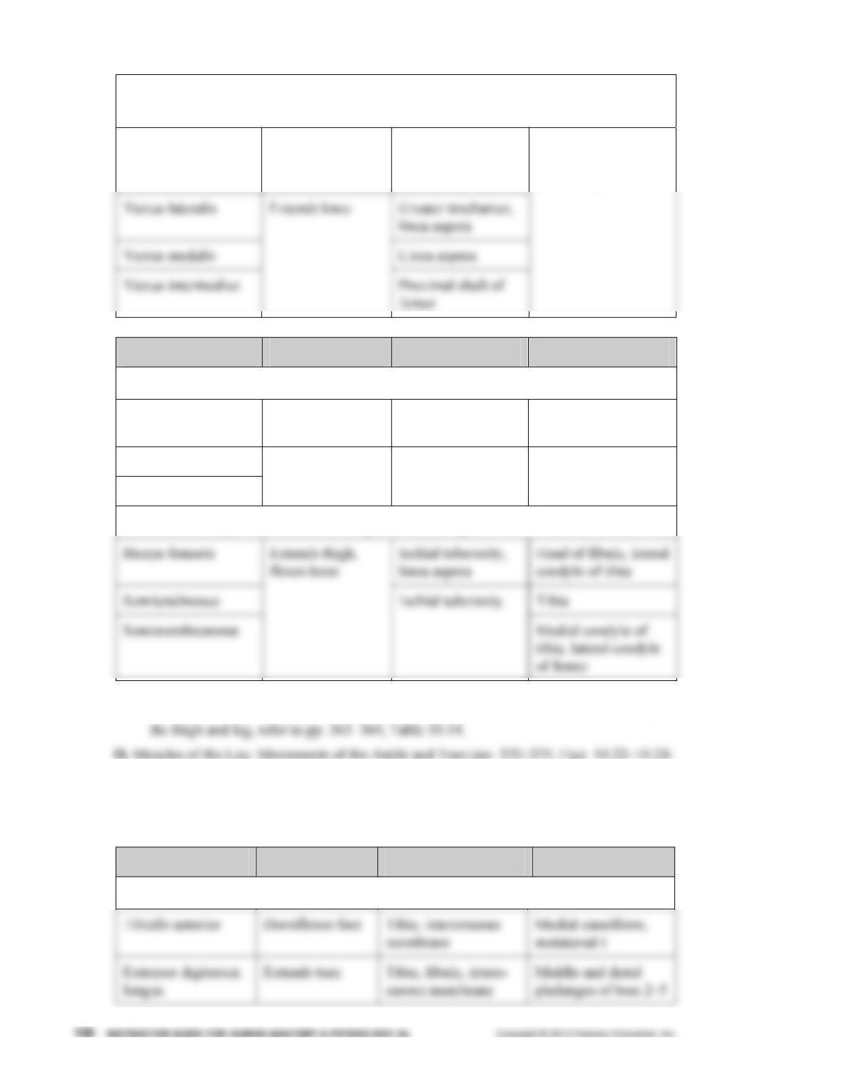
Muscles of the Anterior Compartment: Quadriceps Femoris Group and
Tensor Fasciae Latae
Rectus femoris Extends knee,
flexes thigh
Anterior inferior
iliac spine,
acetabulum
Patella, tibial
tuberosity via
patellar ligament
Muscle Action Origin Insertion
Posterior Muscles
Gluteus maximus Extends thigh Ilium, sacrum,
coccyx
Gluteal tuberosity of
femur, iliotibial tract
Gluteus medius
Gluteus minimus
Abducts, medially
rotates thigh
Ilium Greater trochanter
Posterior Compartment of the Thigh: Hamstrings
2. For a detailed listing of the names, actions, origins, and insertions of muscles acting on
Table 10.15)
1. The leg muscles moving the ankle and toes are divided into three compartments by
deep fascia: anterior, lateral, and posterior.
Muscle Action Origin Insertion
Muscles of the Anterior Compartment
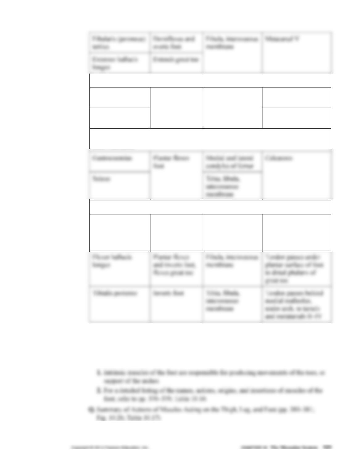
Muscles of the Lateral Compartment
Fibularis (peroneus)
longus
Metatarsal I, medial
cuneiform
Fibularis (peroneus)
brevis
Plantar flexes
and everts foot
Fibula
Metatarsal V
Superficial Muscles of the Posterior Compartment: Triceps Surae
and Plantaris
Deep Muscles of the Posterior Compartment
Flexor digitorum
longus
Plantar flexes
and inverts foot,
flexes toes
Posterior tibia Tendon passes behind
medial malleolus to
distal phalanges of toes
2–5
2. More detailed descriptions of the actions, origins, and insertions of the muscles of the
leg listed above are found on pp. 370–375, Table 10.15.
P. Intrinsic Muscles of the Foot: Toe Movement and Arch Support (pp. 376–379; Fig.
10.25; Table 10.16)
Cross References
Additional information on topics covered in Chapter 10 can be found in the chapters listed below.
1. Chapter 7: Bones of the skull; facial bones; bones of the vertebral column; the bony
thorax; pectoral bones; upper extremity; pelvic girdle; lower extremity
Lecture Hints
1. It is easy for students to treat the muscular (or any other) system as an individual unit,
without relating it to the rest of the body. Emphasize that students should associate any
specific muscle (and its associated synergists, antagonists, etc.) with its fascicle arrange-
ment, origin and insertion sites, and bones involved to “keep sight of the whole picture.”
4. When describing different muscles of the body, try coaxing names from the class by
carefully indicating locations, properties, etc. For example, point to a diagram (or model)
displaying transverse abdominis and ask the class which way the fibers are running. They
should answer: “transversely.” Then ask: “What general area of the body is this muscle
located in?” Answer: “abdominal.” Finally, ask: “What would be a logical name for this
muscle?” Using this approach in class will encourage students to do this type of analysis
when they start learning the information outside of class.
Activities/Demonstrations
1. Audiovisual materials are listed in the Multimedia in the Classroom and Lab section of
this Instructor Guide (p. 387).
3. Obtain a dissected preserved animal, such as a cat or a fetal pig, and exhibit the major
muscle groups.
4. Have students work in pairs as follows: One student should attempt to contract a particu-
lar muscle, while the second student provides resistance to prevent that movement. In this
5. As muscles are being described, project 2x2 slides of cadaver dissection so that students
6. Obtain a 3-D model or chart to illustrate the major human muscle groups.
Critical Thinking/Discussion Topics
1. Why is it necessary for pregnant women to strengthen their “pelvic floor”?
2. What do bones possess that allow them to act as effective levers?
3. What are the most appropriate modes of therapy for a pulled hamstring muscle or pulled
groin muscle?
Library Research Topics
1. Because muscle cells do not regenerate, what methods of treatment are available if a
major group of muscles is lost? What is the status of skeletal muscle transplants?
2. What effect does old age have on skeletal muscle? What type of research is under way
concerning this topic?
List of Figures and Tables
All of the figures in the main text are available in JPEG format, PPT, and labeled & unlabeled
format on the Instructor Resource DVD. All of the figures and tables will also be available in
Transparency Acetate format. For more information, go to www.pearsonhighered.com/educator.
Figure 10.1 Muscle Action
Figure 10.2 Patterns of fascicle arrangement in muscles.
Figure 10.3 Lever systems operating at a mechanical advantage and a mechanical
disadvantage.
Figure 10.14 Superficial muscles of the thorax and shoulder acting on the scapula
and arm.
Figure 10.15 Muscles crossing the shoulder and elbow joints, causing movements of
the arm and forearm, respectively.
Figure 10.16 Muscles of the anterior fascial compartment of the forearm acting on the
right wrist and fingers.
Figure 10.17 Muscles of the posterior fascial compartment of the right forearm acting
on the wrist and fingers.
Table 10.1 Muscles of the Head, Part I: Facial Expression (Figure 10.7)
Table 10.2 Muscles of the Head, Part II: Mastication and Tongue Movement
(Figure 10.8)
Table 10.3 Muscles of the Anterior Neck and Throat: Swallowing (Figure 10.9)
Table 10.5 Deep Muscles of the Thorax: Breathing (Figure 10.11)
Table 10.6 Muscles of the Abdominal Wall: Trunk Movements and Compression of
Abdominal Viscera (Figure 10.12)
Table 10.7 Muscles of the Pelvic Floor and Perineum: Support of Abdominopelvic
Organs (Figure 10.13)
Table 10.9 Muscles Crossing the Shoulder Joint: Movements of the Arm (Humerus)
(Figure 10.15)
Table 10.10 Muscles Crossing the Elbow Joint: Flexion and Extension of the Fore
arm (Figure 10.15)
Table 10.11 Muscles of the Forearm: Movements of the Wrist, Hand, and Fingers
(Figures 10.16 and 10.17)
Table 10.13 Intrinsic Muscles of the Hand: Fine Movements of the Fingers
(Figure 10.19)
Table 10.14 Muscles Crossing the Hip and Knee Joints: Movements of the Thigh and
Leg (Figures 10.20 and 10.21)
Answers to End-of-Chapter Questions
Multiple-Choice and Matching Question answers appear in Appendix H of the main text.
Short Answer Essay Questions
17. Student answers will vary. (p. 320)
a. Location of the muscle—frontalis, occipitalis, zygomaticus
d. Direction of muscle fibers—rectus abdominus, external oblique, superficial transverse
perineus
18. First-class lever: fulcrum between effort and load
19. When the load is far from the fulcrum and the effort is applied near the fulcrum, the
effort applied must be greater than the load to be moved. This type of leverage can be
advantageous because it allows the load to be moved rapidly through a large distance,
with only minimal muscle shortening. (p. 324)
22. See p. 342.
a. The rectus abdominus, external oblique, internal oblique, and transversus abdominus
23. Flexion of the humerus: pectoralis major and deltoid
Extension of the humerus: latissimus dorsi and deltoid
24. a. The extensor carpi radialis longus and brevis are strong wrist flexors. (p. 356)
b. The flexor digitorum profundus is used to flex the distal interphalangeal joints. (p. 355)
25. Piriformis, obturator externus and internus, gemellus, and quadratus femoris are grouped
together, and perform lateral rotation at the hip. (pp. 366, 368)
28. a. Opponens pollicis, flexor pollicis brevis (p. 360)
b. Supinator, abductor pollicis longus (p. 356)
c. Flexor pollicis longus, flexor digitorum profundus (p. 355)
d. Biceps brachii, brachialis (p. 353)
e. Hyoglossus, styloglossus (p. 332)
Critical Thinking and Clinical Application Questions
1. Flexion of the forearm is more difficult when the forearm is pronated because the biceps
brachii, a prime mover of forearm flexion, is unable to act. (p. 353)
2. Levator ani, coccygeus, and external urethral sphincter (p. 344)
Suggested Readings
Agur, A. M., and A. F. Dalley. Grant’s Atlas of Anatomy. 12th ed. Baltimore: Lippincott
Williams & Wilkins, 2009.


