Interactive Physiology
®
Exercise Sheet Answers
Muscular System:
Anatomy Review: Skeletal Muscle Tissue
1.
Muscle Type Cardiac Skeletal Smooth
Shape of Short and Elongated Spindle-
cell branching shaped
2. Tendons
3. fascicles
4. epimysium
perimysium
endomysium
5. 1. Sarcolemma →plasma membrane of muscle
cell
8. A
I
A band: contains both thick and thin
filaments
defined by length of thick filament
10. lines get closer together
decreases or disappears
11. 1. protein fibers that connect thick filaments:
located in the middle of the H zone
2. the distance between two Z lines; the func-
tional unit of muscle contraction
12. myofilament, myofibril, muscle fiber (cell),
3. Voltage-regulated Ca
++
(same as voltage gated)
4. Exocytosis
5. Chemically regulated (chemically gated)
6. depolarization
7. By the enzyme acetylcholinesterase (AChE)
8. sarcolemma, T tubules
9. Ca
++
c. Nicotine
Muscular System: Sliding Filament Theory
1. a. Myosin
b. power stroke
C
5. high-energy
6. backsliding
7. 1. Energize the power stroke
Muscular System: Muscle Metabolism
1. 1. energizing the power stroke of the myosin
head
2. disconnecting the myosin head
3. energizing the calcium ion pump
2. hydrolysis of glycogen stores in muscle
7. Glycolysis
1. 2 ATP
2. 2 pyruvic acid molecules
lactic, anaerobic
8. aerobic
1. blood
2. stored in myoglobin
Krebs cycle, oxidative phosphorylation, 36
10.
Red Slow-Twitch Fibers White Fast-Twitch Fibers
Krebs cycle and oxidative uses glycolysis
phosphorylation
high endurance fatigue rapidly
Muscular System:
Contraction of Motor Units
producing minute contractions of random
motor units
b. muscle loses its tone and becomes flaccid; in
time, it will atrophy
7. medium
large
3. Latent
4. a. a second stimulus applied before complete
relaxation
b. ↓
5.
Order Stage Description
4 Fatigue ↓tension due to ↓ATP
and ↑buildup of lactic acid
6. a. Many
b. Few
7. a. 0.3
b. 0.4
c. 1.5
8. a. Moderate
nutritional and metabolic area
Axon →conductive region; generates an action
potential
3. synapses, dendrites, soma
Nervous System I: Ion Channels
1. Integral proteins
2. 1. Charge
2. Size
b. Chloride
7.
Areas on
Channels the Neuron Type of Potential
Nongated dendrites, resting membrane
cell body, axon potential
8. Found along the axon, Important for action
potential, Opened and closed by gates
10. a. Voltage gated sodium
b. Action
c. Respiratory failure
Nervous System I: Membrane Potential
1.
Ions Intracellular Extracellular
4. a. Concentration gradient
b. Electrical gradient
5. equilibrium, –90
b. neg e. neg
c. pos f. pos
Nervous System I: The Action Potential
1. a. –70, +30
b. Na
, Na
4. 1. Inactivation of voltage-gated Na
+
channels
2. Opening of voltage-gated K
+
channels
5. a. repolarize
b. Out of
c. hyperpolarization
2. Presence of myelin
b. Saltatory
8. a. Multiple sclerosis
c. Too few voltage-gated Na
channels between
the nodes of Ranvier
Nervous System II: Anatomy Review
1. skeletal, cardiac, smooth, glands
2. increase, decrease, decrease, increase, secrete
3. action potential, action potential
4. axodendritic, axosomatic, axoaxonic, chemical
Nervous System II: Ion Channels
1. directly
1. ACh
3. a. hyperpolarize
b. GABA
c. Cl
–
4. a. indirectly
b. first
c. G protein
7. a. ACh
b. Glutamate
c. GABA
8. Glycine
Nervous System II: Synaptic Transmission
1. Voltage-gated Ca
++
2. Ca
++
3. a. Neurotransmitters
7.
Type Found
Nicotinic Neuromuscular junction
Muscarinic excitatory: target organ in most cases
inhibitory: heart and CNS
9. Smooth muscle, cardiac muscle, glands
10. glutamate
11. 1. GABA
Synaptic Potentials and Cellular
Integration
1. Ca
++
, rapid firing of action potentials
2. Ca
++
, an axoaxonic synapse
3. a. graded
b. decay
9.
Action Synaptic
Potential Potential
Function Release neuro- Generate/inhibit
transmitters action potentials
Depolarization/ Depolarizations Both
hyperpolarization only
Magnitude 100 mV Varies with strength
3. a. initiates the depolarizing impulse and sets
the pace for the entire heart
b. link between the SA node and the AV node
c. delay occurs allowing atria to contract
d. link between atria and ventricles
e. convey impulses down the interventricular
7. QRS complex
8. atrial, contraction
9. QRS complex
10. tachycardia
Cardiovascular System:
Cardiac Action Potential
1. Through gap junctions
2. contract
3. fast calcium, into
4. positive
5. decreased, potassium, sodium
6. –40, fast calcium channels
7. potassium, repolarization
8. Na
+
-K
+
ATPase
Cardiovascular System: Cardiac Cycle
1. differences in blood pressure
2.
Chamber Valve Chamber/Vessel
Right ventricle Pulmonary semilunar Pulmonary trunk
Right atrium Tricuspid Right ventricle
3. a. mid to late, diastole
b. atrioventricular, AV
4. Intraventricular pressure is greater than atrial
pressure.
5. Intraventricular pressure is greater than
pressure in the pulmonary trunk and aorta.
one with hypertension must work harder to
eject the same stroke volume.
9. 70
10.
AV Semilunar
Phase Valves Valves
Ventricular filling Open Closed
Isovolumetric contraction Closed Closed
Isovolumetric relaxation Closed Closed
Cardiovascular System: Cardiac Output
1. The amount of blood pumped out by each ven-
tricle in one minute
d. 50 ml
6. CO = HR ×SV
= 75 beats/minute ×70 ml/beat
= 5250 ml/minute or 5.25 L/minute
7. HR SV CO
a. ↑SNS ↑↑↑
b. ↑Venous return ↔↑ ↑
8. An increase in contractility leads to an increase
in the force of contraction.
9. An increase in filling time leads to an increase
in end diastolic volume (Frank Starling
mechanism).
10. 3 bottles (6 liters)
Cardiovascular System:
Anatomy Review: Blood Vessel
2. V contain the lowest pressure
A contain the highest pressure
A has thick tunica media
V thin tunica media
3. 1. elastic arteries
2. muscular arteries
3. arterioles
4. Elastic, aorta
5. elastin, renal, pressure, vasoconstriction
6. arterioles, resistance, increase
Cardiovascular System:
Measuring Blood Pressure
1. pumping action of the heart, resistance
2. millimeters, mmHg
3. laminar
4. pressure wave
5. systolic, contraction, 120
Cardiovascular System:
Factors That Affect Blood Pressure
1. 1. Vessel diameter
2. Blood viscosity
3. Total vessel length
2. 1. Epinephrine
5. A ↓arterial diameter
A ↑total vessel length
B ↑vessel elasticity
B ↓plasma epinephrine
A ↑blood volume
A ↑sympathetic stimulation
6. ↓, ↓
7. ↑, ↑
8. ↓,↑
9. ↓, ↓
2. Heart rate
3. Contractility
b. blood volume
2. Aortic arch, carotid sinus
3. ↑BP →↑impulses →↑PNS and ↓SNS →↓BP
4. Heart →↓heart rate
→↓cardiac output
Blood vessels →vasodilation (increased arterial
10. a. ADH
b. ↑in plasma osmolarity
Cardiovascular System:
Autoregulation and Capillary Dynamics
1. a. Precapillary sphincters
b. shunted, thoroughfare
3. a. more perfusion
b. less perfusion
4. 1. fenestrations
2. clefts
8. a. high
b. into
9. 12 mmHg (34 – 22)
10. a. diffusion
b. fenestrations or clefts
c. fenestrations or clefts
d. fenestrations or clefts
Respiratory System: Anatomy Review
1. External nares of nose, pharynx, primary
bronchi
2. visceral, parietal, pleural, pleural, lubricant
7. 1. Type I
2. Macrophage
3. Type II, decreases
Respiratory System:
Pulmonary Ventilation
1. a. Boyle’s
b. 1. ↑volume→↓pressure
2. ↓volume →↑pressure
2. a. I
b. E
8. constrict, ↑, ↓
9. dilate, ↓, ↑
10. constrict, ↑, ↓
11. ↓, harder
b. 0.3 mmHg, 0.3 mmHg
c. 597 mmHg, 587 mmHg
d. 3.5 mmHg, 3.4 mmHg
3. 440 mmHg
4. 92
5. a. CO
2
is much more soluble in liquid than O
2
b. Henry’s Law
6. 1 surface area and structure of the respiratory
membrane
2. partial pressure gradients
3. matching alveolar air flow to pulmonary
capillary blood flow
9. 1. available surface area
2. partial pressure gradients
3. variable rate of blood flow varies
2
tration in blood.
Respiratory System: Gas Transport
1. 98.5, 1.5
2. 4, 4, Iron in heme
3. cooperative binding (or positive cooperativity)
4. oxyhemoglobin, deoxyhemoglobin
5. 100, 98, 40, 75
9. 7, 23, 70, carbaminohemoglobin
Respiratory System:
Control of Respiration
1. Ventral respiratory group (VRG)
2. a. Central and peripheral chemoreceptors
b. Pons
3. CO
2
4. H
+
2
8. a. Pulmonary stretch receptors (PSRs)
b. inhibition
c. Inflation reflex or Hering-Breuer reflex
9. a. Irritant receptors
b. Remove irritants from the airways by invok-
ing coughing and sneezing.
Urinary System: Anatomy Review
1. 1. kidneys
2. ureters
3. urinary bladder
4. urethra
2. retroperitoneal
3. adrenal, hilus
4. nephrons, cortical nephrons, juxtamedullary
Urinary System: Glomerular Filtration
1. Blood pressure
2. bulk flow, hydrostatic pressure
4. Organic molecules (glucose, amino acids)
4. 60
5. 1. Capsular hydrostatic pressure (15 mmHg)
2. Osmotic pressure of blood (28 mmHg)
6. 17
7. 180 L
8. constrict
9. 1. Myogenic mechanism
Urinary System: Early Filtrate Processing
1. 1. Transcellular through luminal and basolat-
eral membranes (most substances)
2. Paracellular—through tight junctions
2. Increased osmolarity of the interstitium
3. Transport of Na
+
from the cell into the inter-
6. water, NaCl
7. Na
+
, Cl
–
, and K
+
, water
8. Forms and maintains the interstitial osmolarity
gradient
9. Delivers nutrients without altering osmotic
gradient
10. It causes dilution of the filtrate because trans-
port in the ascending loop will be impaired. It
blocks the Na
+
-K
+
-2Cl
–
cotransporter.
Furosemide is a potent loop diuretic.
2. increased potassium
b. the number of sodium-potassium ATPase
pumps
3. Water channels
4. a. ↑, ↓
b. ↑, ↓
b.
Urine Urine
Hydration ADH Osmolarity Volume
Normal Moderate 600 mOsm 1.1 ml/min
Dehydration High 1400 mOsm 0.25 ml/min
Fluid, Electrolyte, and Acid-Base Balance:
Introduction to Body Fluids
1. a. Intestines
b. In urine
2. 1. Maintain body temperature
2. Protective cushioning
3. a. fat tissue
b. 1. Newborns, 73
2. Heavier persons, 40
4. a. Intracellular fluid, 62
b. Interstitial fluid, 30
c. Glucose
6. a. Extracellular cations: Na
+
(K
+
, Ca
++
,
Mg
++
), anions: Cl
–
(proteins, HCO
3–
)
b. Intracellular cations: K
+
(Na
+
, Mg
++
),
anions: proteins, phosphates (Cl
–
, SO
4–
)
5. Acid-base balance
6. Secondary active transport
7. Osmosis
Fluid, Electrolyte, and Acid-Base Balance:
Water Homeostasis
1.
Disturbance Volume Osmolarity Example
2. Thirst
3. Aldosterone
4. Sympathetic nervous system
3. a. Increased plasma osmolarity
b. Increased reabsorption of water
c. ↑volume and ↓plasma osmolarity
3. Decreased blood volume
5. a. Renin
b. Angiotensin converting enzyme (ACE)
c. Increased aldosterone and increased
vasoconstriction
d. Increases the number of sodium-potassium
2. ↓
3. ↓
4. ↑
7. a. decreased ADH secretion
Fluid, Electrolyte, and Acid-Base Balance:
Electrolyte Homeostasis
1. In the urine (some through skin and feces)
2. ion channels, ion pumps, out of, osmosis
3. into, colloid osmotic, out of, hydrostatic
5. increase, water, shrink
6. fluid accumulation
1. Decreased colloid osmotic pressure
(decreased albumin synthesis)
7. sodium, 136, 145, hyponatremia, hypernatremia
8. aldosterone, angiotensin II
9. antidiuretic hormone (ADH)
10. decrease, decrease, 3.5, 5.1
11. acidosis, alkalosis, vomiting or diarrhea
Fluid, Electrolyte, and Acid-Base Balance:
Acid-Base Homeostasis
1. 1. Carbonic acid—bicarbonate buffer system
2. Phosphate buffer system
3. Protein buffer system
5. a. 7.35, 7.45
b. pH > 7.45
c. pH < 7.35
Endocrine System:
Endocrine System Review
1. receptors
2. anterior pituitary, somatomedins or insulin-like
growth factors (IGFs)
5. glucagon, insulin, glucagon
6. ANP (atrial natriuretic peptide), sodium (Na
+
)
7. PTH (parathyroid hormone), calcitriol
10. FSH (follicle-stimulating hormone)
11. Melatonin
12. aldosterone, kidneys, sodium (Na
+
)
13. adrenal medulla, catecholamines, epinephrine,
norepinephrine
Endocrine System:
Biochemistry, Secretion, and
Transport of Hormones
1.
Peptides Amines Steroids
vasopressin (ADH) epinephrine testosterone
2. preprohormones, secretory vesicles, exocytosis,
No carrier required—water soluble (hydrophilic)
Endocrine System:
The Actions of Hormones on Target Cells
1. hormone
1. contraction 4. synthesis
2. secretion 5. breakdown
3. transport
4. a. glucose →glycogen, amino acids →
proteins, fatty acids →triglycerides
b. anabolic
c. increase
d. sympathetic →decrease, parasympathetic →
increase
5. a. 2
b. hyperglycemia
c. saturated, glucosuria
d. osmotic diuretic
e. plasma lipids, ketones
f. ketosis (ketonemia), ketonuria
6. a. steroid, thyroid, in the cell (cytoplasm or
nucleus)
8. regulating metabolic rate
1. alter carbohydrate, lipid, and protein meta-
bolism
2. essential for growth
3. essential for nervous system development
and function
Endocrine System:
The Hypothalamic–Pituitary Axis
2. ventral, anterior, hypophyseal portal veins,
infundibulum
3. Oxytocin, vasopressin (ADH), supraoptic,
paraventricular, posterior, action potential
4. decreasing
5. T
3
and T
4
—negative feedback to TSH in
anterior pituitary, Cortisol—negative feedback
iodine
12. myxedema, lethargy, low BMR, low to normal
heart rate, feeling cold, weight gain
13. primary, goiter
14. hyperthyroidism, thyroid-stimulating
immunoglobulin, goiter
3. Epinephrine,norepinephrine
↑CO
↑ventilation
↑BP
↑plasma levels of glucose, fatty acids, etc.
↑sweating
↓insulin
↑BP
↑blood glucose
↑HR
↑TPR
10. lipophilic, does, inside the cell, cholesterol, 90
minutes
11. Cushing’s disease, ACTH, Cushing’s syndrome
12. Addison’s disease, Cortisol, aldosterone
13. Addison’s disease
P: maintains body defenses
maintains fuel levels
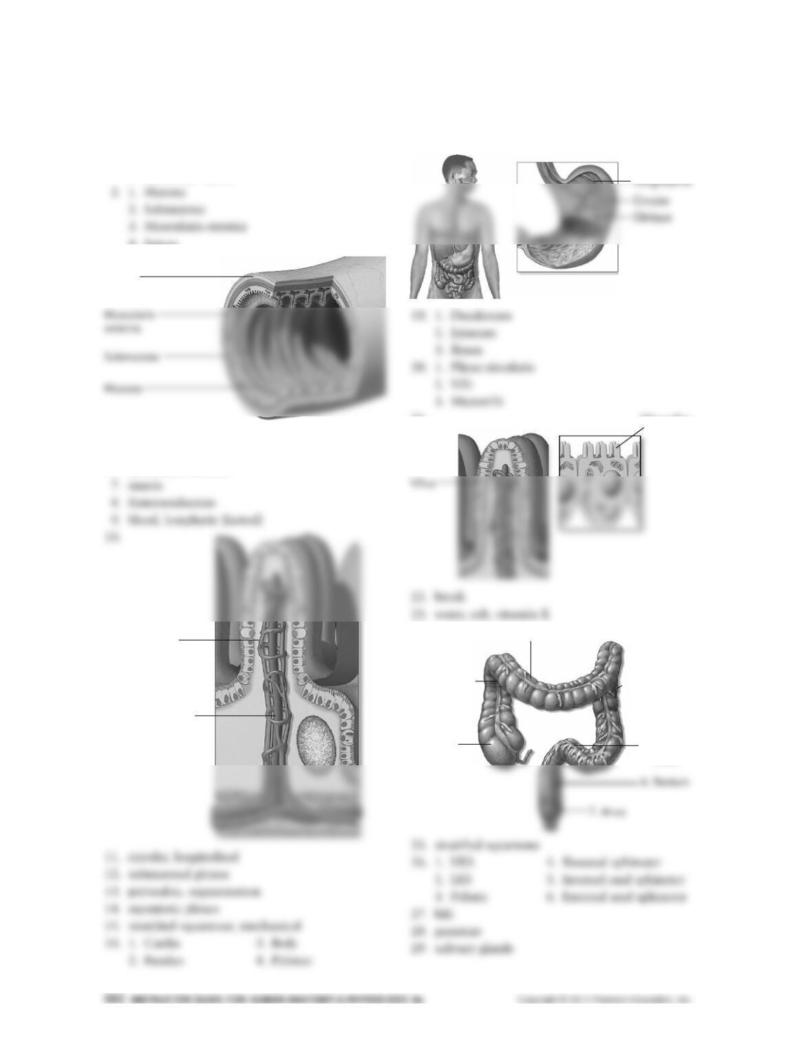
Digestive System: Anatomy Review
1. 1. Digestive (alimentary) tract
2. Accessory organs
4. Serosa
3.
4. epithelium
5. lamina propria
6. muscularis mucosa
17. 1. Circular
2. Longitudinal
3. Oblique
18.
21.
24.
Serosa
Blood capillary
Lymphatic (lacteal)
Microvillus
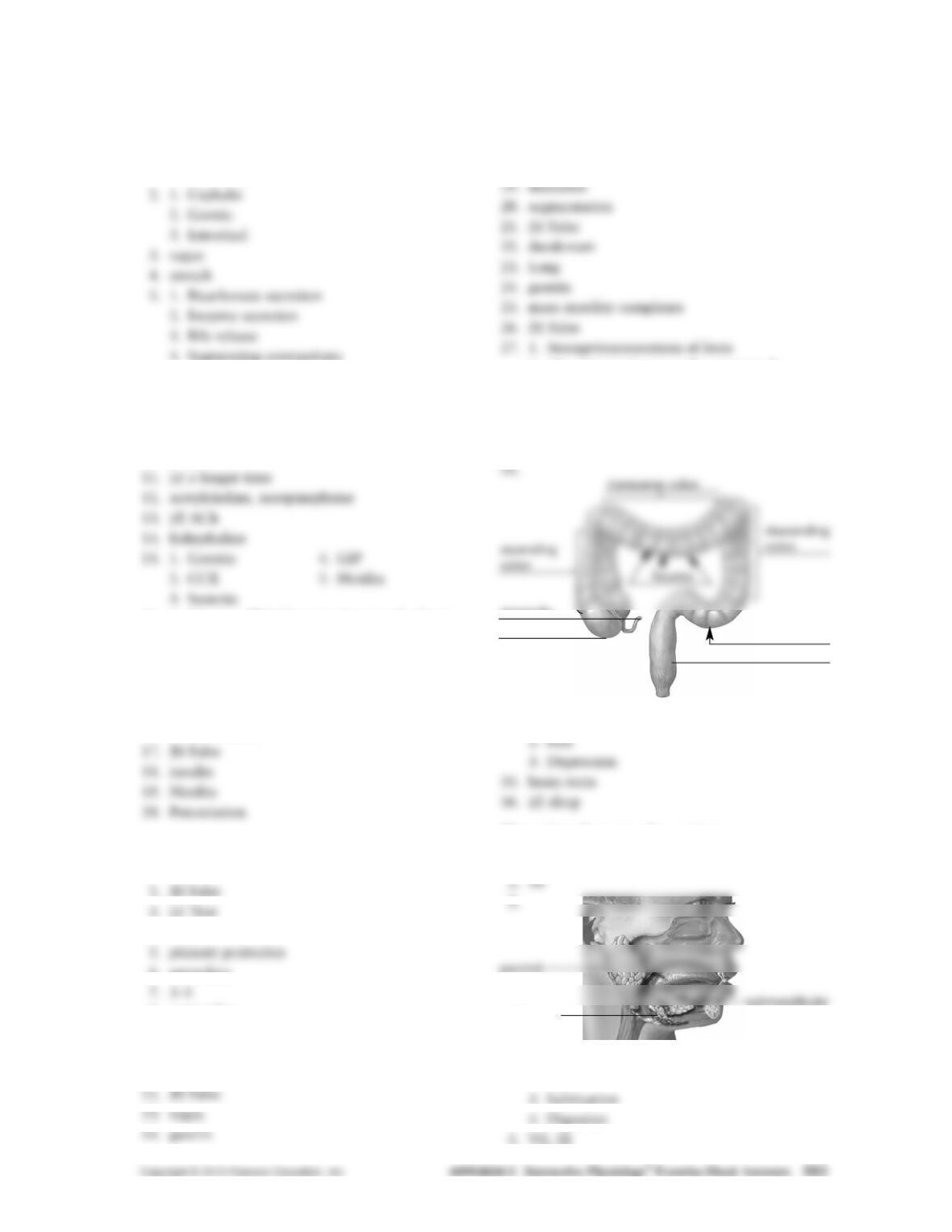
3. Transverse colon
2. Ascending
colon
1. Cecum
4. Descending
colon
5. Sigmoid
colon
Digestive System:
Control of the Digestive System
1. 1. Autonomic nervous system
2. Hormones
4. Segmenting contractions
6. (a) slows gastric emptying
7. vagus, pelvic splanchnic
8. (b) postganglionic
9. submucosal, myenteric
10. long reflexes
16. 1. Causes gallbladder to contract and release
bile
2. Causes pancreas to release digestive
enzymes
3. Inhibits gastric emptying
4. Stimulates growth of pancreas and gallblad-
der mucosa
Digestive System: Motility
1. ingestion
4. trachea
6. secondary
8. pacemaker
9. (e) All of the above
10. Nerves
11. [No response]
15. increases
16. CCK
17. CCK
18. enterogastric
2. Absorption of water, salts, vitamin K
28. haustra
29. mass movements
30. (b) external anal sphincter
31. 150
33. gastroileal
34. 1. Pain
Digestive System: Secretion
1. 0.15
4. 1. Protraction
2. Taste
cecum
appendix
rectum
sigmoid colon
sublingual
6. (a) True
7. Parasympathetic, sympathetic
8.
11. pyloric
12.
13. pyloric
14. 1. Aspirin 2. Alcohol
15. 1.5–2.0
16. (e) All of the above are functions of HCl.
17. intrinsic factor
18. 1. gastrin
20. CCK, secretin
bicarbonate pancreatic juice
22. 1. trypsinogen
2. chymotrypsinogen
3. procarboxypeptidase
23. enterokinase
24. insulin, glucagon
25. 1. bile salts
2. lecithin
3. cholesterol
4. bilirubin
26. brush border
7. (b) False
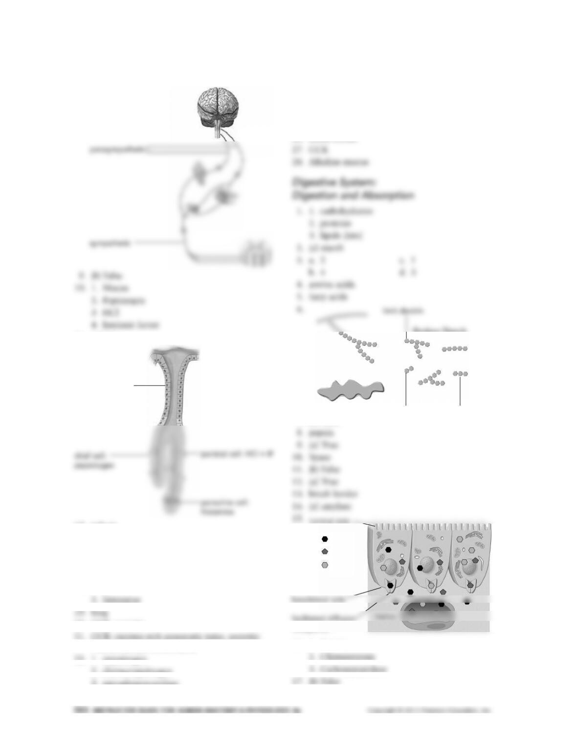
16. 1. Trypsin
mucus neck cells
maltose
Broken Starch
Starch
maltotriose
Amylase
transporter
Galactose
Fructose
Glucose
luminal side
18. 1. Aminopeptidase
2. Dipeptidase
19. 1. Segmentation
Immune System: Immune System Overview
1. 1. To destroy disease-causing organisms
2. To detect and kill abnormal cells such as
cancerous cells
4–5.
Line of Defense Example
Innate external defenses Skin and mucous membranes
(surface barriers)
Innate internal defenses Cells and chemicals in body
fluids
Adaptive defenses T and B cells
4. are systemic
9. antigenic determinant
10. plasma, antibodies
11. Humoral, B
12. Cellular, T
13. Humoral
Immune System: Anatomy Review
1. 1. Specialized immune cells (for example,
leukocytes)
2. Lymphoid organs and tissues (for example,
bone marrow)
3–4.
Name of Leukocyte Description
Basophil Large granules hide lobed nucleus
Stains blue/purple
5. a. neutrophils (blood) and macrophages (tissue)
7. a. lymph nodes d. appendix
b. spleen e. tonsils
c. Peyer’s patches
8. 1. Lymphatic vessels
2. Lymph
3. Lymph nodes
9. 3
10. lymphedema
16. B, T, cortex
17. spleen
18. pathogens, aged erythrocytes and platelets
platelets and breakdown products of erythrocytes
immune system
20. thymus
The thymus decreases in size and activity.
Immune System: Innate Host Defenses
1. 1. Surface barriers or innate external defenses
(for example, skin and mucous membranes)
3. 1. Keratin
3. Antimicrobial proteins
6. Neutrophil, Macrophage, Neutrophil
7. mannose, Toll-like (TLR)
8. 1. They ingest the pathogen
2. They release chemicals that mobilize other
cells of the innate and adaptive immune
system
11. Opsonization
1. Antibodies
2. Complement proteins
12. T cells
13. Natural killer, absence
14. cytotoxic, apoptosis
17. viral infection of the cell
18. inhibit viral replication, viral RNA, viral
proteins
19. a. mark cells for phagocytosis
b. promote inflammation
c. kill some bacteria by themselves
21. inflammation
opsonization
22. 1. inflammation
b. prostaglandins and kinins
c. cytokines
29. pyrogens, fever
a. most pathogens do not grow as well at
higher temperatures
b. fever causes the liver and spleen to sequester
iron and zinc
c. regulation of activation—clonal expansion
d. memory
2. self-antigens, specificity
3. antigenic determinants, lymphatic antigen,
antigenic determinant
4. antibody
5. major histocompatibility complex (MHC)
6. clones, clonal selection
7. 100 million
1. Generate a viable lymphocyte antigen
receptor
2. Survive a series of practical exams
10. antigen receptors, medulla
11. MHC, positive, apoptosis
12. self-antigens, negative, self-tolerant
14. TSH receptors
myelin in the nervous system
red blood cells
15. 1. Infection with a pathogen that has antigens
2. intact skin, mucous membranes 25. 1. Vasodilation
2. Increased vascular permeability
19. memory, secondary, faster, greater
20. vaccinations
Immune System: Humoral Immunity
1. B lymphocytes, immunoglobulins or gamma
globulins
5. bound
complement
opsonin
dimer, pentamer
8. IgA
IgM
IgM
IgA
IgA
IgM
13. Antihistamines
14. anaphylaxis
15. naïve B, B
16. 1. phagocytosis 3. agglutination
2. lysis 4. neutralization
17. 1. extracellular
2. secondary lymphoid organs
24. when your body makes antibodies in response
to an antigen
when you receive antibodies from another
person or animal
antibodies passed from mother to baby in
breast milk
injection of antibodies for rabies
b. Promote inflammation
c. Trigger apoptosis
d. Promote activation of immune cells
e. Help defend against viruses
5. CD4 cells: helper T, regulatory T, class II
CD8 cells: cytotoxic, class I
6. helper T
7. MHC, MHC, rejection
8. Cytotoxic T, class I, endogenous, foreign
9. a. Dendritic cells
both CD4 and CD8
both MHC I and MHC II
13. dendritic, CD8
14. 1. T-cell receptors bind to MHC proteins bear-
ing antigens.
2. Other co-stimulatory molecules bind to the
antigen-presenting cell.
20. gamma, macrophages, cytotoxic, 4, 5, B
21. cell-to-cell, cytokines, autoimmune
17. clonal expansion
1. Effector 2. Memory
18. plasma cells, primary
encountering antigen in the environment
(for example, cold)
vaccination