The Muscular System
Laboratory I: Extrinsic Muscles of the Thoracic Limb and
related areas
QUESTION ( PAGE CURRENTLY P. 7-8 )
vertebrae. What muscles would the needle encounter after it
penetrated the skin?
A: After penetration of the skin, the needle would pass
through the subcutaneous fascia and then the trapezius
CHAPTER
7
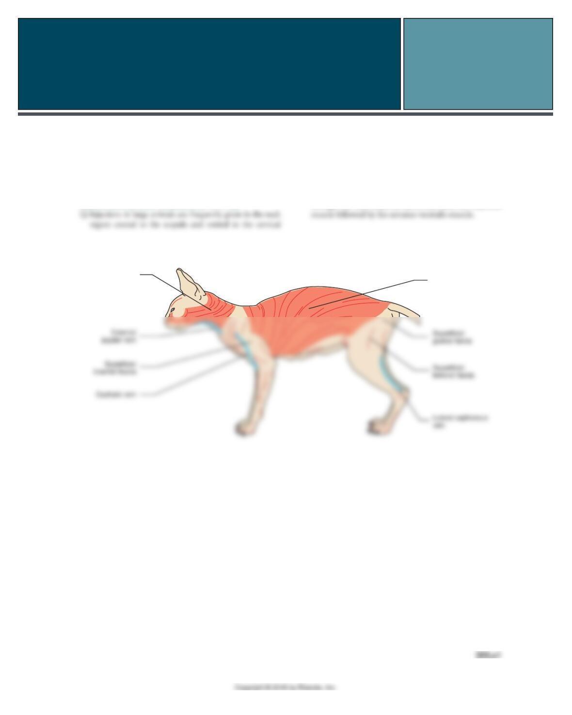
EXERCISE 1. IDENTIFY AND COLOR MUSCLES IN THE CAT
Platysma
muscle
Cutaneous trunci
muscle
CHAPTER 7 The Muscular System
205.e2
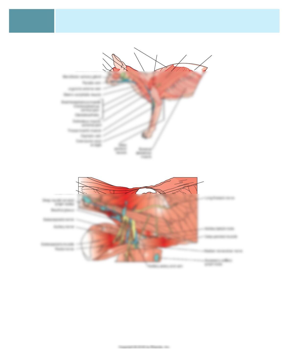
Cervical part
Thoracic part
Trapezius muscle:
Deltoid muscle:
spinous part Latissimus
dorsi muscle
Thoracolumbar
fascia
Omotransversarius muscle
Temporalis muscle
Parotid salivary gland
Masseter muscle
Thoracic portion of
serratus ventralis
Cervical portion of
serratus ventralis
Superficial cervical
lymph nodes
205.e3
CHAPTER 7 The Muscular System
EXERCISE 2. IDENTIFY MUSCLE NAME, ORIGIN, INSERTION, AND ACTION
MUSCLE ORIGIN INSERTION ACTION
1. Superfi cial pectoral Cranial sternum Greater tubercle of humerus Adducts thoracic limb
Deep pectoral Sternum Greater tubercle of humerus Adducts and pulls limb caudally
Brachiocephalicus 5. Clavicular intersection Ulna, mastoid, dorsal neck Pulls limb forward, extends
shoulder
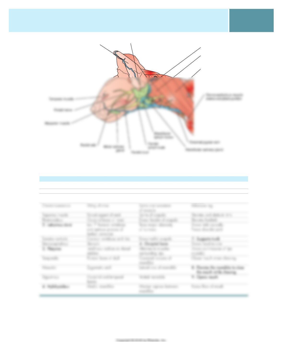
Parotid lymph node
Auricular cartilage Parotid salivary gland
Cleidocephalicus muscle
Sternocephalicus muscle
sterno-occipitalis portion
CHAPTER 7 The Muscular System
205.e4
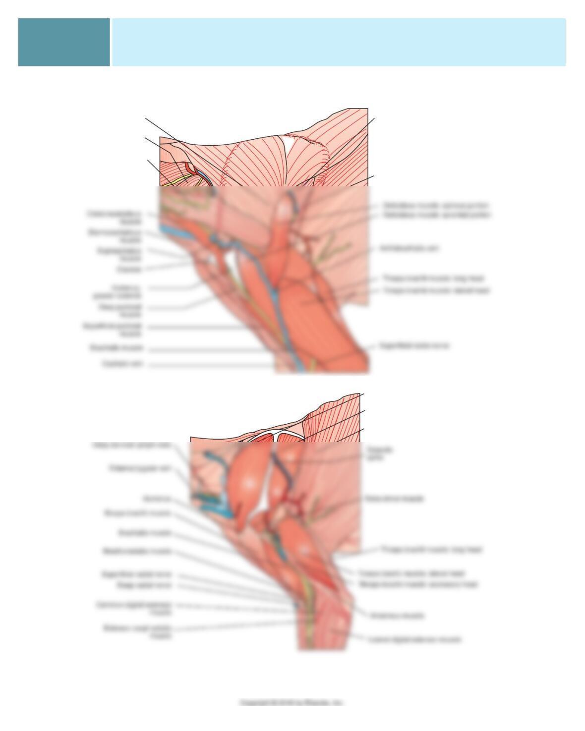
EXERCISE 3. IDENTIFY AND COLOR INTRINSIC MUSCLES
Scapula, spine
Cervical portion
serratus ventralis
Omotransversarius
muscle
Infraspinatus muscle
Teres major muscle
Supraspinatus
muscle
Infraspinatus muscle
Latissimus dorsi muscle
205.e5
CHAPTER 7 The Muscular System
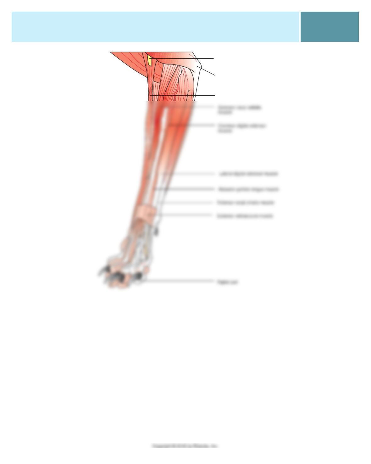
Brachioradialis muscle
Ulna, olecranon process
Superficial radial nerve
CHAPTER 7 The Muscular System
205.e6
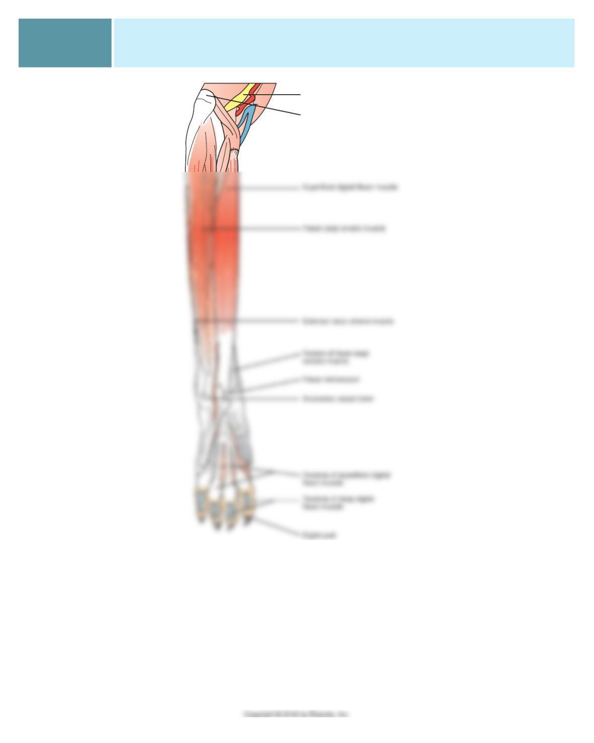
Ulna, olecranon process
Ulnar nerve
205.e7
CHAPTER 7 The Muscular System
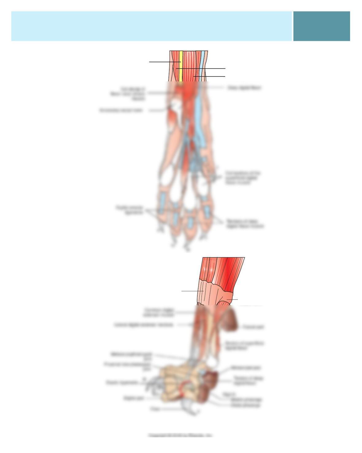
Median nerve
Deep digital flexor muscle:
Deep digital flexor
Deep digital flexor
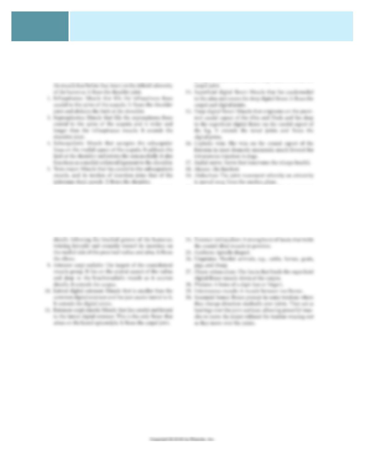
Extensor retinaculum
Accessory
carpal bone
CHAPTER 7 The Muscular System
205.e8
EXERCISE 4. IDENTIFY AND DESCRIBE
MUSCLES AND TERMS
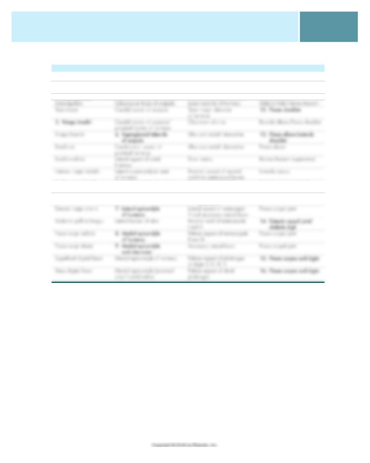
1. Deltoideus: Muscle that consists of two parts that join and
act in common. The scapular portion covers a majority of
the infraspinatus muscle located below it. The two parts of
6. Triceps brachii: Muscle that makes up the large muscle
mass on the caudal aspect of the humerus. It extends the
elbow and fl exes the shoulder.
7. Biceps brachii: Muscle located on the craniomedial
aspect of the humerus. It consists of only one head in
animals. It fl exes the elbow and extends the shoulder.
8. Brachialis: Muscle located on the lateral side of the hu-
merus. After its origin on the lateral humerus, it courses
12. Abductor pollicis longus: Flat triangular muscle that lies
primarily in the groove between the radius and ulna.
It extends the carpal joint and abducts the digit.
13. Flexor carpi ulnaris: The most caudal of this muscle
group; lies next to the extensor carpi ulnaris. It fl exes the
20. Adduction: The joint movement whereby an extremity
is moved toward the median plane.
21. Circumduction: A joint motion whereby the distal end
of an extremity moves in a circle.
22. Flexion: The joint movement that decreases the angle
between two bones.
23. Extension: The joint movement that increases the angle
between two bones.
205.e9
CHAPTER 7 The Muscular System
EXERCISE 5. IDENTIFY MUSCLE NAME, ORIGIN, INSERTION, AND ACTION
MUSCLE ORIGIN INSERTION ACTION
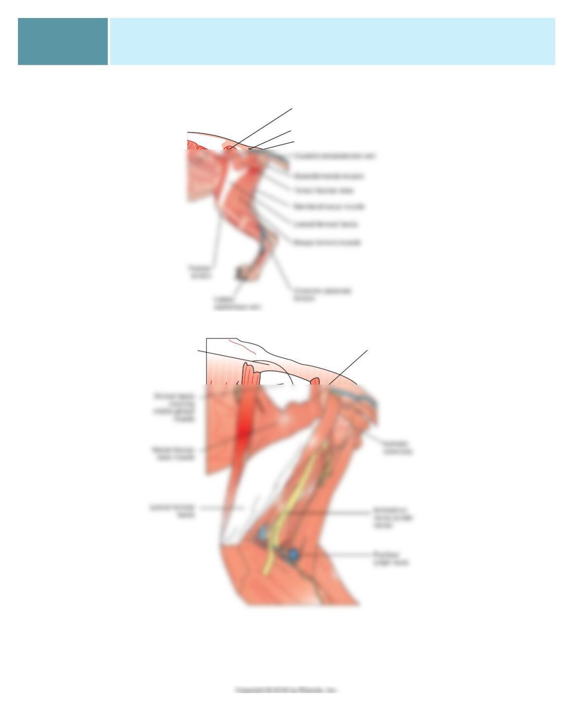
1. Deltoideus 5. Spine/acromion of scapula Deltoid tuberosity of humerus Flexes shoulder
Infraspinatus Infraspinous fossa of scapula Greater tubercle of humerus 11. Flexes shoulder/
abducts limb
2. Supraspinatus Supraspinous fossa of scapula Greater tubercle of humerus Extends shoulder
4. Common digital
extensor Lateral epicondyle of humerus Dorsal surface of distal
phalanges digits II, III, IV, V Extends carpus and digits
Lateral digital extensor Lateral epicondyle of humerus 10. Proximal ends of digits
II, III, IV, V Extends digital joints
CHAPTER 7 The Muscular System
205.e10
EXERCISE 6. IDENTIFY AND COLOR MUSCLES OF THE PELVIC LIMB
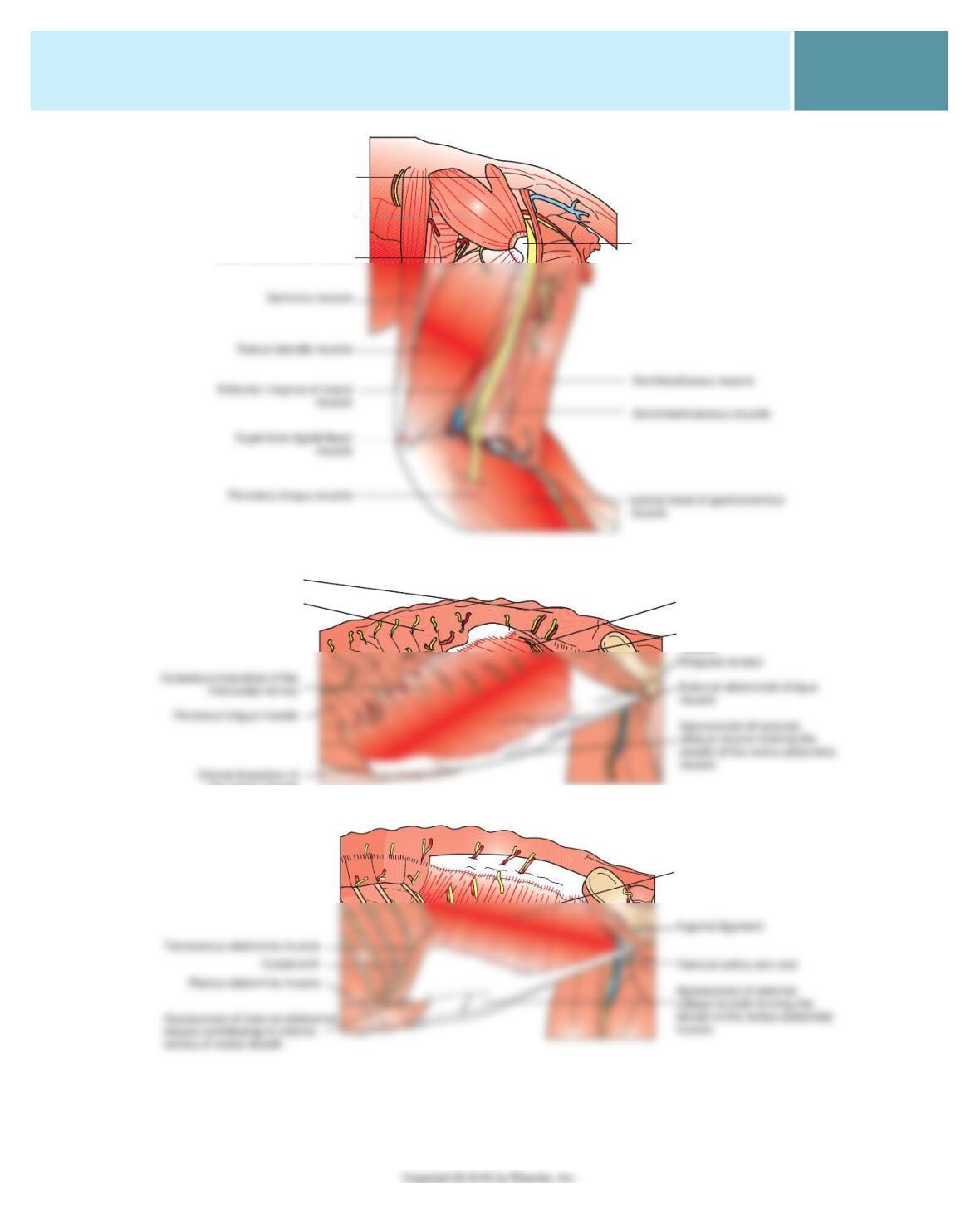
Internal abdominal
oblique muscle
Sartorius muscle
Gluteal fascia
Ilium, tuber
sacrale
Superficial
gluteal muscle
205.e11
CHAPTER 7 The Muscular System
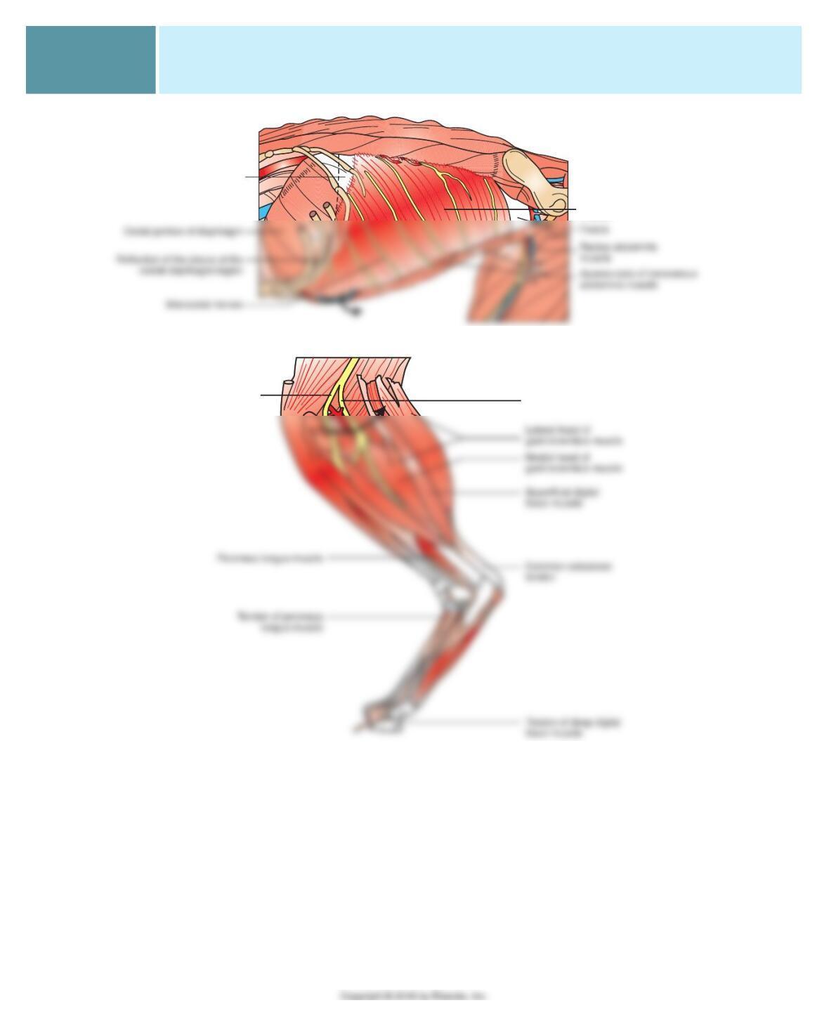
Superficial gluteal muscle
Middle gluteal muscle
Tensor fasciae latae muscle
Femur, greater trochanter
Abdominal artery
Internal abdominal oblique
muscle
Dorsocaudal portion of the
serratus ventralis muscle
Thoracolumbar fascia
the rectus sheath
Internal abdominal oblique
muscle
CHAPTER 7 The Muscular System
205.e12
Transversus abdominis
muscle
Fascia
Tibial nerve
Peroneal nerve
205.e13
CHAPTER 7 The Muscular System
EXERCISE 7. IDENTIFY MUSCLE NAME, ORIGIN, INSERTION, AND ACTION
MUSCLE ORIGIN INSERTION ACTION
1. Tensor fasciae latae
muscle Cranial wing of ilium Lateral femoral fascia Flexes hip/extends stifl e
Superfi cial gluteal muscle Sacrum and coccygeal
vertebrae 13. Greater trochanter
of femur Abducts limb/extends hip
Middle gluteal muscle Lateral ilium Greater trochanter of femur Abducts limb/extends hip
4. Quadriceps femoris Ilium/proximal femur Tibial tuberosity 21. Extends stifl e/fl exes hip
Cranial tibial muscle Cranial tibial border 15. Proximal metatarsus Flexes tarsus
Long digital extensor muscle 8. Extensor fossa of femur Extensor processes of distal
phalanges Extends digits/fl exes tarsus/
extends stifl e
5. Peroneus longus muscle Proximal tibia and fi bula Fourth tarsal bone/plantar
aspect of metatarsus Flexes tarsus
Gastrocnemius muscle 9. Medial/lateral
supracondylar
tuberosities of femur
Proximal surface of tuber
calcanei Extends tarsus/fl exes stifl e
CHAPTER 7 The Muscular System
205.e14
EXERCISE 8. CLINICAL THINKING CHALLENGE
1. Excitability—they can respond to a stimulus such as a
nerve impulse, 2) contractibility—they shorten in length
when stimulated, 3) extensibility—they will stretch when
respiration.
6. It offers several advantages over other sites. Excellent expo-
sure of abdominal organs, easy closure, and few sensory
nerves. Nearly all abdominal organs and tissues can be
reached through a ventral midline incision. Also, because all
of the abdominal muscles come together at the linea alba,
an incision through it opens the abdomen in one cut. When
it is time to close the abdomen, one layer of sutures
(stitches) in the linea alba can effectively and securely close
9. (1) The latent phase, (2) the contracting phase, and
(3) the relaxation phase.
10. When the animal dies, lack of oxygen to the cells
causes normal activities and barriers within the cells
11. Cardiac muscle cells are much smaller than skeletal
muscle cells and have only one nucleus per cell. They are
not shaped like the long, thin fi bers of skeletal muscle.
They are longer than they are wide and often have mul-
tiple branches. They are securely attached to each other
end to end to form intricate, branching networks of
cells. The fi rm, end-to-end attachments between cardiac
muscle cells are visible under the microscope as dark,
transverse lines between the cells.