362e.1
The Respiratory System
EXERCISE 1. LABEL THE STRUCTURES IN THE
SKULL AND NECK
1. Nasal passage
2. Tongue
EXERCISE 2. LABEL THE INDICATED
STRUCTURES
1. Larynx
2. Tracheal rings
EXERCISE 3. LABEL THE STRUCTURES
OF THE RESPIRATORY TRACT
1. Nostrils or nares
2. Nasal passage
3. Larynx
EXERCISE 4. LABEL THE STRUCTURES
IN THE THORAX OF THE CAT
(Indicate right or left where applicable.)
1. Diaphragm
EXERCISE 5. LABEL THE STRUCTURES
IN THE RESPIRATORY TRACT OF THE CAT
(Indicate right or left where applicable.)
1. Trachea
3. Diaphragm
4. Left lung
5. Right lung
4. Cranial lobe of the right lung
5. Primary bronchus
6. Accessory lobe of the right lung
7. Bifurcation of the trachea
8 . T r a c h e a
9. Caudal lobe of the left lung
10. Cranial lobe of the left lung
EXERCISE 7. IS THIS THE RIGHT OR LEFT SIDE
OF THE CAT? WHY?
The right side: (1) because three distinct lobes of the right
EXERCISE 8. IDENTIFY THE LOBES OF THE
LUNG
(Indicate right, left, and lung lobe names where applicable.)
6. Right caudal lobe
CHAPTER
13
CHAPTER 13 The Respiratory System
362.e2
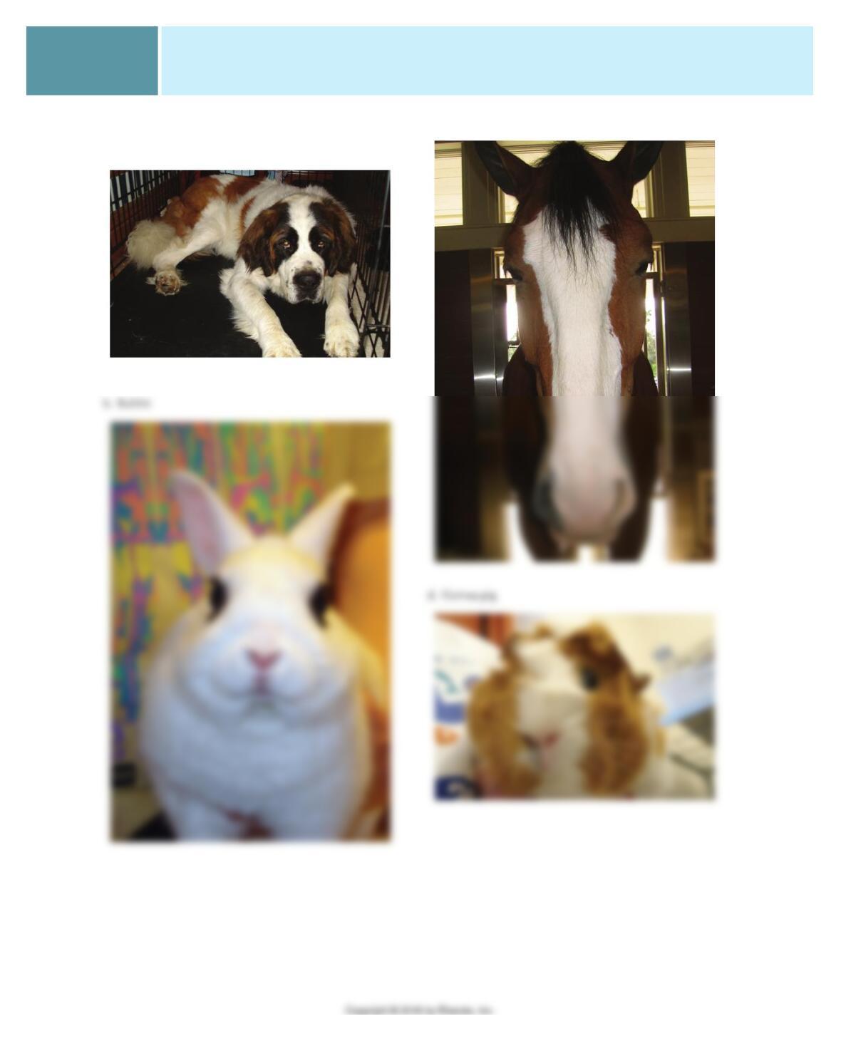
EXERCISE 9. NAME THAT NOSE—COMMON
ANIMALS
a. Dog (Saint Bernard)
c. Horse (Clydesdale)
362.e3
CHAPTER 13 The Respiratory System
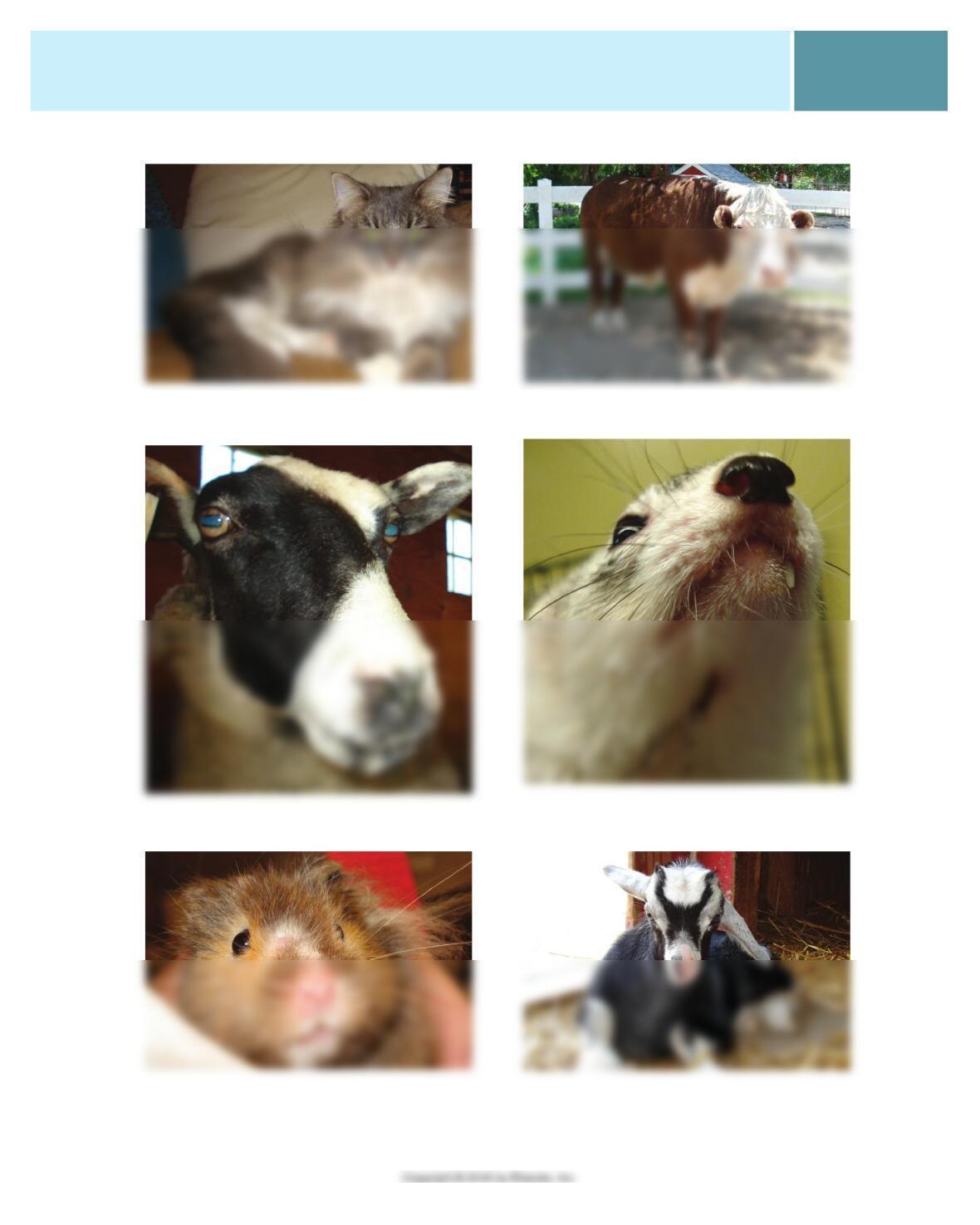
e . C a t h. Cow (dwarf Hereford steer)
f. Sheep
g. Hamster
i . F e r r e t
j . G o a t
CHAPTER 13 The Respiratory System
362.e4
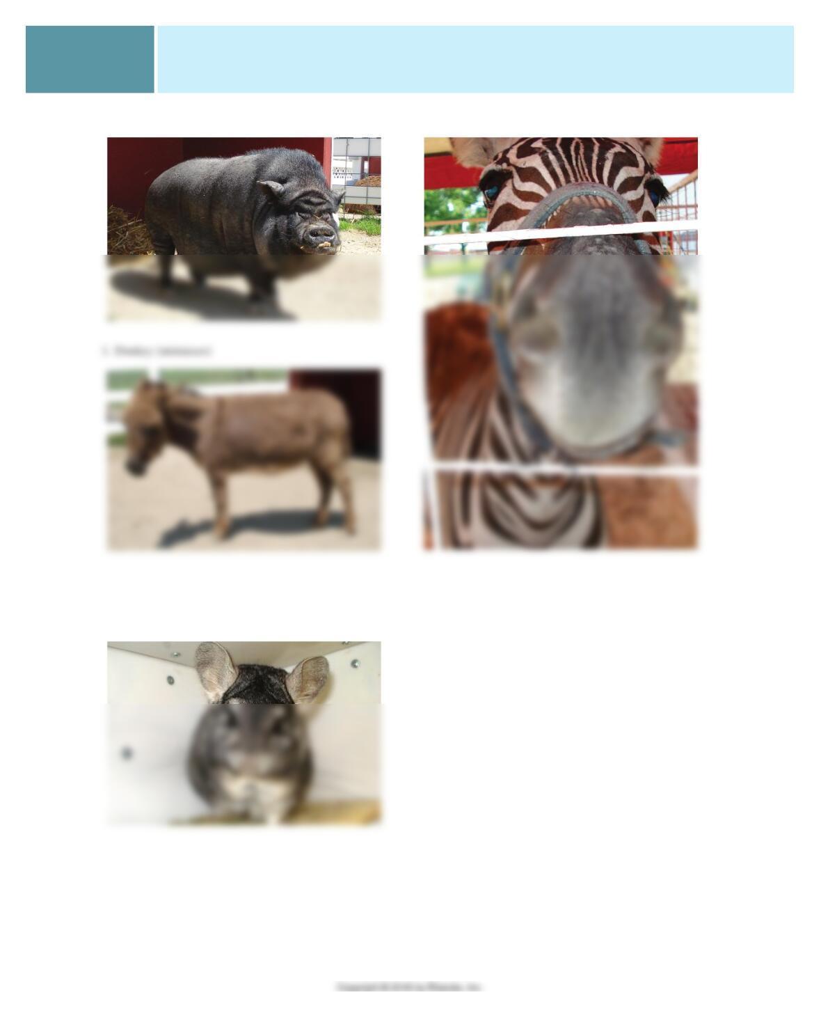
k. Pig (Vietnamese pot bellied pig) b . Z e b r a
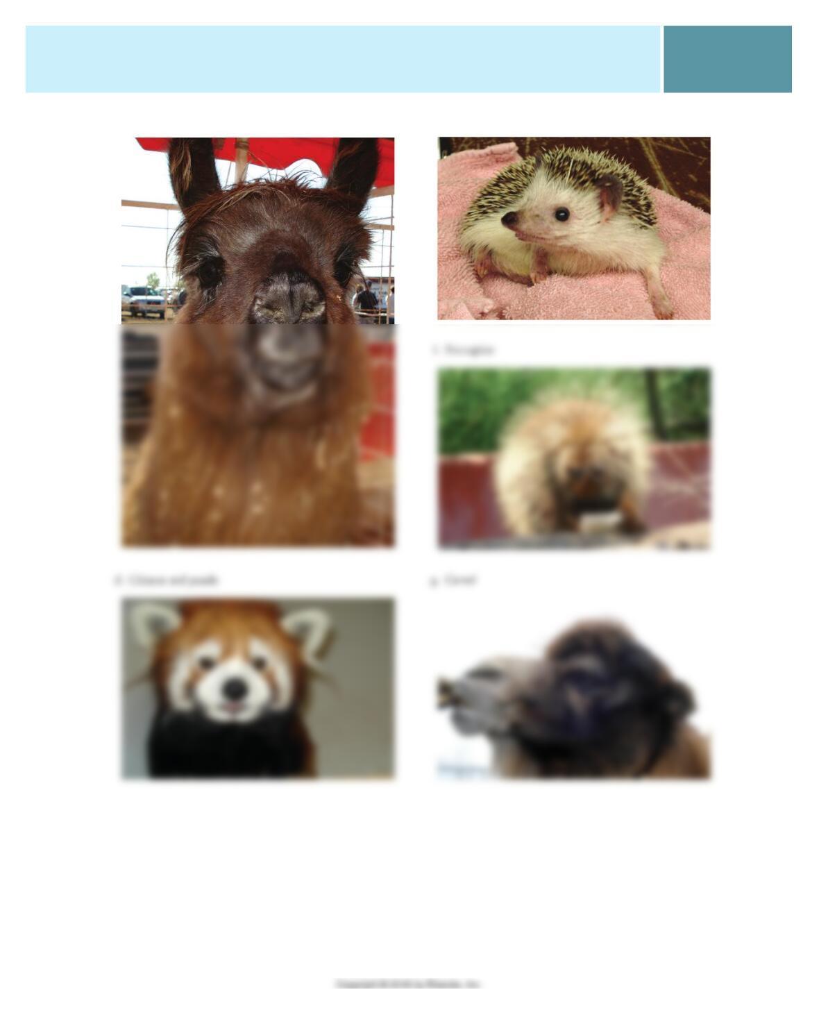
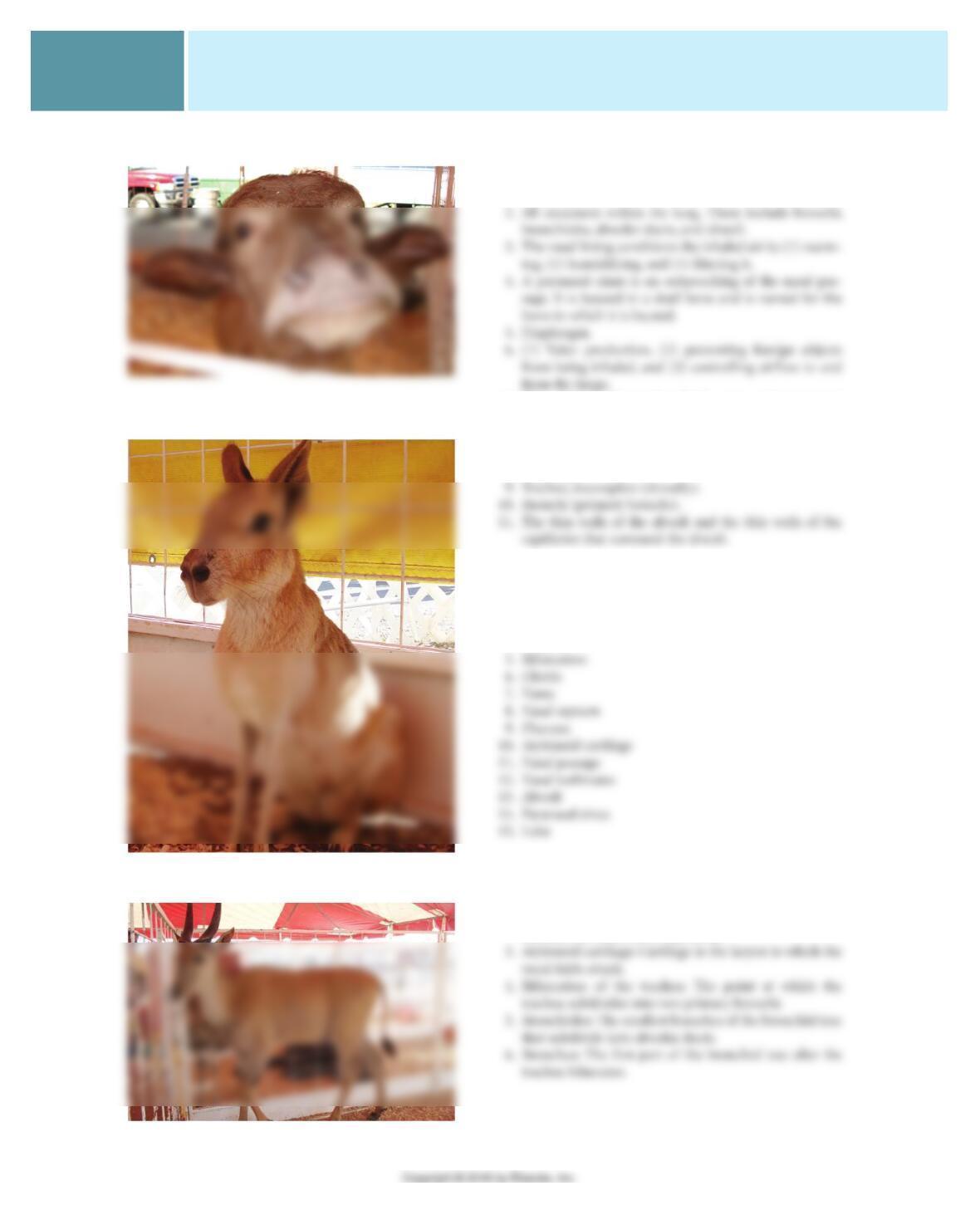
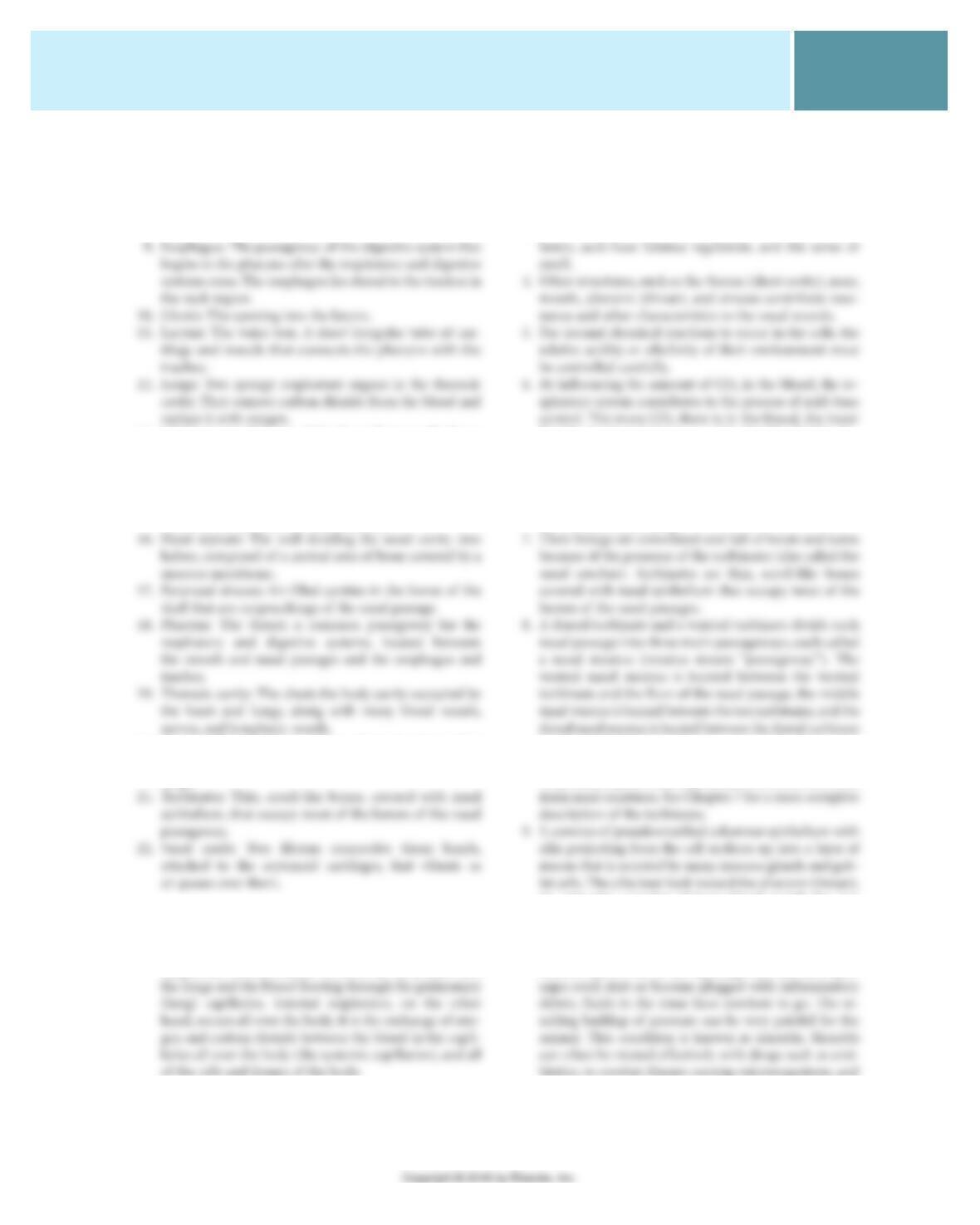
EXERCISE 10. NAME THAT NOSE—EXOTIC
ANIMALS
a. Chinchilla
362.e5
CHAPTER 13 The Respiratory System
e . H e d g e h o g c. Llama
CHAPTER 13 The Respiratory System
362.e6
h. Water buffalo
j. Antelope (eland)
i. Patagonian cavy
EXERCISE 11. CONNECT THE ANATOMIC DOTS
1. All structures between the nostrils and the entrance to
the lungs. These include the nares, nasal passages, phar-
ynx, larynx, and trachea.
7. The epiglottis leaves the glottis open and uncovered
when the animal is breathing. When the animal swal-
lows, the epiglottis covers the glottis to prevent food/
saliva from entering the larynx.
8. Esophagus.
EXERCISE 12. WHO AM I?
1. Vocal cords or vocal folds
2 . T r a c h e a
3. Diaphragm
4. Larynx
EXERCISE 13. DEFINE CLINICAL TERMS
1. Alveolar sacs: Clusters of alveoli at the ends of the alveo-
lar ducts arranged like bunches of grapes.
2. Alveoli: Microscopic, thin-walled sacs in the lung sur-
rounded by networks of capillaries.
362.e7
CHAPTER 13 The Respiratory System
7. Diaphragm: The thin, dome-shaped muscular structure
that forms the boundary between the thoracic and
abdominal cavities.
8. Epiglottis: The cartilage of the larynx that projects
forward from the ventral portion of the larynx. It is the
“trapdoor” of the larynx.
13. Mediastinum: The area of the thorax between the lungs
that contains the trachea, esophagus, blood vessels,
nerves, and lymph structures.
14. Nares: The nostrils.
15. Nasal passages: The passageways that conduct air
between the nostrils and the pharynx.
20. Trachea: The windpipe; a thin-walled tube descending
from the larynx to the bronchi that carries air to the
lungs.
EXERCISE 14. CLINICAL CHALLENGE
1. External respiration and internal respiration. External
respiration occurs in the lungs. It is the exchange of
oxygen and carbon dioxide between the air inhaled into
2. It is the means by which the body’s cells receive the oxy-
gen they need and get rid of the waste carbon dioxide
they produce. Without external respiration, there would
be no oxygen in the blood for the cells to absorb, and no
way for the cells to get rid of their carbon dioxide.
3. These include voice production, body temperature regu-
the blood pH and the more acidic the blood (CO
2
dis-
solves in the plasma to form carbonic acid [H
2
CO
3
]).
The respiratory system can alter the CO
2
content of the
blood by adjusting the volume and rate of breathing. We
describe this process more completely in the section on
the control of breathing.
and the roof of the nasal passage. A small fourth meatus,
called the common nasal meatus, is located on either side
of the nasal septum. It is continuous with the other three
An extensive complex of large blood vessels lies just
beneath the nasal epithelium.
10. The sinuses can be clinically signifi cant if they become
infl amed and swollen as a result of allergies, infections,
tumors, and so on. If the openings into the nasal pas-
CHAPTER 13 The Respiratory System
362.e8
decongestants, to reduce the swelling in the lining of the
sinus. However, in some severe cases, a hole must be
drilled surgically into an infl amed sinus to allow fl uid to
drain from it.
11. Because the pharynx is a common passageway that must
allow both breathing and swallowing, delicate refl exes
come into play to control the actions of the muscles
around the pharynx. Breathing is easy; the pharynx just
12. Aside from its role as part of the upper airway, the larynx
has three main functions: voice production, prevention
of foreign material being inhaled, and control of airfl ow
to and from the lungs.
13. The purpose of coughing is usually to clear mucus and
other matter from the lower respiratory passages. The
15. Actually, it is a paralysis of the muscles that tighten the
arytenoid cartilage and vocal cord on one side (usually
the left) of the larynx. The result is that the affected
vocal cord just “fl aps in the wind” as the animal breathes.
It usually does not cause a problem when the animal is
at rest, but when the animal exercises and breathes heav-
ily, the paralyzed vocal cord partially obstructs the glot-
tis each time the animal inhales. This produces the
characteristic “roaring” sound as the animal breathes
and makes it diffi cult for the animal to get enough air.
18. The structures of the lungs develop along with the rest of
the fetus but, until birth, the alveoli do not expand into
their saclike shapes. The fetal lungs have a solid consistency,
much like liver. If a piece of lung from a fetus that has never
breathed air is dropped into water, it will sink. Once an
animal is born and takes its fi rst breaths, the lungs expand,
and surfactant in the alveolar fl uid prevents the expanded
alveoli from collapsing again. In those fi rst few moments
aided by some of the muscles of the shoulder, neck, and
chest that attach to the rib cage.
20. The internal intercostal muscles are located between the
ribs. Their fi bers run at right angles to those of the ex-
ternal intercostals. When the internal intercostal muscles
contract, they rotate the ribs backward, which decreases
expiration. No matter how hard an animal tries, the
lungs cannot be completely emptied of air. The residual
volume always remains.
22. (British scientist) John Dalton’s law states that the total
pressure of a mixture of gases is the sum of the pressures of
each individual gas. It holds true for gases dissolved in
liquids. The amount of a particular gas that dissolves in
liquid exposed to a gaseous environment is determined
by the partial pressure of the gas in the gaseous environ-
ment. So the partial pressure of O
2
and CO
2
in the blood
362.e9
CHAPTER 13 The Respiratory System
the alveolar air into the blood of the alveolar capillaries?
It is moving from an area of high concentration (100
mm Hg in the alveolar air) to an area of low concentra-
24. A cough is a protective reflex that is stimulated by ir–
ritation or foreign matter in the trachea or bronchi. A
sneeze is similar to a cough, but the irritation origi-