Module 6: Film Mounting, Normal Anatomy, and Interpretation Basics 13
TEACH Lesson Plan
IANNUCCI AND HOWERTON: Dental Radiography: Principles and Techniques, 5th Edition
Short Answer
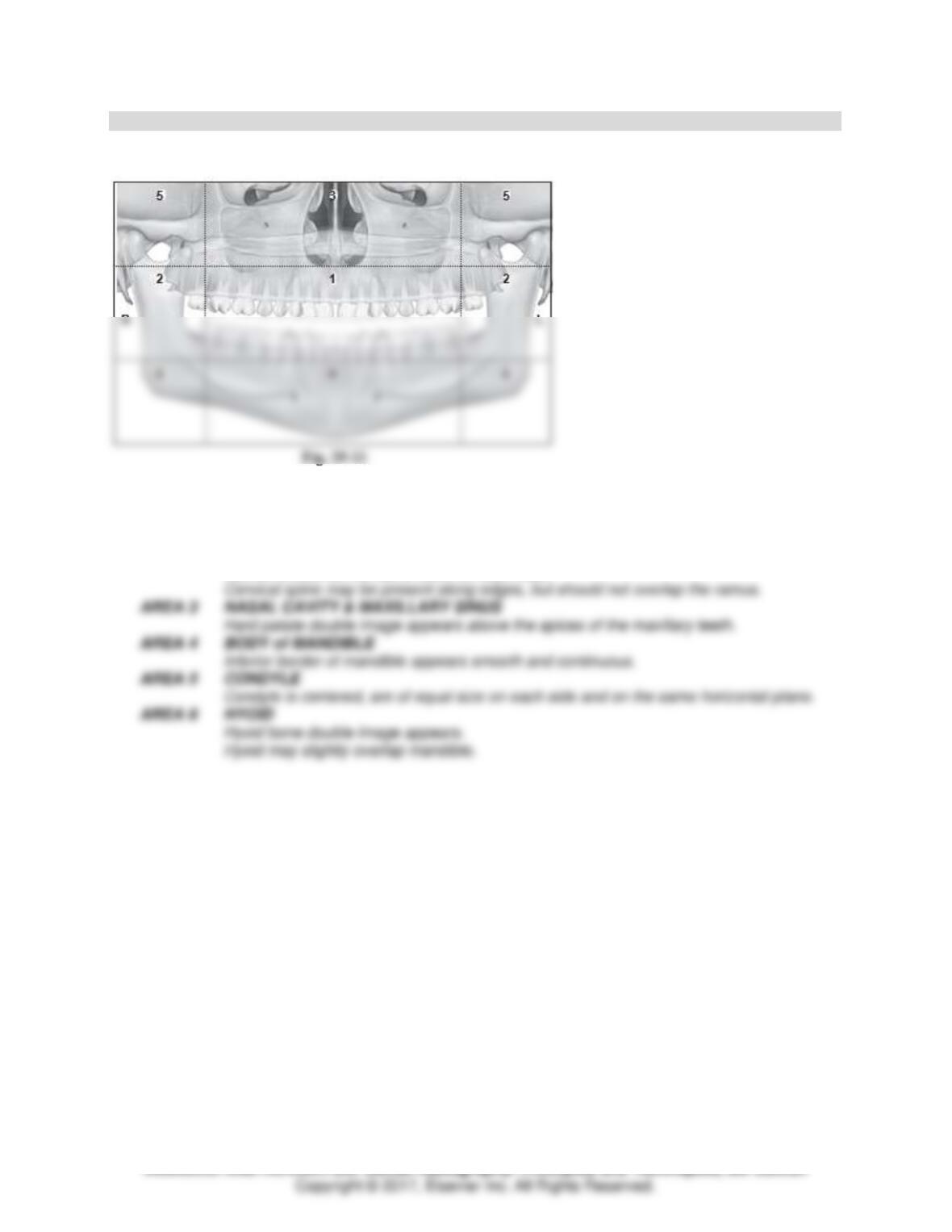
For each of the six areas identified on Fig. 29-11, describe the features that should be visible to
determine the diagnostic quality of the panoramic image. (See Box 22-1 in text.)
Diagnostic Panoramic Image Features
AREA 1 DENTITION
Teeth are arranged in a smile-like curve.
Crowns and apices of all teeth visible.
AREA 2 RAMUS & CERVICAL SPINE
Ramus should be the same width on each side.
Module 6: Film Mounting, Normal Anatomy, and Interpretation Basics 14
TEACH Lesson Plan
IANNUCCI AND HOWERTON: Dental Radiography: Principles and Techniques, 5th Edition
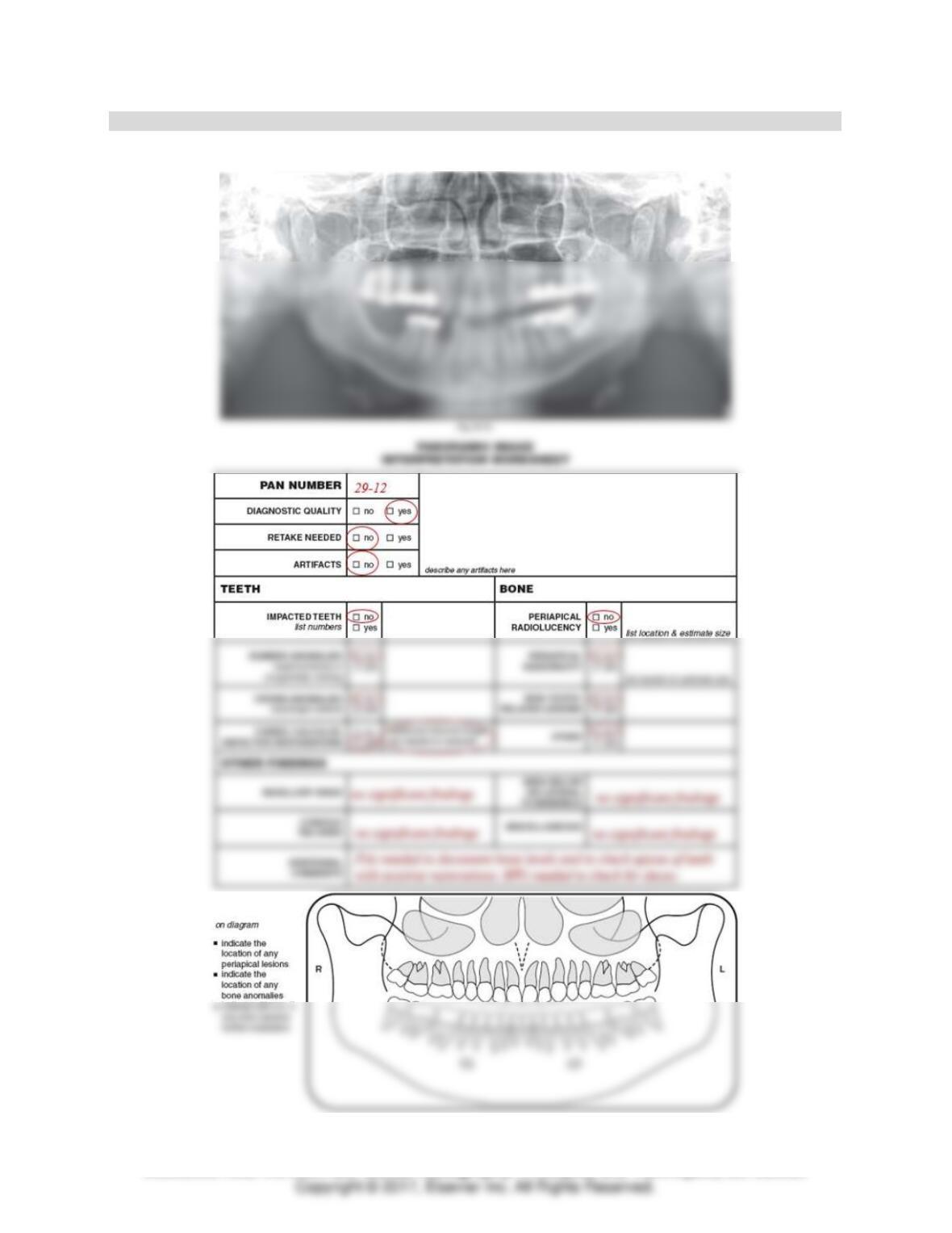
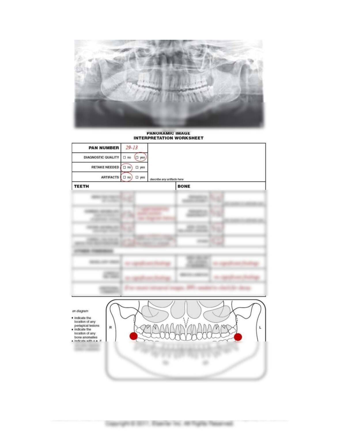
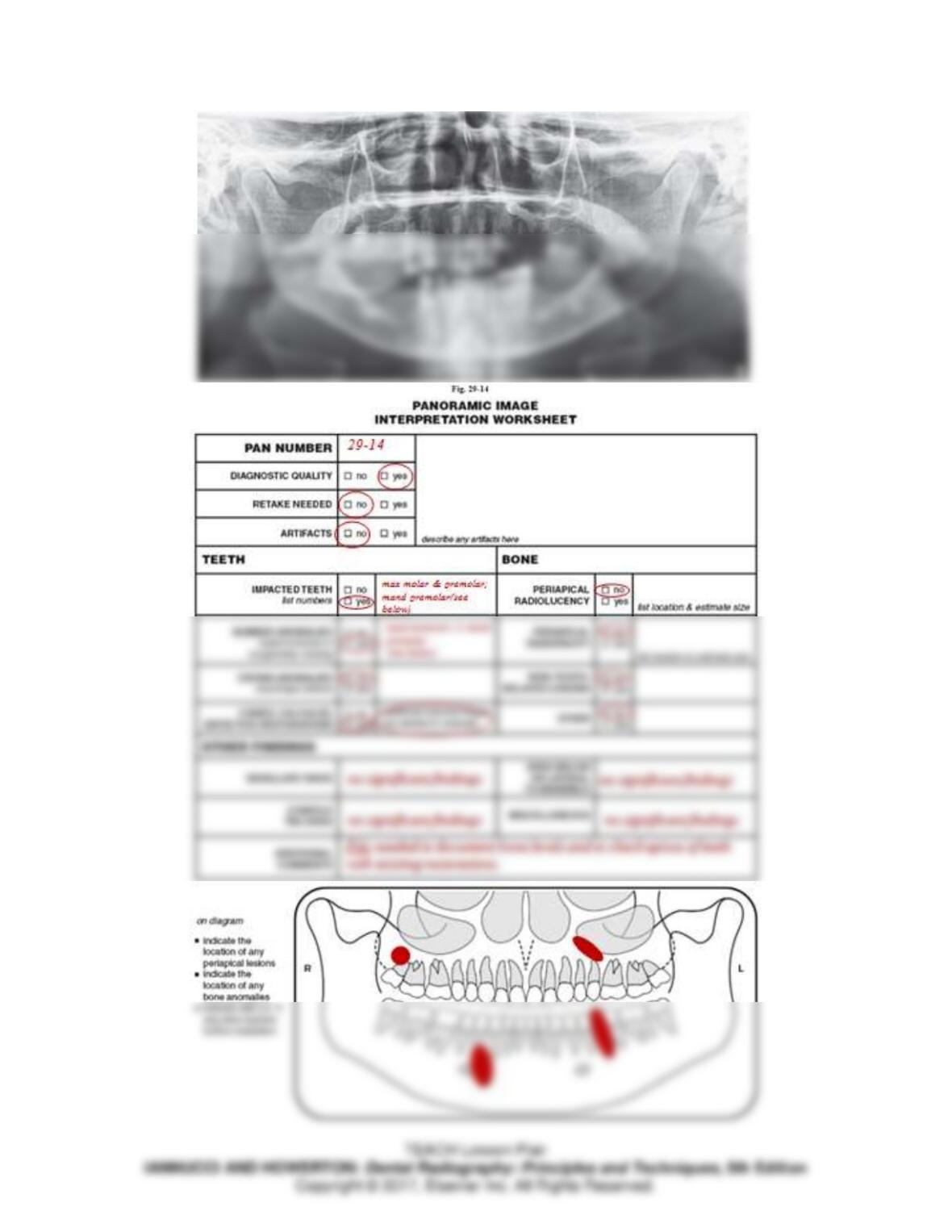
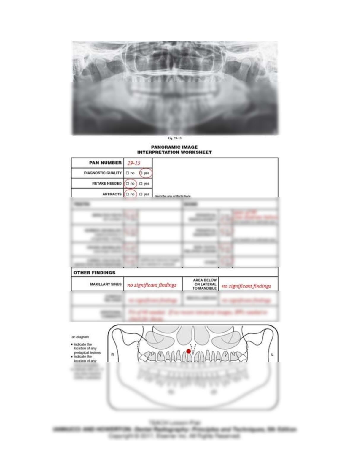
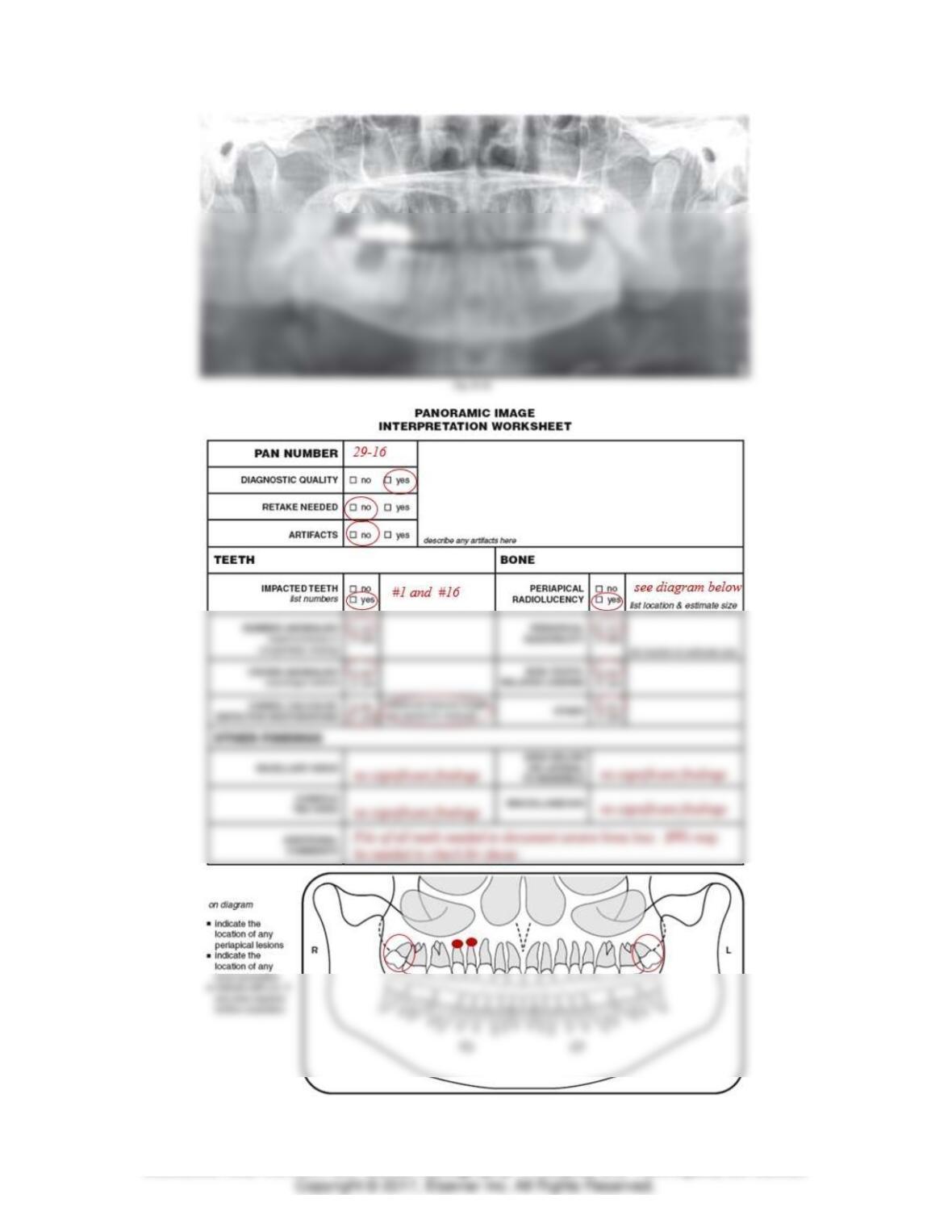
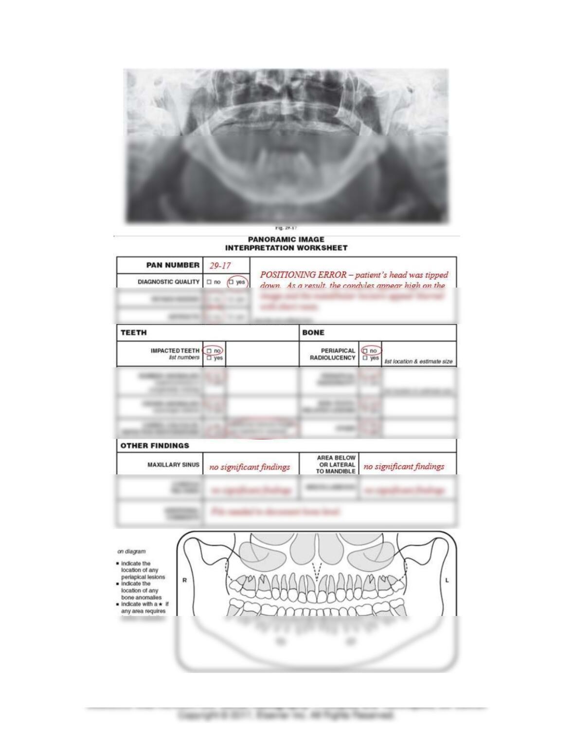
Panoramic Image Interpretation Exercises
For each panoramic image provided, complete a Panoramic Image Interpretation Worksheet. For each
panoramic image, practice identifying normal anatomy.
Module 6: Film Mounting, Normal Anatomy, and Interpretation Basics 15
TEACH Lesson Plan
IANNUCCI AND HOWERTON: Dental Radiography: Principles and Techniques, 5th Edition
Module 6: Film Mounting, Normal Anatomy, and Interpretation Basics 16
Module 6: Film Mounting, Normal Anatomy, and Interpretation Basics 17
Module 6: Film Mounting, Normal Anatomy, and Interpretation Basics 18
TEACH Lesson Plan
IANNUCCI AND HOWERTON: Dental Radiography: Principles and Techniques, 5th Edition
Module 6: Film Mounting, Normal Anatomy, and Interpretation Basics 19
TEACH Lesson Plan
Module 6: Film Mounting, Normal Anatomy, and Interpretation Basics 20
TEACH Lesson Plan
IANNUCCI AND HOWERTON: Dental Radiography: Principles and Techniques, 5th Edition
CHAPTER 30: INTRODUCTION TO INTERPRETATION
Ordering
Number the following in order of the recommended viewing sequence for interpretation of images.
1. 8 bite-wings – left
2. 7 bite-wings – right
True or False
1. F The documentation of dental image interpretation is optional.
Short Answer
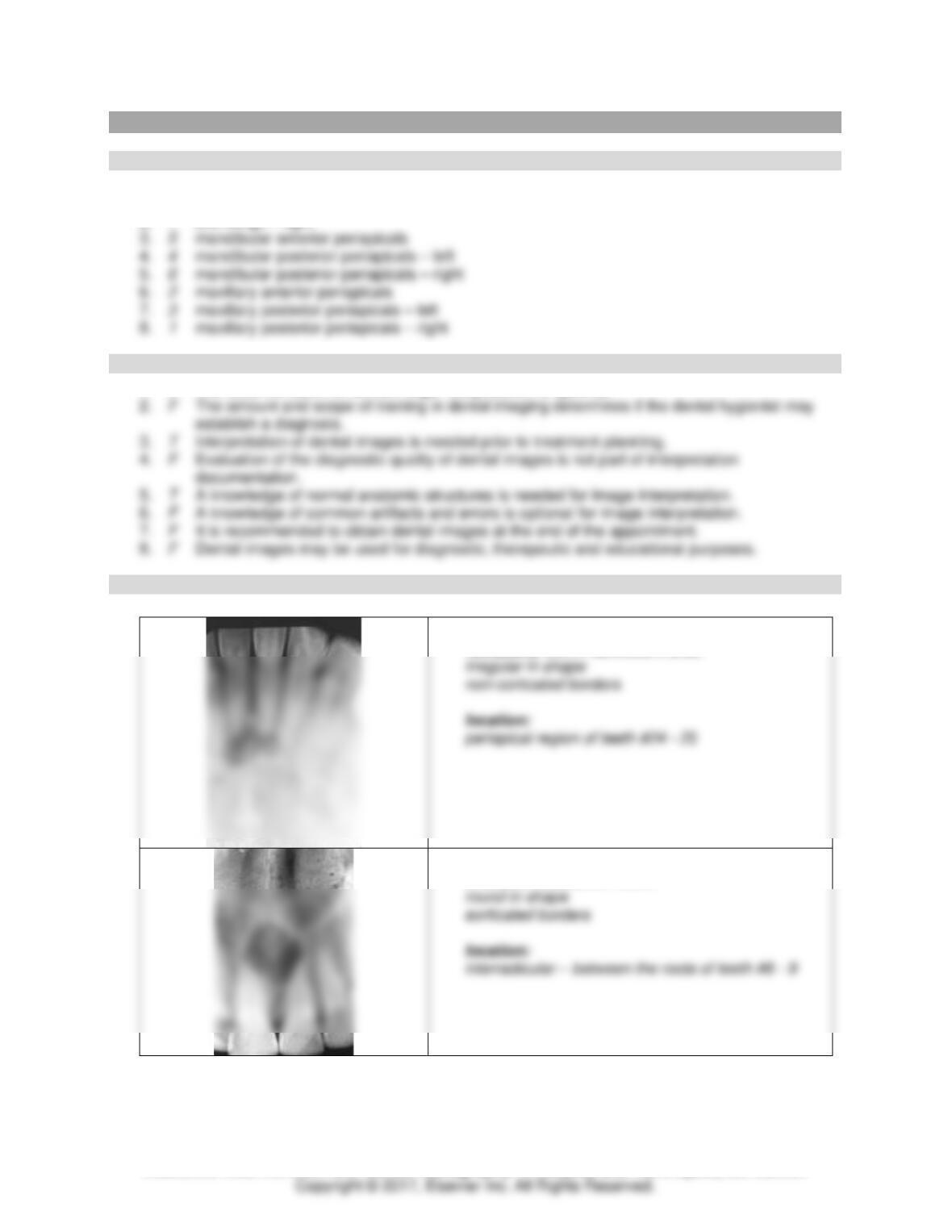
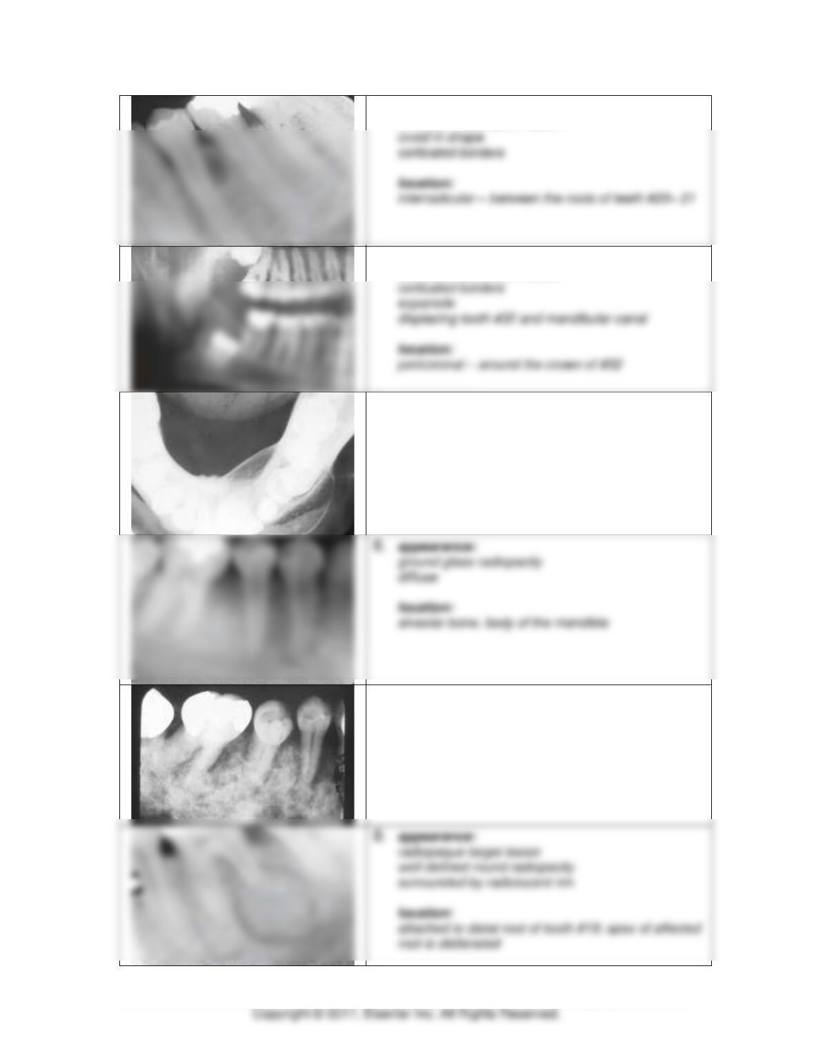
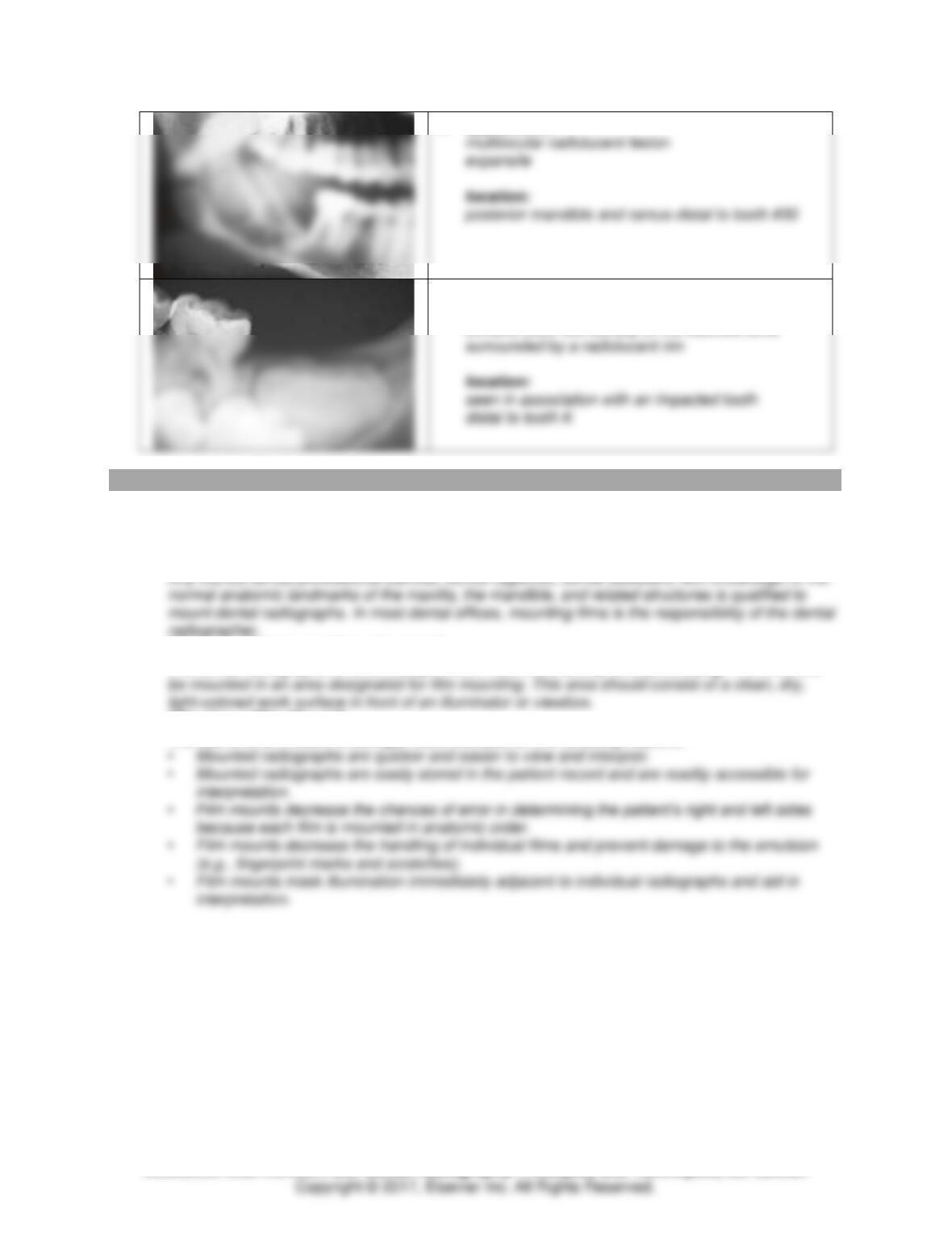
For each image, describe the appearance and location of the lesion or condition.
1. appearance:
radiopacity within radiolucent area
2. appearance:
unilocular radiolucent lesion
Module 6: Film Mounting, Normal Anatomy, and Interpretation Basics 21
TEACH Lesson Plan
IANNUCCI AND HOWERTON: Dental Radiography: Principles and Techniques, 5th Edition
3. appearance:
unilocular radiolucent lesion
4. appearance:
unilocular radiolucent lesion
5. appearance:
mixed lucent-opaque lesion
expansile with corticated borders
location:
body of the mandible
causing expansion of the buccal and lingual cortical
plates of bone
7. appearance:
mixed lucent-opaque lesion
diffuse cotton wool appearance
location:
alveolar bone, body of mandible
Module 6: Film Mounting, Normal Anatomy, and Interpretation Basics 22
TEACH Lesson Plan
IANNUCCI AND HOWERTON: Dental Radiography: Principles and Techniques, 5th Edition
9. appearance:
10. appearance:
well-defined radiopacity
exhibits tooth-like density of the calcified area
MODULE 2: CRITICAL THINKING QUESTIONS
1. A new assistant has started working in your office. It is your job to teach and instruct your new co–
worker on who, when, where, and why images are mounted. What important details will you
share with the assistant?
Who Mounts Films?
When and Where are Films Mounted?
The dental radiographer should always mount films immediately after processing. Films should
Why Use a Film Mount?
The use of a film mount is strongly recommended for the following reasons:
Module 6: Film Mounting, Normal Anatomy, and Interpretation Basics 23
TEACH Lesson Plan
IANNUCCI AND HOWERTON: Dental Radiography: Principles and Techniques, 5th Edition
2. A student at the local university has come into your office for a career day. She is interested in
becoming a part of the dental team and would like to see what each job entails and what it is like
to be in the dental profession for a day. As you are explaining to her the daily routine of your
office, she asks why you can interpret images but not diagnose. How can you answer the student
correctly?
Who Interprets Images?
Interpretation versus Diagnosis
In the dental setting, the terms interpretation and diagnosis are often confused; it is important to
3. After viewing a radiolucent periapical lesion on a dental image, what should the dental
radiographer record in the patient’s record?
In order to properly document a lesion seen on a dental image, the lesion must be described in
preferred unit of measurement is the millimeter or centimeter.
4. In dental imaging, a number of different terms can be used to describe the appearance, location,
and size of a lesion. Why would you want to utilize descriptive terminology to discuss and
describe what is seen on a dental image?
Descriptive terminology allows dental professionals to intelligently describe and discuss what is