
TEACH Lesson Plan
IANNUCCI AND HOWERTON: Dental Radiography: Principles and Techniques, 5th Edition
Module 5 Digital Imaging Basics and
Normal Anatomy Basics
CHAPTER 27: NORMAL ANATOMY: INTRAORAL IMAGES
Normal Anatomy Crossword Puzzle 1
Across
2. inferior border
Down
1. angle
Normal Anatomy Crossword Puzzle 2
Across
1. tubercle
3. mylohyoid ridge
Down
2. lamina dura
3. mastoid process

Module 5: Digital Imaging Basics and Normal Anatomy Basics 2
TEACH Lesson Plan
IANNUCCI AND HOWERTON: Dental Radiography: Principles and Techniques, 5th Edition
Identification
Radiolucent
Radiopaque
Radiolucent
Radiopaque
GENERAL TERMS
BONY LANDMARKS of MANDIBLE
1
✓
canal
32
✓
coronoid process
2
✓
canal – borders of
33
✓
external oblique ridge
3
✓
cortical bone
34
✓
genial tubercles
5
fossa
36
✓
internal oblique ridge
6
✓
process
37
✓
lingual foramen
7
✓
ridge
38
✓
mandibular canal
8
✓
septum
39
✓
mental foramen
9
sinus
40
✓
mental fossa
10
✓
spine
41
✓
mental ridge
11
suture
42
✓
mylohyoid ridge
12
✓
tubercle
43
✓
nutrient canals
13
✓
tuberosity
44
✓
submandibular fossa
BONY LANDMARKS of MAXILLA
NORMAL TOOTH ANATOMY
14
✓
anterior nasal spine
45
✓
alveolar crest – anterior
15
✓
hamular process
46
✓
alveolar crest – posterior
16
incisive foramen
47
✓
dentin
17
✓
inferior nasal conchae
48
✓
dentino-enamel junction
18
✓
inverted Y
49
✓
enamel
19
lateral fossa
50
✓
lamina dura
20
maxillary sinus
51
✓
periodontal ligament space
21
✓
maxillary sinus floor
52
✓
pulp cavity
22
maxillary sinus nutrient canals
SOFT TISSUE
23
✓
maxillary sinus septa
53
✓
lip line
24
✓
maxillary tuberosity
54
✓
nasolabial groove
25
median palatal suture
55
✓
nose
26
nasal cavity
27
✓
nasal cavity floor
28
✓
nasal septum
29
superior foramina of incisive canal
30
✓
zygoma
31
✓
zygomatic process of maxilla

Module 5: Digital Imaging Basics and Normal Anatomy Basics 3
TEACH Lesson Plan
IANNUCCI AND HOWERTON: Dental Radiography: Principles and Techniques, 5th Edition
Copyright © 2017, Elsevier Inc. All Rights Reserved.
Identify each item listed as appearing on a maxillary image, a mandibular image, or on both. Place a
check mark (✔) in the appropriate column or columns.
Found on
Maxillary
Images
Found on
Mandibular
Images
1
✓
✓
alveolar crest
9
✓
inferior nasal conchae
10
✓
internal oblique ridge
11
✓
inverted Y
12
✓
✓
lamina dura
13
✓
lateral fossa
14
✓
lingual foramen
15
✓
mandibular canal
16
✓
maxillary sinus
17
✓
maxillary sinus floor
18
✓
maxillary sinus nutrient canals
19
✓
maxillary sinus septa
20
✓
maxillary tuberosity
21
✓
median palatal suture
22
✓
mental foramen
23
✓
mental fossa
24
mental ridge
✓
25
✓
mylohyoid ridge
26
✓
nasal cavity
27
✓
nasal cavity floor
28
✓
nasal septum
29
✓
nasolabial groove
31
✓
✓
nutrient canals
32
✓
✓
periodontal ligament space
33
✓
submandibular fossa
34
✓
superior foramina of incisive
35
✓
zygoma
36
✓
zygomatic process of maxilla
✓
anterior nasal spine
3
✓
coronoid process
4
✓
external oblique ridge
5
✓
genial tubercles
6
✓
hamular process
7
✓
incisive foramen
✓
inferior border of mandible
Module 5: Digital Imaging Basics and Normal Anatomy Basics 4
TEACH Lesson Plan
IANNUCCI AND HOWERTON: Dental Radiography: Principles and Techniques, 5th Edition
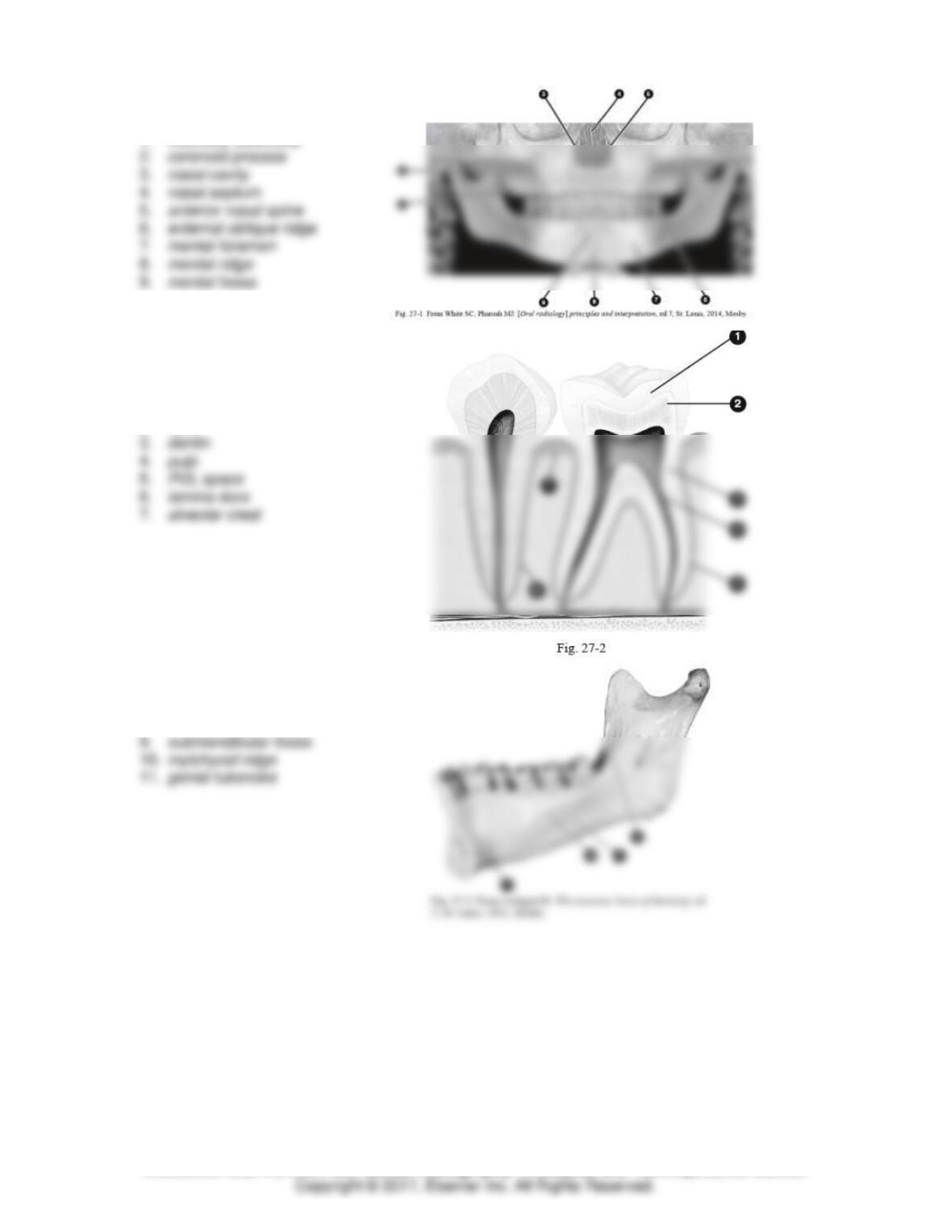
Identify each structure in Figure 27–1.
1. maxillary tuberosity
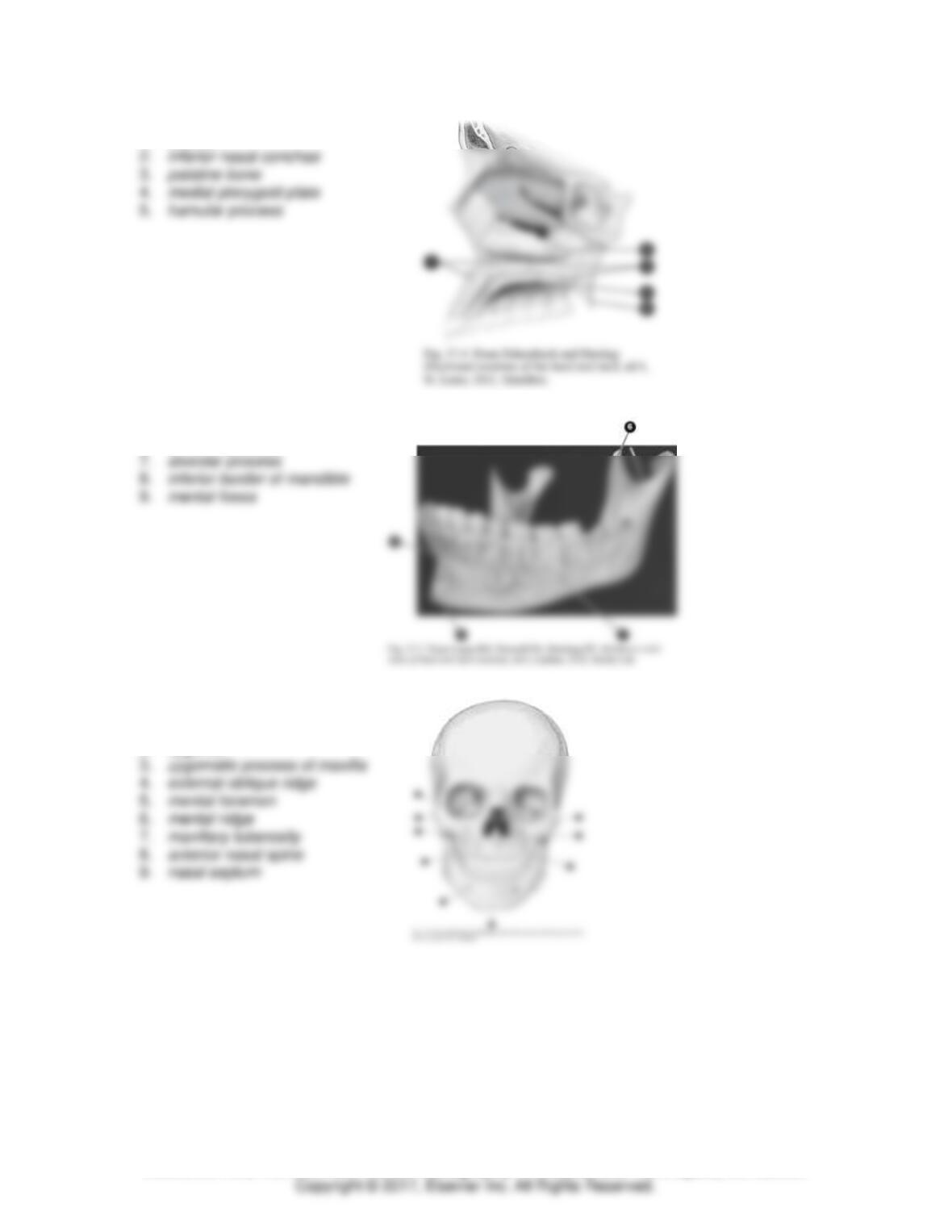
Identify each structure in Figure 27–2.
1. enamel
2. DEJ
Identify each structure in Figure 27–3.
8. internal oblique ridge
Module 5: Digital Imaging Basics and Normal Anatomy Basics 5
TEACH Lesson Plan
IANNUCCI AND HOWERTON: Dental Radiography: Principles and Techniques, 5th Edition
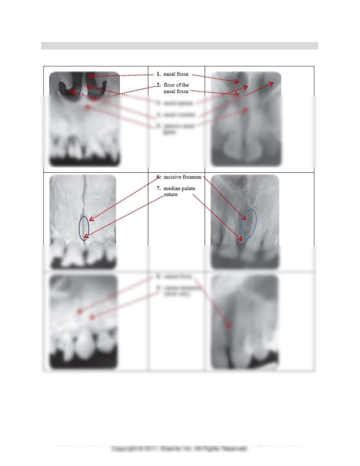
Identify each structure in Figure 27–4.
1. maxilla
Identify each structure in Figure 27-5.
6. coronoid process
Identify each structure in Figure 27–6.
1. inferior nasal conchae
2. zygoma
Module 5: Digital Imaging Basics and Normal Anatomy Basics 6
TEACH Lesson Plan
IANNUCCI AND HOWERTON: Dental Radiography: Principles and Techniques, 5th Edition
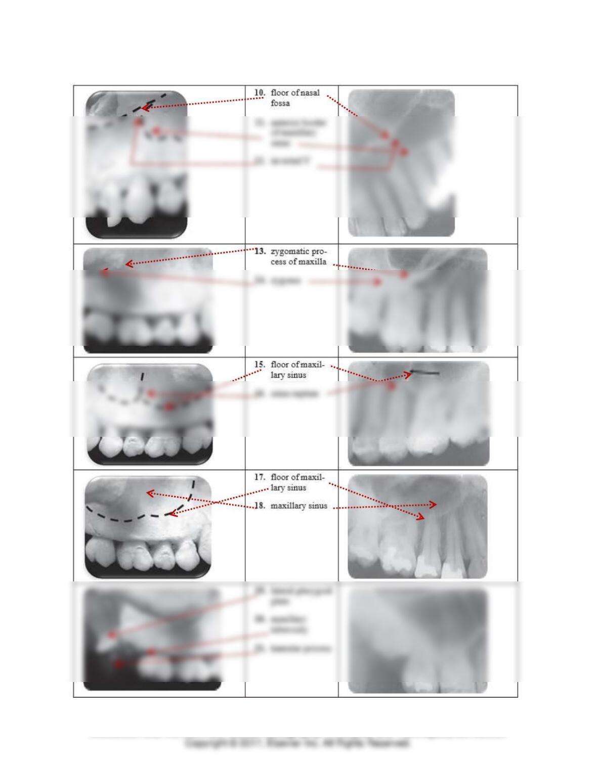
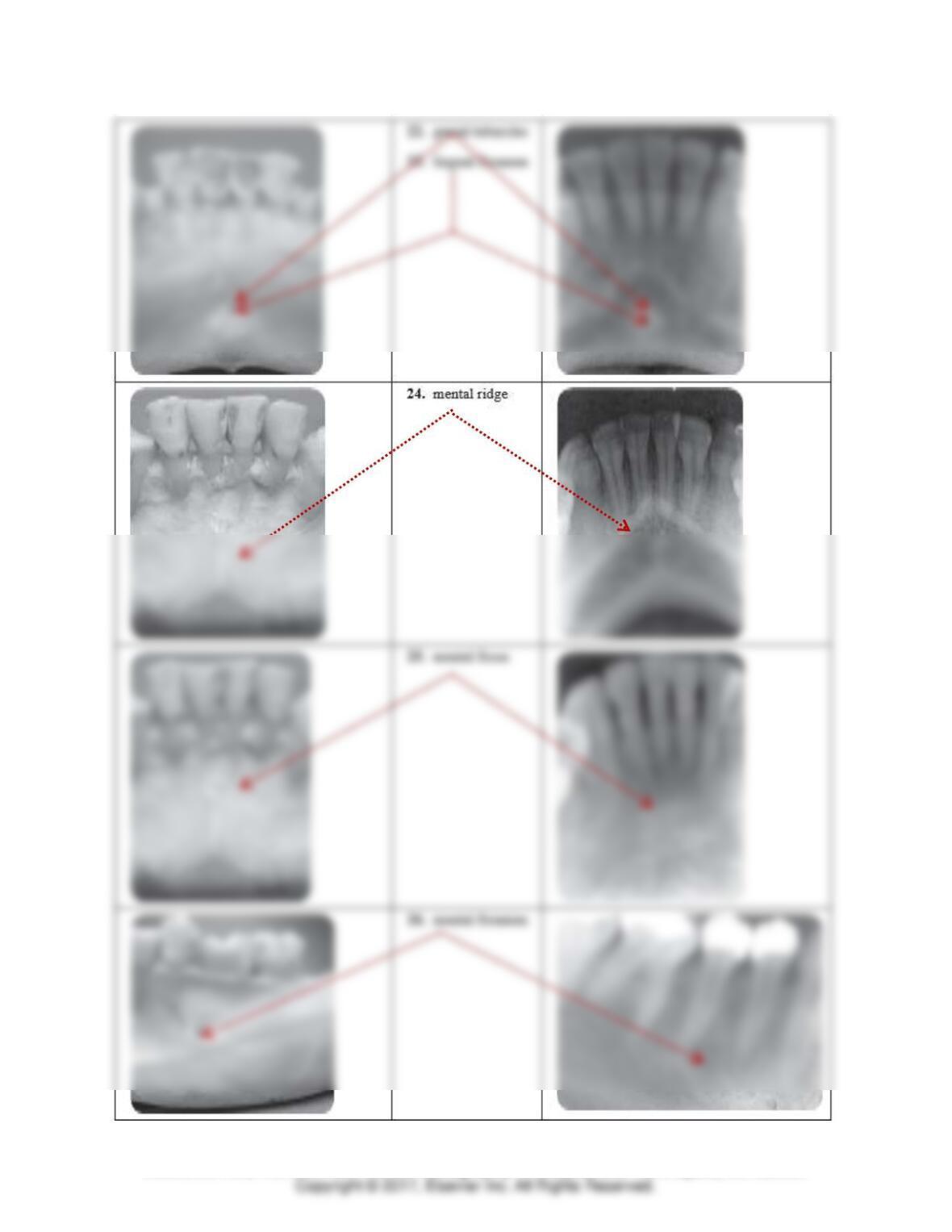
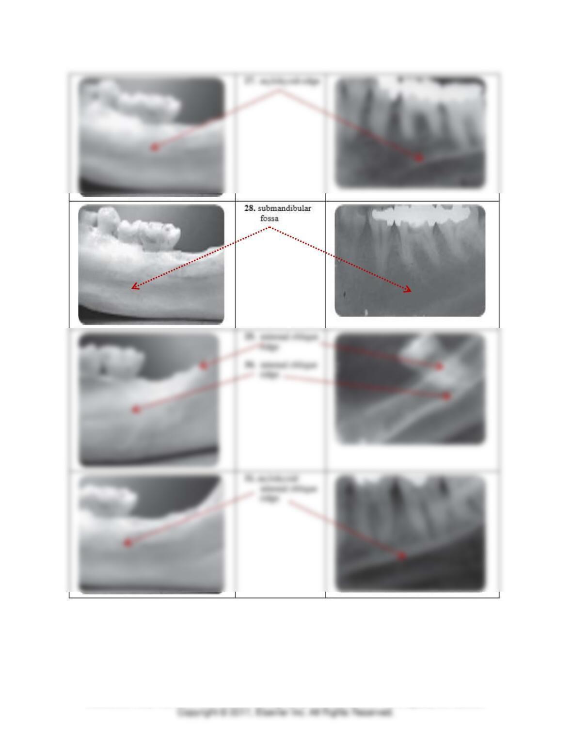
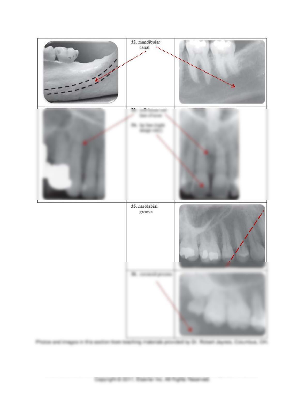
Labeling Exercise
Draw an arrow from each numbered item to the correct area on both the skull photo and the intraoral
image.
Module 5: Digital Imaging Basics and Normal Anatomy Basics 7
TEACH Lesson Plan
IANNUCCI AND HOWERTON: Dental Radiography: Principles and Techniques, 5th Edition
Module 5: Digital Imaging Basics and Normal Anatomy Basics 8
TEACH Lesson Plan
IANNUCCI AND HOWERTON: Dental Radiography: Principles and Techniques, 5th Edition
Module 5: Digital Imaging Basics and Normal Anatomy Basics 9
TEACH Lesson Plan
IANNUCCI AND HOWERTON: Dental Radiography: Principles and Techniques, 5th Edition
Module 5: Digital Imaging Basics and Normal Anatomy Basics 10
TEACH Lesson Plan
IANNUCCI AND HOWERTON: Dental Radiography: Principles and Techniques, 5th Edition
Module 5: Digital Imaging Basics and Normal Anatomy Basics 11
TEACH Lesson Plan
IANNUCCI AND HOWERTON: Dental Radiography: Principles and Techniques, 5th Edition
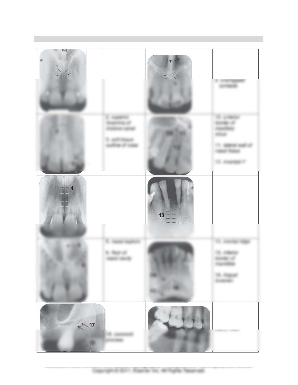
Identification
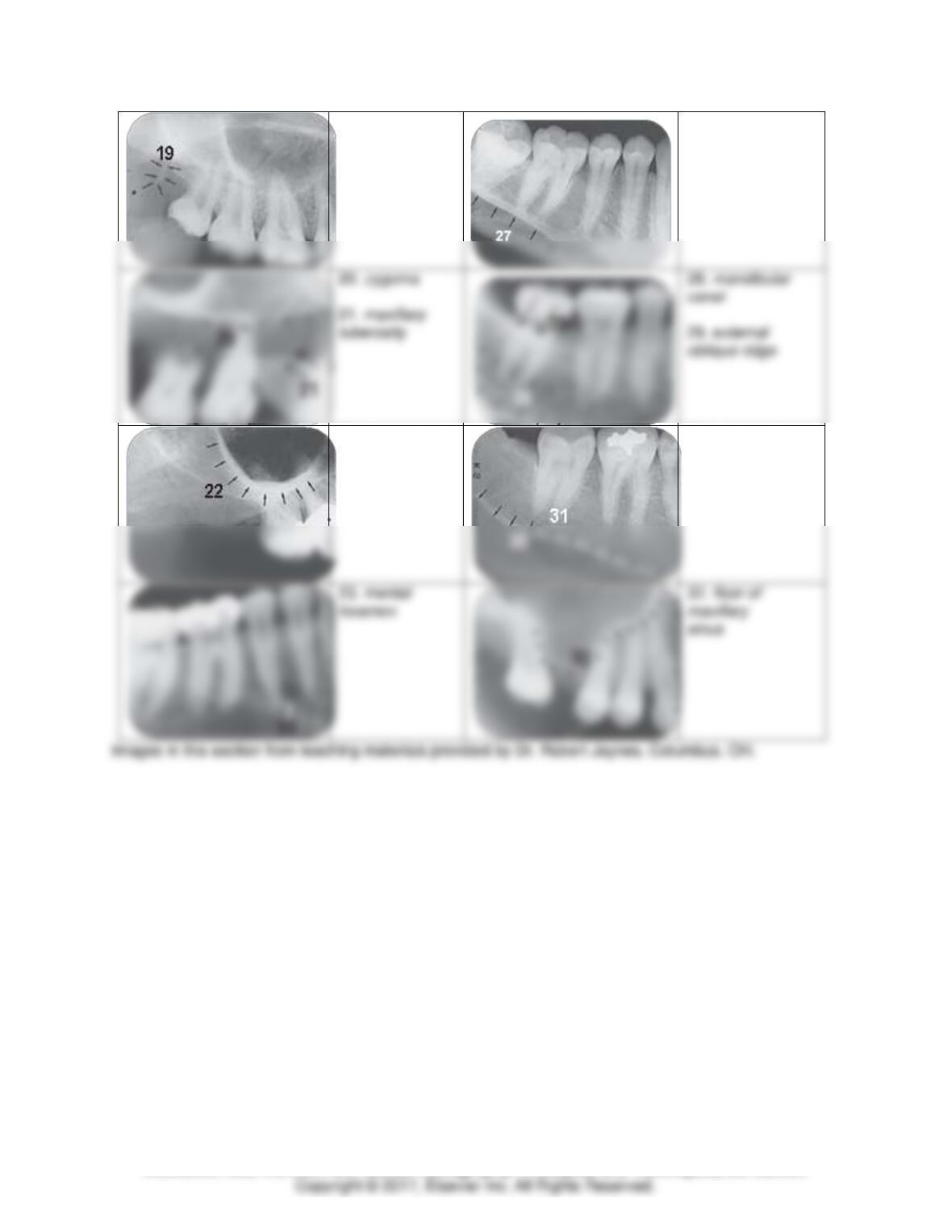
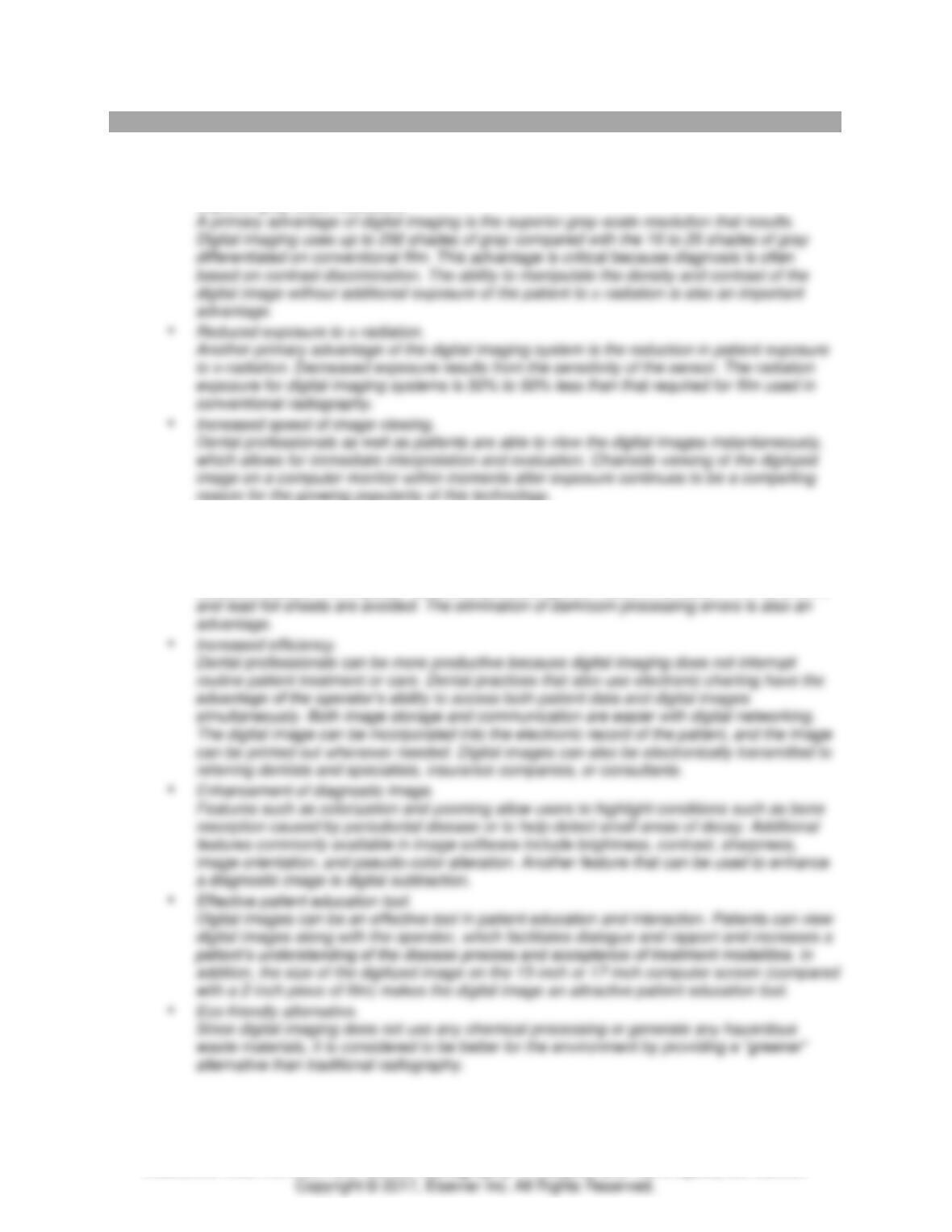
Identify each of the numbered structures.
1. incisive
foramen
7. nasal conchae
8. anterior nasal
spine
4. median
palatal suture
13. nutrient
canal
17. posterior
wall of
maxillary
sinus
24(A). enamel
25(B). dentin
Module 5: Digital Imaging Basics and Normal Anatomy Basics 12
TEACH Lesson Plan
IANNUCCI AND HOWERTON: Dental Radiography: Principles and Techniques, 5th Edition
19. hamular
process
27. mylohyoid
ridge
23. mental
32. floor of
22. zygomatic
process of
maxilla
30. submandibular
fossa
31. mylohyoid
ridge
Module 5: Digital Imaging Basics and Normal Anatomy Basics 13
TEACH Lesson Plan
IANNUCCI AND HOWERTON: Dental Radiography: Principles and Techniques, 5th Edition
MODULE 5: CRITICAL THINKING QUESTIONS
1. If you were helping to plan a new practice, which type of imaging system (digital versus film–
based) would you choose and why?
Digital imaging has the following advantages:
• Superior gray-scale resolution.
• Lower equipment and film cost.
Long-term digital imaging eliminates the need for purchasing conventional film, costly
processing equipment, and processing solutions. With digital imaging, darkroom and
processing solutions and maintenance are unnecessary. Also, environmental costs are
reduced because the disposal hazards of processing chemicals, silver salts in film emulsion,

Module 5: Digital Imaging Basics and Normal Anatomy Basics 14
TEACH Lesson Plan
IANNUCCI AND HOWERTON: Dental Radiography: Principles and Techniques, 5th Edition
2. A patient in your office needs to be referred for three-dimensional imaging in order to begin her
implant treatment planning. How will you explain the importance of obtaining three-dimensional
imaging in terms the patient can understand?
Three-dimensional imaging provides the dental professional with a more complete
3. Why is it important for dental professionals to learn about anatomy of the skull? Give examples of
how normal anatomy knowledge is needed in everyday practice.
Each normal anatomic landmark seen on a dental image corresponds to what is seen on the
PANORAMIC IMAGES
• Knowledge of normal anatomic landmarks is necessary to interpret panoramic images;