Module 4: Technique Basics 13
TEACH Lesson Plan
IANNUCCI AND HOWERTON: Dental Radiography: Principles and Techniques, 5th Edition
Cause
Correction
Exposure/Technique Error
12.
DROPPED RECEPTOR
CORNER
The edge of the receptor
To prevent a dropped
receptor corner, make certain
that the edge of the receptor
CHAPTER 21: CHAPTER 21: OCCLUSAL AND LOCALIZATION TECHNIQUES
Completion and Short Answer
Occlusal Technique
1. Define occlusal. Refers to the chewing surfaces of posterior teeth.
2. What size receptor is used for an adult occlusal projection? Size 4
5. List 5 of the 10 uses for the occlusal projection:
a. To locate retained roots of extracted teeth
b. To locate supernumerary (extra), unerupted, or impacted teeth
j. To measure changes in the size and shape of the maxilla or the mandible
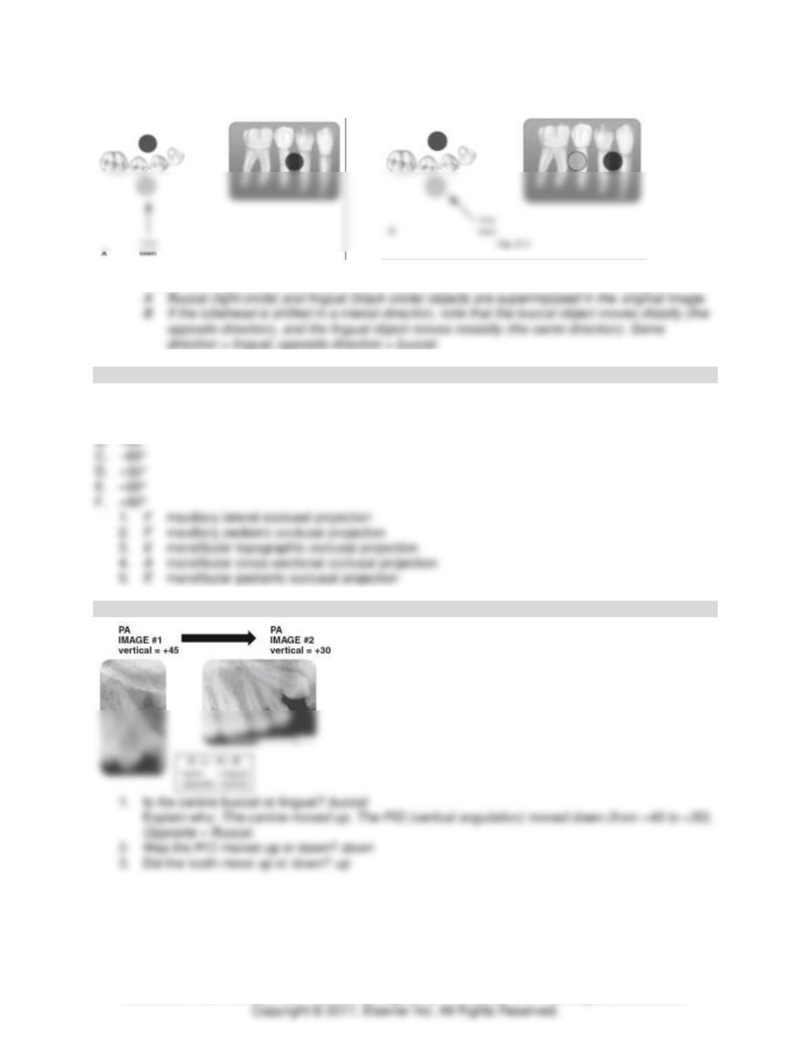
6. Identify the projection in Figure 21-1.
Module 4: Technique Basics 14
TEACH Lesson Plan
IANNUCCI AND HOWERTON: Dental Radiography: Principles and Techniques, 5th Edition
Basic Object Rule
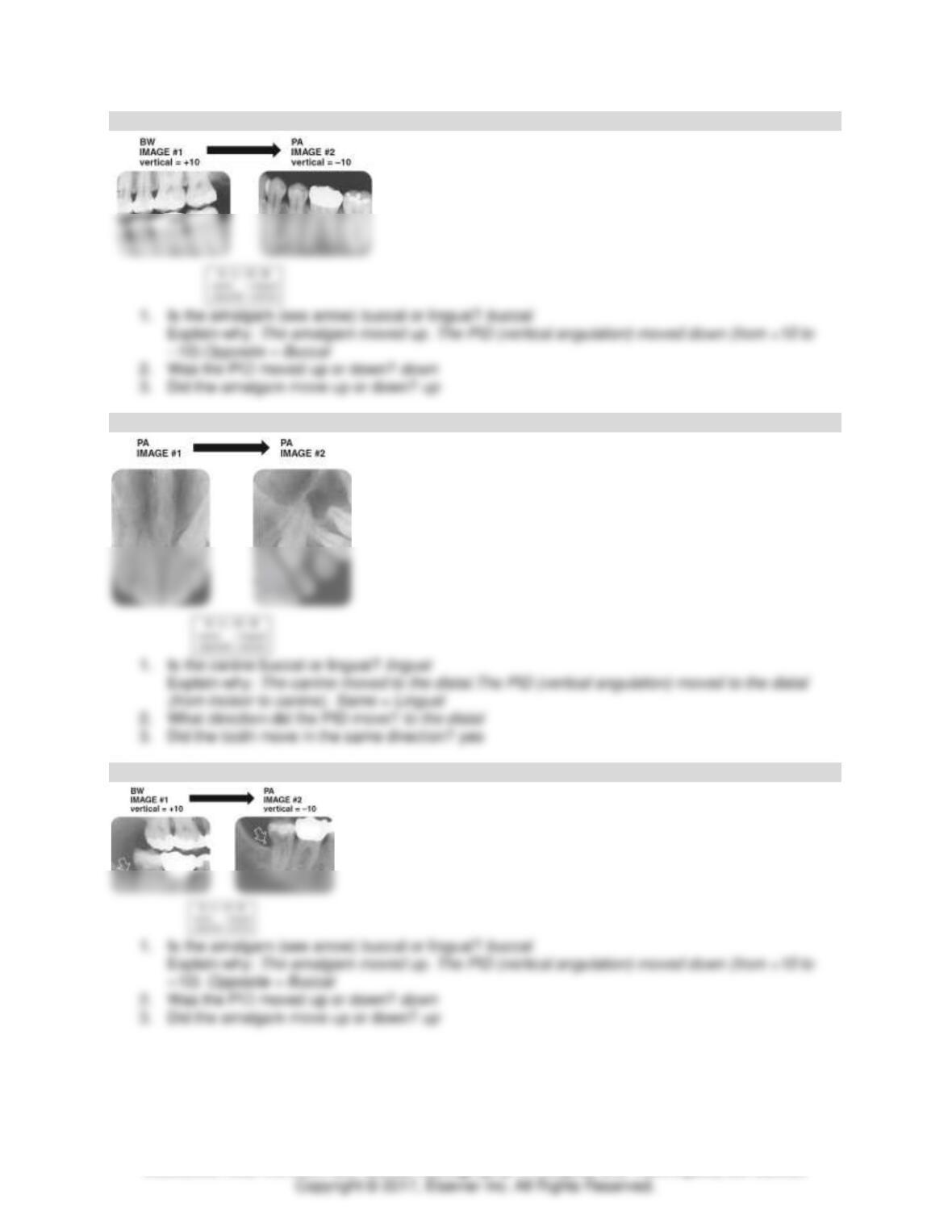
1. Using the diagram in Figure 21-2, describe the buccal object rule.
Buccal and lingual objects shift positions when the direction of the x-ray beam is changed.
Matching
Vertical Angulation
Some choices may be used more than once, and some choices may not be used.
A. 90°
Buccal Object Rule: Exercise 1
Module 4: Technique Basics 15
TEACH Lesson Plan
IANNUCCI AND HOWERTON: Dental Radiography: Principles and Techniques, 5th Edition
Buccal Object Rule: Exercise 2
Buccal Object Rule: Exercise 3
Buccal Object Rule: Exercise 4
Module 4: Technique Basics 16
TEACH Lesson Plan
IANNUCCI AND HOWERTON: Dental Radiography: Principles and Techniques, 5th Edition
CHAPTER 22: PANORAMIC IMAGING
Identification and Short Answer
Panoramic Image #1
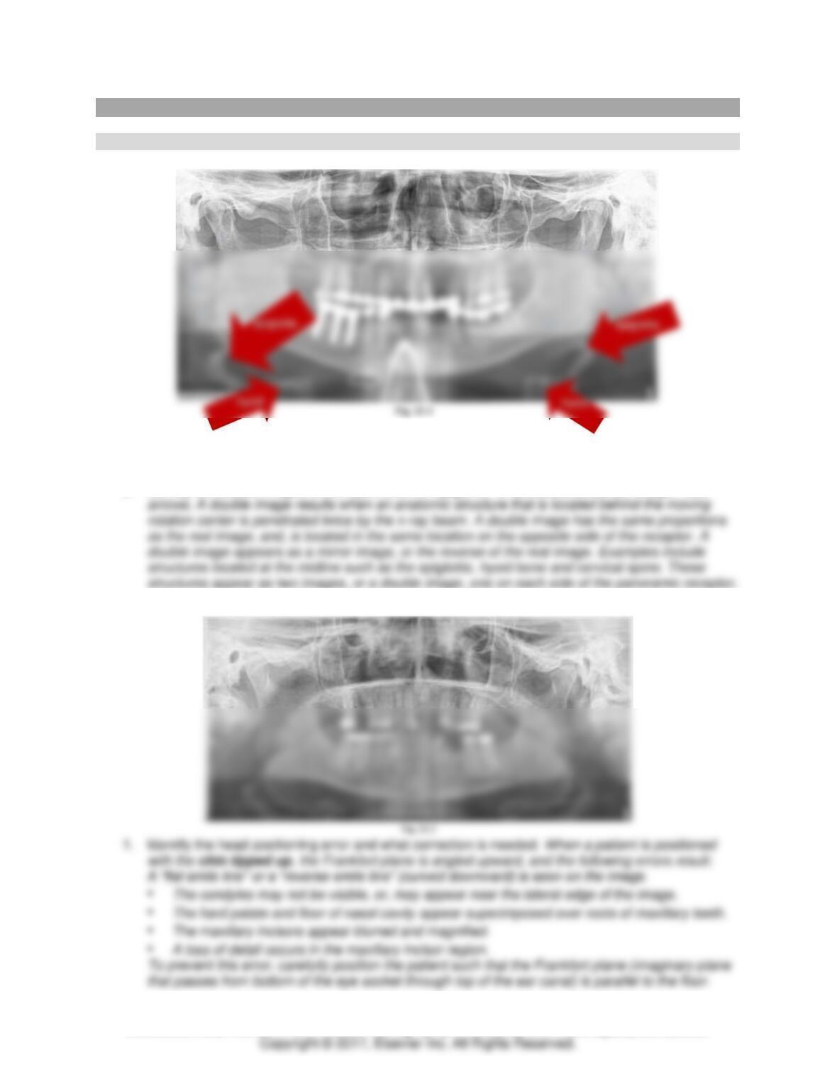
1. Identify the radiopacity (arrow). How can this error be avoided? The radiopacity is a necklace.
Avoid this error by requesting that the patient remove all radiodense objects from the head and
neck region prior to exposing the image.
2. Identify the double images of the hyoid and the epiglottis. What causes a double image? See
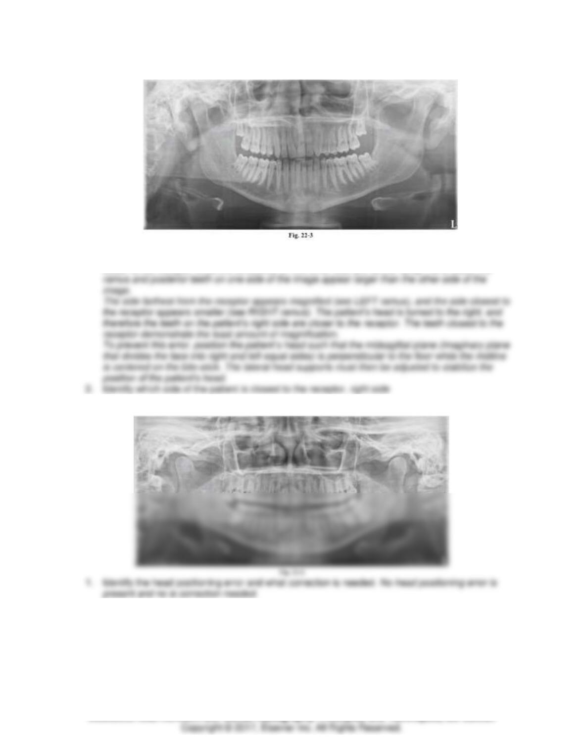
Panoramic Image #2
Module 4: Technique Basics 17
TEACH Lesson Plan
IANNUCCI AND HOWERTON: Dental Radiography: Principles and Techniques, 5th Edition
Panoramic Image #3
1. Identify the head positioning error and what correction is needed. When a patient’s head is
turned slightly to one side and not centered on the biteblock, the structures on one side are
closer to the receptor while the structures on the other side are farther away. As a result, the
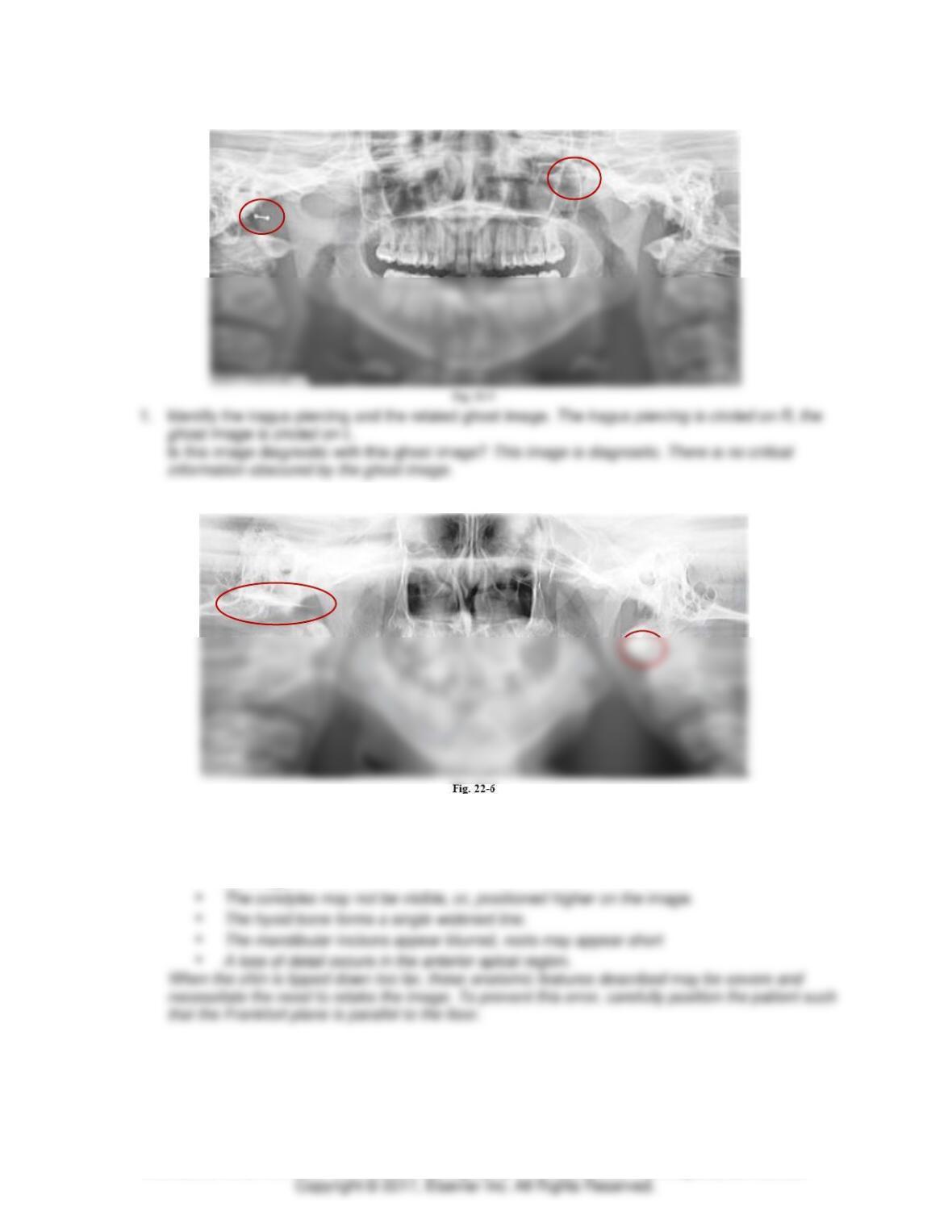
Panoramic Image #4
Module 4: Technique Basics 18
TEACH Lesson Plan
IANNUCCI AND HOWERTON: Dental Radiography: Principles and Techniques, 5th Edition
Panoramic Image #5
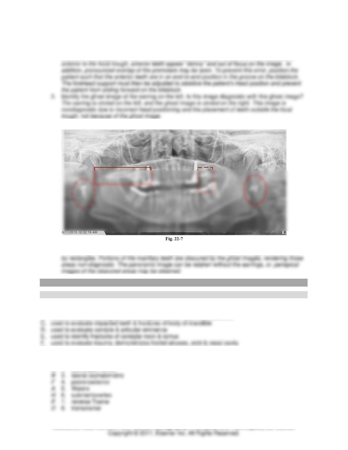
Panoramic Image #6
1. Identify the head positioning error and what correction is needed. When the patient is positioned
such that the chin is too low or is tipped down, the Frankfort plane is angled downward, and
the following errors result:
• An “exaggerated smile line” or “Jack-O–Lantern” appearance (curved upward) is seen on
the image.
Module 4: Technique Basics 19
TEACH Lesson Plan
IANNUCCI AND HOWERTON: Dental Radiography: Principles and Techniques, 5th Edition
2. Identify the focal trough error and what correction is needed. If the patient is positioned such that
the anterior teeth are not positioned in the focal trough, as indicated by the groove in the
biteblock, teeth appear blurred. If the patient’s teeth are too far forward on the biteblock or
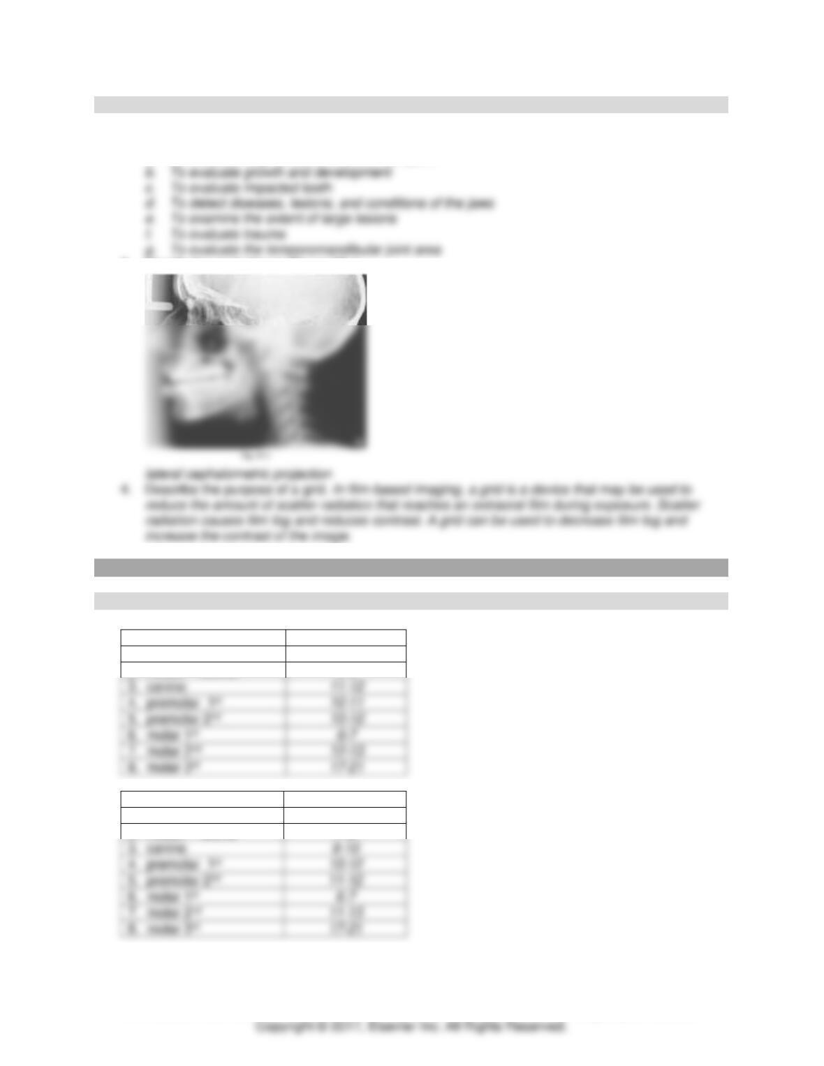
Panoramic Image #7
1. Identify the earrings and related ghost images. Is this image diagnostic with these ghost images?
The earrings are identified by circles on the right and left sides. The ghost images are identified
CHAPTER 23: EXTRAORAL IMAGING
Matching
Extraoral Projections
A. used to evaluate maxillary sinus area
B. used to evaluate facial growth & development; often used in orthodontics
G. used to evaluate impacted teeth and fractures that extend to the ramus
H. used to identify the position of the condyles; used for zygomatic fractures
C 1. lateral jaw, body
G 2. lateral jaw, ramus
Module 4: Technique Basics 20
TEACH Lesson Plan
IANNUCCI AND HOWERTON: Dental Radiography: Principles and Techniques, 5th Edition
Short Answer
1. Define extraoral. outside the mouth
2. List 5 uses for extraoral images:
a. To evaluate large areas of the skull and jaws
3. Identify the projection in Figure 23-1.
CHAPTER 24: IMAGING OF PATIENTS WITH SPECIAL NEEDS
Identification
Eruption of Permanent Teeth
Maxillary
Age at Eruption
1. incisor – central
7-8
2. incisor – lateral
8-9
3. canine
6-7
Mandibular
Age at Eruption
1. incisor – central
6-7
2. incisor – lateral
7-8
3. canine
6-7
Module 4: Technique Basics 21
TEACH Lesson Plan
IANNUCCI AND HOWERTON: Dental Radiography: Principles and Techniques, 5th Edition
What permanent tooth replaces each tooth listed?
1. D 7
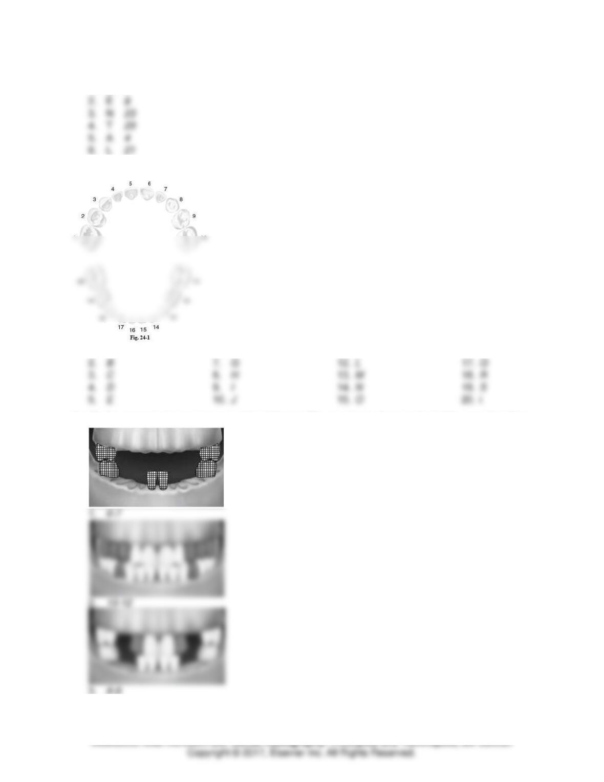
Label each primary tooth in Figure 24–1.
1. A
6. F
11. K
16. P
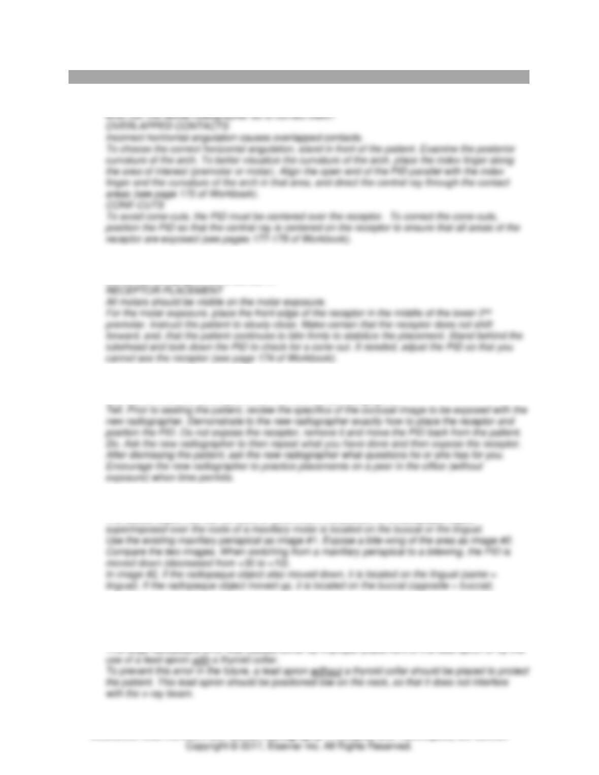
Identify the approximate age for each of the following. The erupting teeth are indicated by cross-hatching.
3. 8-9
Module 4: Technique Basics 22
TEACH Lesson Plan
IANNUCCI AND HOWERTON: Dental Radiography: Principles and Techniques, 5th Edition
MODULE 4: CRITICAL THINKING QUESTIONS
1. After exposing and processing images, the dental radiographer notices that all four of the bite-
wing images have overlapped contact areas and cone–cuts. Why did these errors happen, and
2. After exposing bite-wing images, the dental radiographer notices that both molar exposures do
not show the necessary teeth for a diagnostic image. Why did this error happen, and what can
the dental radiographer do to correct it?
3. You are teaching the new dental radiographer in your dental office how to take occlusal images.
What is your plan for training her?
Use the tell-show-do technique to train the new dental radiographer.
4. A radiopaque object is noted on maxillary molar periapical superimposed over the roots. Using
the buccal object rule, how can you determine if the object is buccal or lingual?
The buccal object rule can be used to determine if the position of a radiopaque object
5. A 28-year-old woman came to the office for a panoramic image. All procedural steps were
followed for panoramic exposure, but upon processing, the image showed a large radiopaque
area concealing a portion of the mandible. What caused this error? What must be done in the
future to prevent it?
Module 4: Technique Basics 23
TEACH Lesson Plan
IANNUCCI AND HOWERTON: Dental Radiography: Principles and Techniques, 5th Edition
6. A 22-year-old man came into the office with pain in the jaw joint. He complained about discomfort
in both ears and about having trouble opening and closing his mouth. The patient also said that
he hears popping and clicking sounds when he eats. What imaging procedure is recommended
for this type of issue?
The patient is experiencing discomfort associated with his temporomandibular joints (TMJ).
7. A patient comes into the office for a dental examination. He is hearing impaired and is in a
wheelchair. Dental images must be taken today, and the dental radiographer is worried about a
communication barrier and how the images can be taken while the patient is in his wheelchair.
The patient has come to the office without a caregiver but can read lips. How should the dental
radiographer handle this situation?
The dental radiographer will need to speak clearly and directly to the patient without a mask on so
8. Your next patient is a boy with Down syndrome. Describe how you will prepare for this patient
with special needs, and identify what techniques you can use to make him feel more comfortable
and have a positive experience.
Patients with Down syndrome, have a chromosomal defect that commonly results in certain