TEACH Lesson Plan
IANNUCCI AND HOWERTON: Dental Radiography: Principles and Techniques, 5th Edition
Module 3 Radiographer and Technique
Basics
CHAPTER 11: DENTAL IMAGES AND THE DENTAL RADIOGRAPHER
Short Answer
Dental Images
1. Briefly summarize the importance and uses of dental images.
Dental images are a necessary component of comprehensive patient care and can be used as
follows:
• to detect lesions, diseases, and conditions of the teeth and surrounding structures that
cannot be identified clinically
Dental Radiographer
Describe your commitment to each of the following professional goals:
1. Patient protection: Patient protection must be a top priority and a primary concern of the dental
radiographer. Whenever the dental radiographer performs imaging procedures on patients, the
2. Operator protection: Operator protection must also be a primary concern for the dental
radiographer. To avoid occupational exposure to x-radiation, the dental radiographer must always
3. Patient education: Patient education is another priority for the dental radiographer. The dental
Module 3: Radiographer and Technique Basics 2
TEACH Lesson Plan
IANNUCCI AND HOWERTON: Dental Radiography: Principles and Techniques, 5th Edition
4. Operator competence: Operator competence must always be a concern of the dental
5. Operator efficiency: The dental radiographer must be committed to performing assigned duties in
6. Production of quality images: The dental radiographer must be committed to producing high–
CHAPTER 12: PATIENT RELATIONS AND THE DENTAL RADIOGRAPHER
Identification
Communication Skills
For each of the following, place an X next to words that are associated with negative images in the dental
setting.
1. restore
2. X cut
For each of the following, place an X next to appropriate communication messages.
11. checking the time on your watch while the patient is talking
Short Answer
Communication Skills
Briefly describe why each of the following phrases should be avoided in the dental setting.
1. “Sorry ’bout that.”
Module 3: Radiographer and Technique Basics 3
TEACH Lesson Plan
IANNUCCI AND HOWERTON: Dental Radiography: Principles and Techniques, 5th Edition
4. “I don’t know.”
This phrase can be interpreted as “it is up to you to figure it out”. When you do not know
something, the appropriate response is “I don’t know, but let me find out for you.”
Chairside Mannger and Patient Relations
1. Briefly describe the chairside manner that is appropriate in the dental setting.
2. Briefly describe the attitude that is appropriate in the dental setting.
CHAPTER 13: PATIENT EDUCATION AND THE DENTAL RADIOGRAPHER
Short Answer
Importance of Patient Education
1. Briefly describe the importance of patient education concerning x-rays. Comprehensive dental
health education is one of the greatest services that a dental professional can provide to the
Common Questions and Answers
You must be able to answer each of these questions as detailed in Chapter 13 of the text.
2. How often do I need dental x-ray images? The first step to limiting the amount of radiation that
Module 3: Radiographer and Technique Basics 4
TEACH Lesson Plan
IANNUCCI AND HOWERTON: Dental Radiography: Principles and Techniques, 5th Edition
3. Can I refuse x-rays and be treated without the images? No. When you refuse the prescribed
5. How do you limit my exposure to x-rays? Because no amount of radiation is considered safe,
strict guidelines are followed to limit the amount of x-radiation. For example, the dentist custom–
6. Why do you use a lead apron? A lead apron and a thyroid collar are used to protect reproductive,
7. Should I avoid dental x-ray exposure during pregnancy? When a lead apron is used during dental
imaging procedures, the amount of radiation received in the gonadal region is nearly zero. No
8. Why do you leave the room when x-rays are used? When you are exposed to x-rays, you receive
9. Are dental x-rays safe? All x-rays are harmful to living tissue. The term safe is defined as “no
Module 3: Radiographer and Technique Basics 5
TEACH Lesson Plan
IANNUCCI AND HOWERTON: Dental Radiography: Principles and Techniques, 5th Edition
10. Will dental x-ray exposure cause cancer? Not a single recorded case of a patient developing
11. What are the advantages to using digital imaging? Digital imaging requires less exposure to
12. Are there risks associated with digital imaging? Because radiation is involved, a certain amount
13. Who owns my dental x-ray images? All your dental records, including the dental images, are the
CHAPTER 14: LEGAL ISSUES AND THE DENTAL RADIOGRAPHER
Basic Terminology Crossword Puzzle
Down
1. self-determination
Short Answer
Legal Issues and the Dental Patient
1. Briefly discuss the ownership and retention of dental images. Legally, dental images are the
property of the dentist, even though the patient or an insurance company may have paid for them.
The basis for this ownership of dental images is that images are indispensable to the dentist as
part of the patient’s record.
Module 3: Radiographer and Technique Basics 6
TEACH Lesson Plan
IANNUCCI AND HOWERTON: Dental Radiography: Principles and Techniques, 5th Edition
2. Briefly discuss patients who refuse dental image. Some patients may refuse exposure of dental
imaging procedures. When this occurs, the dentist must carefully consider the situation. The
3. List the four essential elements of informed consent.
a. purpose of the procedure and who will perform it
4. In regards to dental imaging, list the four essential elements of documentation.
a. informed consent
True or False
CHAPTER 15: INFECTION CONTROL AND THE DENTAL RADIOGRAPHER
Basic Terminology Crossword Puzzle
Across
1. noncritical
Down
2. occupational
Module 3: Radiographer and Technique Basics 7
TEACH Lesson Plan
IANNUCCI AND HOWERTON: Dental Radiography: Principles and Techniques, 5th Edition
Copyright © 2017, Elsevier Inc. All Rights Reserved.
Identify and Describe
Infection Control Procedures
For each photo in Figure 15-1, describe what you see as it pertains to infection control in dental imaging.
1. a film is sealed inside barrier envelope; a barrier envelope protects the film packet from saliva
Short Answer
Infection Control Procedures
1. List the five indications for hand hygiene.
a. before and after treating each patient (e.g., before glove placement and after glove removal)
e. when hands are visibly soiled or contaminated
2. List the three types of hand hygiene that may be used in dental imaging.
CHAPTER 16: INTRODUCTION TO DENTAL IMAGING EXAMINATIONS
Matching
Basic Terminology
A. bite-wing receptor
B. dentulous
I. occlusal receptor
J. occlusion
K. periapical receptor
E 1. receptor placed inside of the mouth
D 2. receptor placed outside of the mouth
Module 2: Equipment, Film, and Processing Basics 8
TEACH Lesson Plan
IANNUCCI AND HOWERTON: Dental Radiography: Principles and Techniques, 5th Edition
Short Answer
Prescribing of Dental Images
1. Review Table 5-1 in Chapter 5 of your textbook. For each example below, list the dental images
that are needed.
a. Adult ‒ new patient
Individualized ordering of images covering all tooth bearing areas; may consist of a
2. List the five criteria for a diagnostic intraoral image.
a. Dental images must display optimal density, contrast, definition, and detail.
b. Dental images must display the least amount of distortion possible; images must be of the
CHAPTER 17: PARALLELING TECHNIQUE
Basic Terminology Crossword Puzzle
Across
Down
Drawing Exercise
Basic Concepts
Indicate the 90 angle formed by each of the following by drawing a ■ on the diagram below.
1. biting surface of bite block — and — long axis of tooth
Module 3: Radiographer and Technique Basics 9
TEACH Lesson Plan
IANNUCCI AND HOWERTON: Dental Radiography: Principles and Techniques, 5th Edition
Multiple Choice
Parallel vs. Perpendicular
1. The receptor is parallel / perpendicular to the CR.
RINN Devices
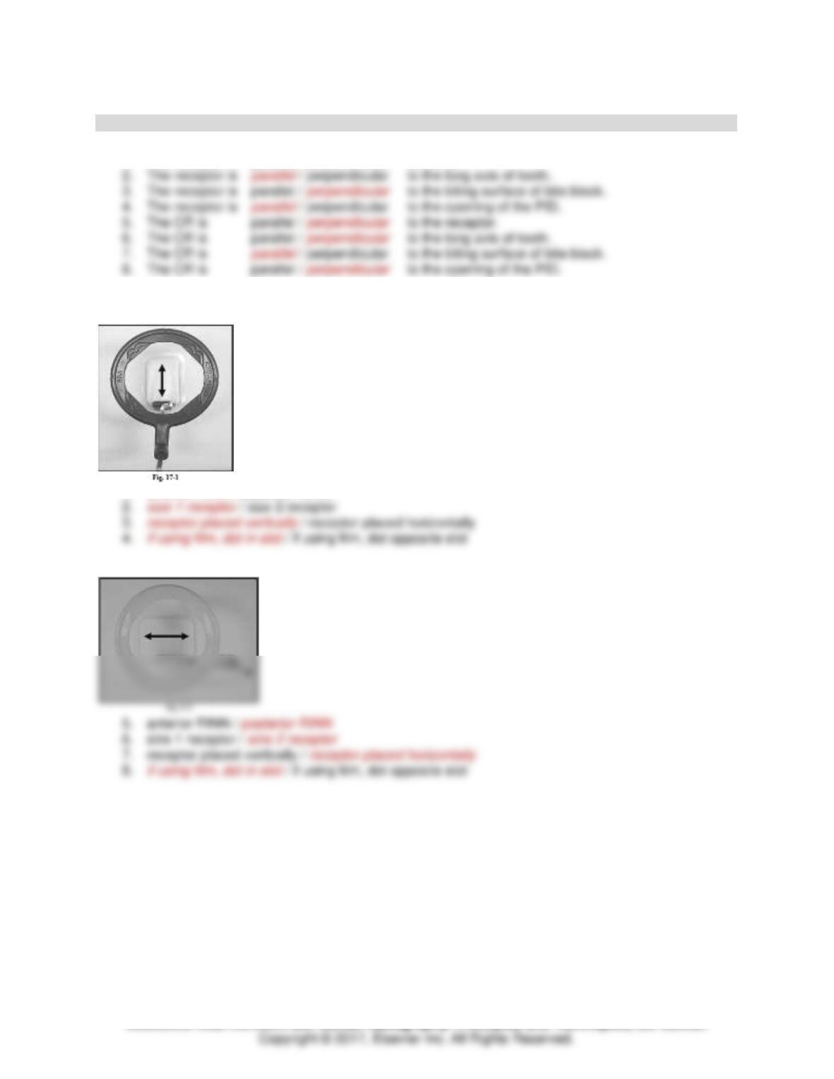
Select the correct answers for Figure 17–1.
1. anterior RINN / posterior RINN
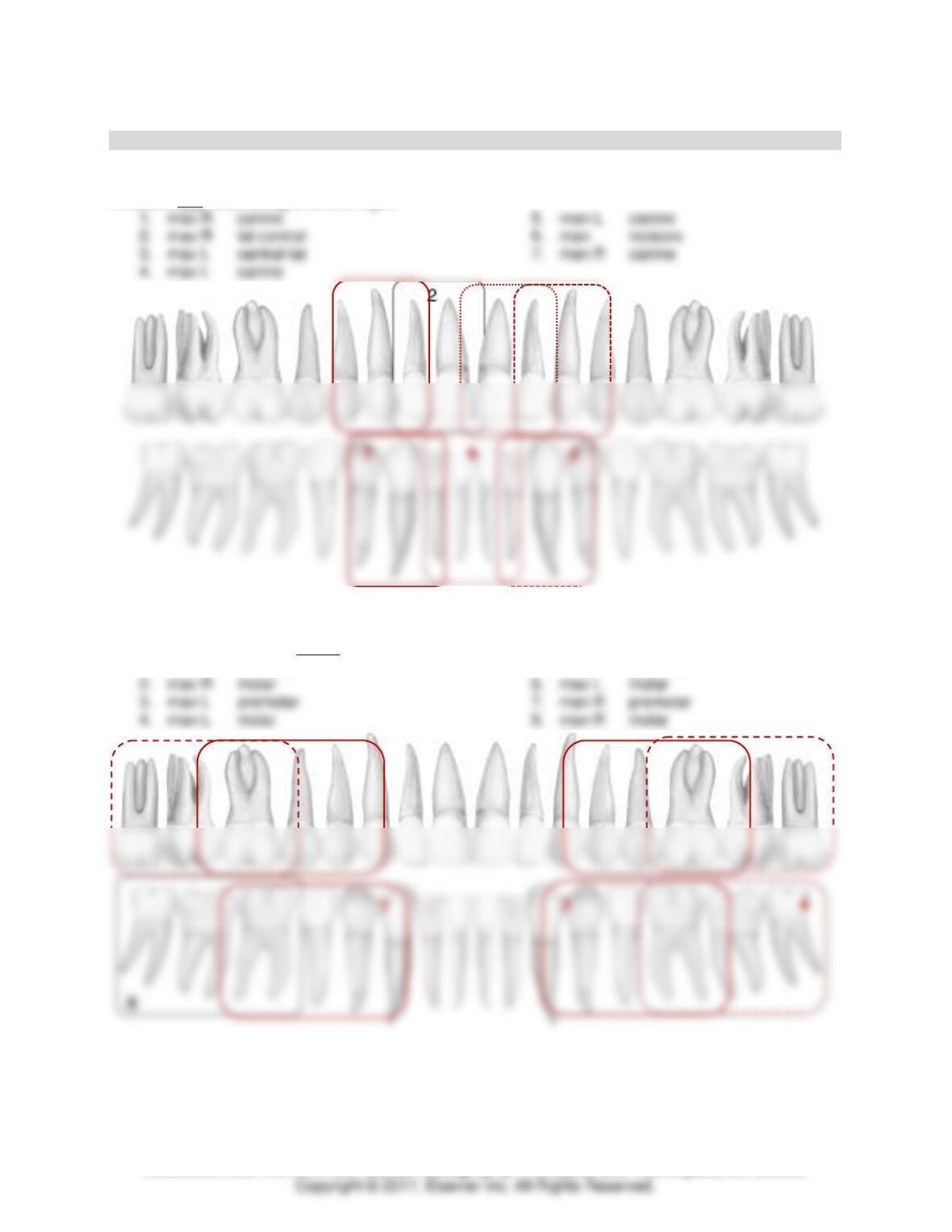
Select the correct answers for Figure 17–2.
Module 3: Radiographer and Technique Basics 10
TEACH Lesson Plan
IANNUCCI AND HOWERTON: Dental Radiography: Principles and Techniques, 5th Edition
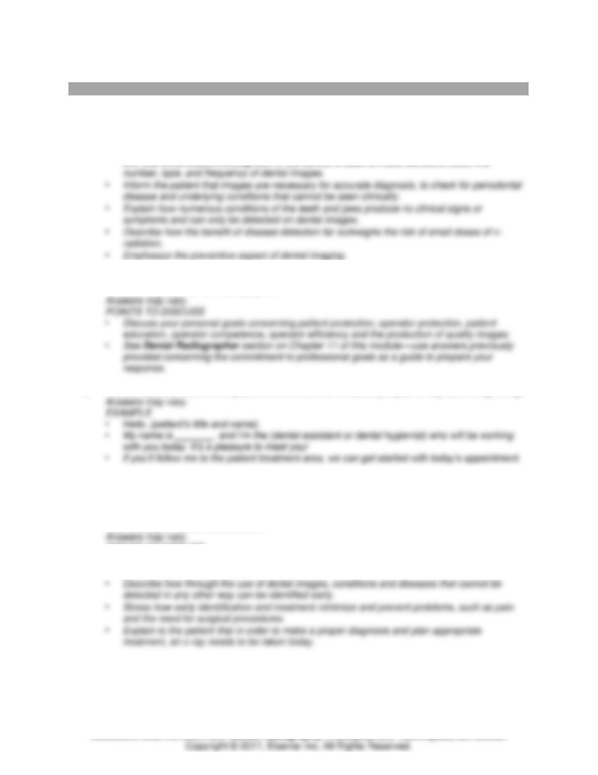
Periapical Receptor Placement Drawing Exercise
Anterior Exposure Sequence
Draw each periapical receptor placement; label with numbers indicated below (see example labeled 2).
Anteriors first. Start on right, end on right.
Posterior Anterior Exposure Sequence
Draw each periapical receptor placement; label with numbers indicated below (see example labeled 8).
Posteriors next—premolar before molar. Start on right, end on right.
1. max R premolar
5. max L premolar
1
5
6
2
1
3
4

Module 3: Radiographer and Technique Basics 11
TEACH Lesson Plan
IANNUCCI AND HOWERTON: Dental Radiography: Principles and Techniques, 5th Edition
Periapical Assessment: Exercise 1
Evaluate each image. Mark each item that is correct with a checkmark. If possible, review Series 1 on
Evolve for optimum viewing.
MAXILLARY PERIAPICALS
Molar
___ front edge of is in
middle of 2nd premolar
Premolar
___ front edge of is in
middle of canine
Canine
✓
canine centered
✓
canine visible
Incisor
✓
lateral and central
centered
MANDIBULAR PERIAPICALS
Molar
✓
front edge of is in
middle of 2nd premolar
Premolar
✓
front edge of is in
middle of canine
Canine
✓
canine centered
Incisor
✓
midline centered

Module 3: Radiographer and Technique Basics 12
TEACH Lesson Plan
Periapical Assessment: Exercise 2
Evaluate each image. Mark each item that is correct with a checkmark. If possible, review Series 2 on
Evolve for optimum viewing.
MAXILLARY PERIAPICALS
Molar
___ front edge of is in
middle of 2nd premolar
Premolar
___ front edge of is in
middle of canine
parallel
Canine
✓
canine centered
✓
canine visible
Incisor
___ lateral and central
centered
MANDIBULAR PERIAPICALS
Molar
___ front edge of is in
middle of 2nd premolar
✓
all molars visible
Premolar
✓
front edge of is in
middle of canine
✓
all premolars visible
Canine
✓
canine centered
✓
canine visible
✓
2-3 mm around
Incisor
___ midline centered
___ all incisors visible
✓
2-3 mm around

Module 3: Radiographer and Technique Basics 13
TEACH Lesson Plan
IANNUCCI AND HOWERTON: Dental Radiography: Principles and Techniques, 5th Edition
Periapical Assessment: Exercise 3
Evaluate each image. Mark each item that is correct with a checkmark. If possible, review Series 3 on
Evolve for optimum viewing.
MAXILLARY PERIAPICALS
Molar
___ front edge of is in
middle of 2nd premolar
✓
all molars visible
Premolar
✓
front edge of is in
middle of canine
✓
all premolars visible
parallel
Canine
___ canine
centered
✓
canine visible
Incisor
✓
lateral and central
centered
✓
lateral and central
MANDIBULAR PERIAPICALS
parallel
Molar
___ front edge of is in
middle of 2nd premolar
✓
all molars visible
Premolar
✓
front edge of is in
middle of canine
✓
all premolars visible
Canine
___ canine
centered
✓
canine visible
Incisor
___ midline centered
___ all incisors visible
Module 3: Radiographer and Technique Basics 14
TEACH Lesson Plan
IANNUCCI AND HOWERTON: Dental Radiography: Principles and Techniques, 5th Edition
MODULE 3: CRITICAL THINKING QUESTIONS
1. A new patient tells you, “This sure does seem like a lot of x–rays to have taken in one visit.” How
would you respond to the patient?
Answers may vary.
POINTS TO DISCUSS
• Explain that the prescribing of dental images is based on the individual needs of the patient
2. Your employer asks you what you plan to do to improve your imaging skills. Discuss your goals
for becoming a competent radiographer.
3. You are about to meet a new patient for the first time. What do you plan to say as a first greeting?
4. A patient has been a regular in your dental office for 3 years, getting exams and cleanings every
year. The patient calls your office and states he has a toothache. You inform the patient that he
must come to the office for an evaluation by the dentist. When the patient arrives you notice there
are entries made in the progress notes that this patient will not allow dental images to be taken.
How would you handle this situation?
POINTS TO DISCUSS
• Explain that dental images are always prescribed to benefit the patient; and that the primary
benefit is disease detection.
Module 3: Radiographer and Technique Basics 15
TEACH Lesson Plan
IANNUCCI AND HOWERTON: Dental Radiography: Principles and Techniques, 5th Edition
5. A new patient comes into your office for a check-up. He has come to your office because he was
very unhappy that the previous dentist would not give him a copy of his dental images. The
previous dentist said that the patient had no rights to the images. The patient is very upset and
feels misled. What can you do to help this patient?
POINTS TO DISCUSS
• Explain that the dental record is an official document that belongs to the practice where
treatment was rendered.
6. A new patient observes you placing plastic barriers. The patient tells you, “Isn’t this overkill? I
don’t have any diseases.” How would you respond to this patient about infection control?
POINTS TO DISCUSS
• Explain that the majority of patients don’t appear to have any major communicable diseases.
• Inform the patient that when you place a receptor in the mouth there is contact with saliva.
7. You are about to take a complete mouth series on a new patient. The patient asks you “How is
the decision made to take all of these x–rays? Does every patient have this done?” How would
you respond to this patient?
POINTS TO DISCUSS
• Explain that the prescribing of dental images is based on the individual needs of the patient
8. A patient comes into the clinic needing a full series of images. You will use the paralleling
technique on her, but she has a large palatal torus. How will you modify your technique to
accommodate this torus?
When using the paralleling technique, maxillary and mandibular tori can cause problems with