Chapter 5
Protein Purification and Characterization Techniques
SUMMARY
Section 5.1
To begin the process of purification, proteins are released from cells using
Section 5.2
Column chromatography refers to several common techniques for purification of
proteins
In gel filtration chromatography, proteins are separated by size
In ion exchange chromatography molecules with a specific charge are selectively
bound to a column, separated from proteins that don’t bind, and then eluted.
Section 5.3
Electrophoresis separates molecules on a gel medium by passing electrical
current through the gel.
Section 5.4
The amino acid sequence of a protein can be determined using a multi-step
process.
First, the protein is hydrolyzed into its constituent amino acids and the
2 Chapter 5
Section 5.5
Many techniques are used to separate and identify proteins.
Enzyme-linked immunoabsorbent assays (ELISA) is a simple technique that
involves screening proteins samples using a plate with wells in it that have an
Section 5.6
The proteome is the complete complement of proteins in a cell, and
proteomics is the study of all of these proteins.
Structural proteomics offers a detailed analysis of the structure of the proteins
being produced.
determine the nature of the proteome.
LECTURE NOTES
This chapter will be most beneficial to students who are or will take a laboratory course
in conjunction with the lecture course, but does contain interesting material regardless.
Protein Purification and Characterization Techniques 3
LECTURE OUTLINE
I. Protein purification
A. Isolation of proteins from cells
1. Cell fractionation
2. Salting out
B. Column chromatography
1. Basic concepts – stationary vs. mobile phases
C. Electrophoresis
1. Agarose & polyacrylamide gels
3. Isoelectric focusing
II. Primary structure determination
A. Hydrolysis of proteins – HPLC analysis
B. Sequence-specific cleavage
C. Edman degradation
III. Protein Identification Techniques
A. ELISA
IV. Proteomics
4 Chapter 5
ANSWERS TO PROBLEMS
5.1 Extracting Pure Proteins from Cells
1. Using a blender, a Potter-Elvejhem homogenizer, or a sonicator.
2. If you needed to maintain the structural integrity of the subcellular organelles, a
Potter-Elvejhem homogenizer would be better because it is more gentle. The
4. Their amino acid content and arrangements make some proteins more soluble
than others. A protein with more highly polar amino acids on the surface is more
soluble than one with more hydrophobic ones on the surface.
5. First homogenize the liver cells using a Potter-Elvejhem homogenizer. Then spin
the homogenate at 500 × g to sediment the unbroken cells and nuclei. Centrifuge
the supernatant at 15,000 × g and collect the pellet, which contains the
mitochondria.
6. No, peroxisomes and mitochondria have overlapping sedimentation
characteristics. Other techniques, such as sucrose-gradient centrifugation, would
the enzyme from the membrane.
9. Tables exist to tell you how many grams of ammonium sulfate [(NH4)2SO4] to add
to get a certain percent saturation. A good plan would be to take the homogenate
and add enough ammonium sulfate to yield a 20% saturated solution. Let the
sample sit for 15 minutes on ice and then centrifuge. Separate the supernatant
from the precipitate. Assay both for the protein you are working with. Add more
ammonium sulfate to the supernatant to arrive at a 40% saturated solution and
Protein Purification and Characterization Techniques 5
5.2 Column Chromatography
11.
(a) Size.
12. The largest proteins elute first; the smallest elute last. Larger proteins are
excluded from the interior of the gel bead so they have less available column
space to travel. Essentially, they travel a shorter distance and elute first.
13. A compound can be eluted by raising the salt concentration or by adding a
14. A compound can be eluted by raising the salt concentration or by changing the
15. Raising the salt concentration is relatively safe. Most proteins will elute this way,
and, if the protein is an enzyme, it will still be active. If necessary, the salt can be
16. The basis of most resins is agarose, cellulose, dextran, or polyacrylamide.
17. See Figure 5.7.
18. Within the fractionation range of a gel-filtration column, molecules elute with a
19. Both proteins would elute in the void volume together and would not be
separated.
20. Yes, the -amylase would come out in the void volume, but the bovine serum
albumin would be included in the column bead and would elute more slowly.
21. In most chromatography systems, the ligands and solvents are polar. In reverse
phase HPLC, a solution of nonpolar compounds is put through a column that has
22. Ion Exchange Chromatography is a specific type of separation based on net
charge of the molecules being separated. The term HPLC refers to
6 Chapter 5
23. Set up an anion-exchange column, such as Q-Sepharose (quaternary amine).
24. Use a cation-exchange column, such as CM-Sepharose, and run it at pH 6.
Protein X will have a positive charge and will stick to the column.
25. With a quaternary amine, the column resin always has a net positive charge, and
26. The easiest way would be to use a sucrose gradient to separate the mitochondria
from the peroxisomes first. Then break open the mitochondria via harsh
homogenization or sonication, and then centrifuge the mitochondria. The pellet
27. Glutamic acid will be eluted first because the column pH is close to its pI. Leucine
and lysine will be positively charged and will stick to the column. To elute leucine,
raise the pH to around 6. To elute lysine, raise the pH to around 11.
28. A nonpolar mobile solvent will move the nonpolar amino acids fastest, so
phenylalanine will be the first to elute, followed by glycine and then glutamic acid.
30. A protein solution from an ammonium sulfate preparation is passed over a gel–
5.3 Electrophoresis
31. Size, shape, and charge.
34. DNA is the molecule most often separated on agarose electrophoresis, although
proteins can also be separated.
35. Those with the highest charge/mass ratio would move the fastest. There are
36. Sodium dodecyl-sulfate polyacrylamide gel-electrophoresis. With SDS–PAGE,
Protein Purification and Characterization Techniques 7
37. SDS binds to the protein in a constant ratio of 1.4 g SDS per gram of protein. It
38. In a polyacrylamide gel used for gel-filtration chromatography, the larger proteins
can travel around the beads, thereby having a shorter path to travel and
therefore eluting faster. With electrophoresis, the proteins are forced to go
5.4 Determining the Primary Structure of a Protein
40. The Edman degradation will give the identity of the N-terminal amino acid in its
first cycle, so doing a separate experiment is not necessary.
41. It might tell you if the protein were pure or if there were subunits.
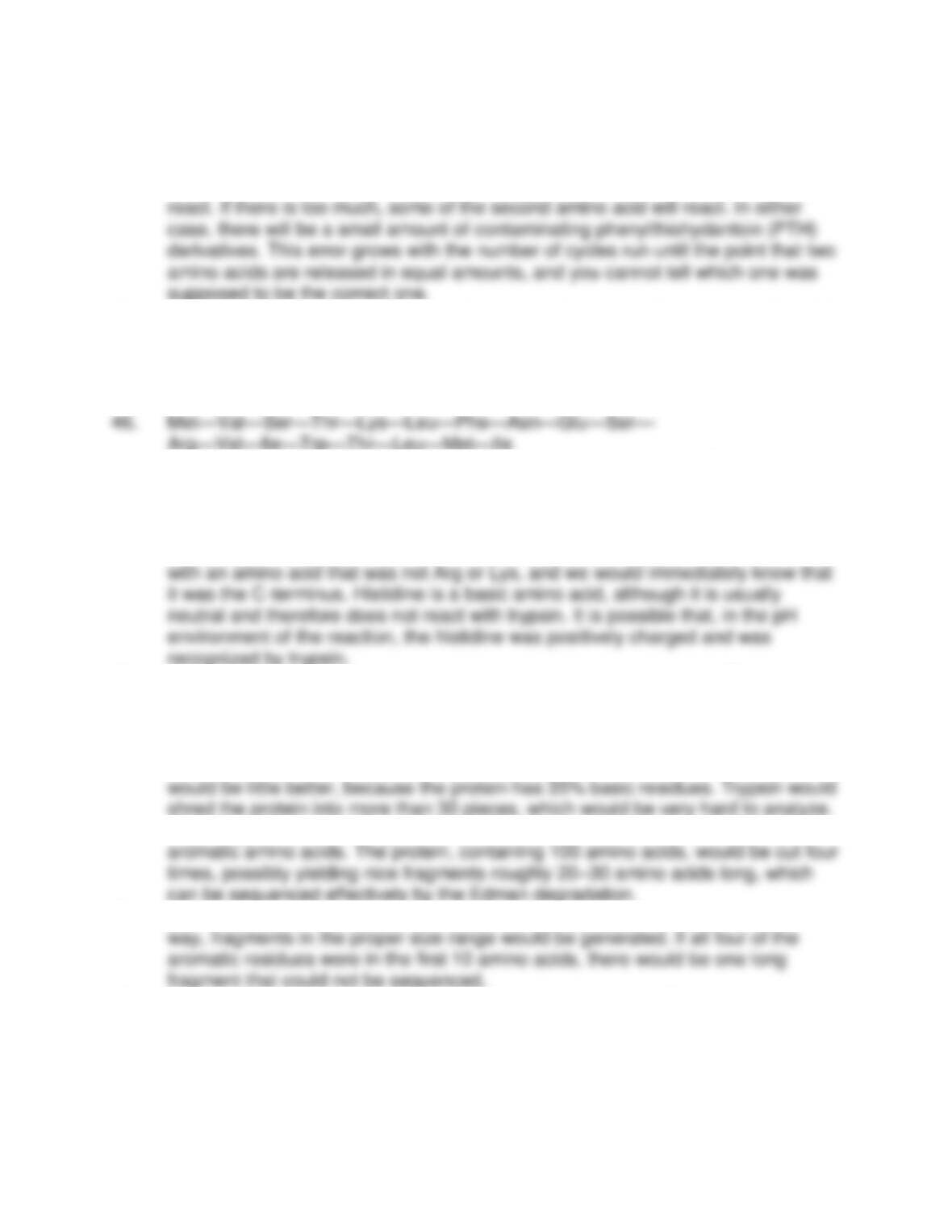
42.
8 Chapter 5
43. The amount of Edman reagent must exactly match the amount of N–termini in the
first reaction. If there is too little Edman reagent, some of the N–termini will not
44. In the first cycle, the first and second amino acids from the N-terminal end would
be reacted and released as PTH derivatives. You would get a double signal and
not know which one was the true N-terminus.
45. Val—Leu—Gly—Met—Ser—Arg—Asn—Thr—Trp—Met—
Ile—Lys—Gly—Tyr—Met—Gln—Phe
47. It is possible that your protein is not pure and needs additional purification steps
to arrive at a single polypeptide. It is also possible that the protein has subunits,
so multiple polypeptide chains could be yielding the contradictory results.
48. There are two fragments that have C-termini that are not lysine or arginine, which
is what trypsin is specific for. Normally there would be only one fragment ending
49. It would tell you a relative concentration of the various amino acids. This is
important because it would help you plan your sequencing experiment better. For
example, if you had a protein whose composition showed no aromatic amino
acids, it would be a waste of time to use a chymotrypsin digestion.
50. Cyanogen bromide would be useless, because there is no methionine. Trypsin
51. Chymotrypsin would be a good choice. There are more than four residues of
52. It would work best if the aromatic residues were spread out in the protein. In that
53. Electrospray Ionization (ESI-MS) and Matrix-Assisted Laser Desorption
Ionization– Time of Flight (MALDI-TOF MS).
54. MALDI-TOF MS is very sensitive and very accurate. Attomole (10–18) quantities of
a molecule can be detected.
Protein Purification and Characterization Techniques 9
5.5 Protein Identification Techniques
55. ELISA is based on antibody-protein interactions. Specific antibodies, called
primary antibodies, are put into microtiter plates to localize target proteins. A
56. A primary antibody is specific for a target protein that a researcher is looking for.
A secondary antibody will react with the primary antibody. The secondary
antibody carries the tag that makes the complex visible.
57. The protein-antibody complexes can be seen based on the nature of the tag
58. The first step in a western blot is the separation of proteins via electrophoresis.
The next step takes the gel from the electrophoresis and transfers the proteins
59. Western blot got its name as a humorous derivation from the original blotting
technique called Southern blotting. The original blotting technique was for DNA
60. The advantages of an ELISA would be ease of use, low cost, and ready
availability to any researcher. The disadvantages are that, compared to a
61. Proteins are transferred to nitrocellulose because all of the protein ends up
layered on top of a very thin membrane. This means that small volumes of the
62. There are thousands of primary antibodies that can be purchased commercially.
Other research requires a new primary antibody to be created. The process of
attaching an enzyme, a fluorescent marker, or a radioactive compound is a long
and difficult task. If every primary antibody had to be tagged, it would be a
daunting exercise for the company trying to do it. Instead, a company can
10 Chapter 5
5.6 Proteomics
63. Proteomics is the systematic analysis of an organism’s complete complement of
proteins, or its proteome. Just as we learned the basic dogma of molecular
64. The bait protein is constructed to have a particular affinity tag. The bait protein
interacts with cell proteins of interest and then binds to an affinity column via the
tag. In this way, the cell proteins of interest can be found and isolated.
65. There are many assumptions behind the experiment described in the
Biochemical Connections on page135. One must assume that the nature of the