IM 7-1
LABORATORY MANUAL AND WORKBOOK FOR BIOLOGICAL
ANTHROPOLOGY INSTRUCTOR’S MANUAL
Lab 7: Bioarchaeology and Forensic Anthropology
ANSWERS TO LAB 7 CONCEPT REVIEW QUESTIONS
1. Bioarchaeology is defined as the study of skeletal remains from archaeological
4. C. After skeletal remains have been identified as human, usually the next step is to
6. Any of the following are acceptable for aging juvenile skeletons (requiring specific
bones):
IM 7-2
7. Either of the following is acceptable for aging adult skeletons (requiring specific bones):
GUIDE & ANSWERS TO LAB 7 EXERCISES
Exercise 1: Animal or Human? (5 minutes)
IM 7-3
For this exercise, you could use materials from your own collection to supplement or replace the
images of Part A: two femurs (A is human, B is chimpanzee) and Part B: two crania (A is
chihuahua, B is fetal human) provided in the lab Image Library. [Note: The final trait may be
mentioned if students have already covered Lab 14.]
Review the skeletal material provided by your instructor (or the image in the lab Image Library)
and answer the questions below.
PART A
1. Which of the skeletal elements below belongs to a human?
2. Describe one trait that helped you make this distinction:
PART B
1. Which of the skulls is a human?
2. Describe one trait that helped you make this distinction:
Exercise 2: Minimum Number of Individuals (10 to 15 minutes)
For this exercise, you could use materials from your own collection to supplement or replace the
image provided.
Refer to the skeletal material provided by your instructor (or the mystery assemblage in the lab
Image Library) to answer the questions below.
1. List the bones depicted. (Be as specific as possible, including the side of the body that
the bone is from, if applicable.)
Exercise 3: Aging (10 to 15 minutes)
For this exercise, you could use materials from your own collection to supplement or replace the
images provided.
Review the skeletal material provided by your instructor (or the images in the text or lab Image
Library) to answer the questions below.
1. Examine the X-ray of a juvenile upper and lower jaw in Figure 7.12 and compare it to
the dental eruption age stages in Figure 7.13. What is the approximate age of this
individual?
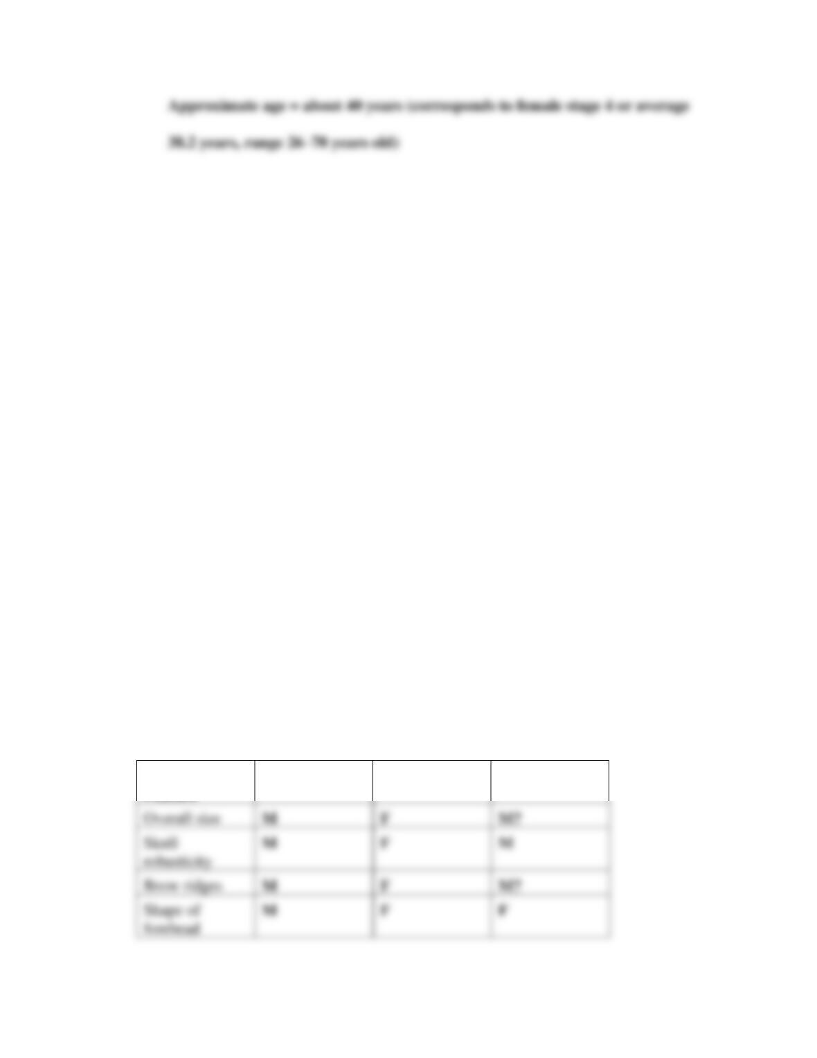
2. Examine the skeletal material, which is from a female and shows the symphyseal face
of the pubic bone (see lab Image Library). Compare this to the Suchey–Brooks age
stages provided in Figure 7.14. What is the approximate age of this individual?
IM 7-5
Exercise 4: Sexing (10 to 15 minutes)
For this exercise, you could use materials from your own collection to supplement or replace the
images provided of human male and female crania (Part A) and pelvises (Part B). For Part C,
students will need access to multiple humeri, femurs, scapulae, and calipers for taking
measurements. If some or all of these materials are unavailable, you may provide students with
the measurements given in the charts here, and have them estimate sex based on this
information.
Review the skeletal material provided by your instructor (or the images in the lab Image Library)
to complete the following exercise.
PART A
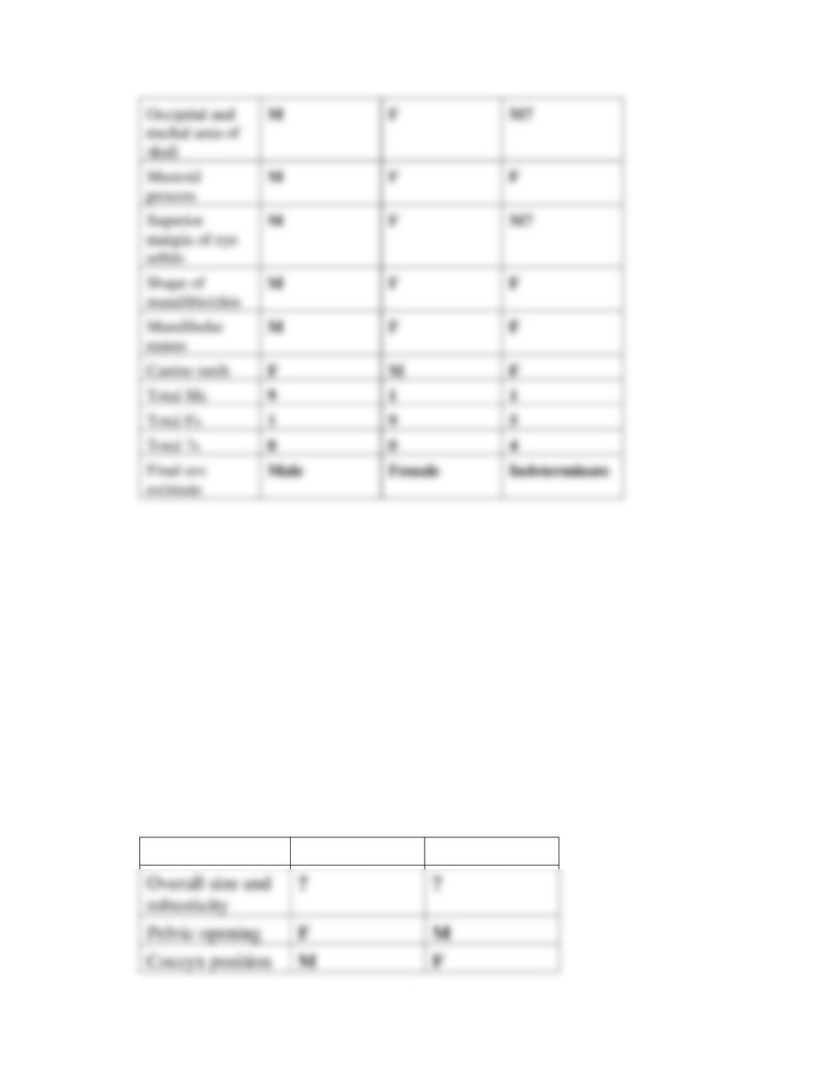
Estimate the sex of the three crania using the table below. For each cranial feature on the list,
score it as either M, F, or ? (if you are unsure or the feature is not visible). Compare the crania
with one another, other crania you have in the classroom, or the crania depicted in Figure 7.6.
Once you have completed the table, tally the total number of Ms and Fs and determine the
estimated sex.
Cranial
Feature
Cranium A
Cranium B
Cranium C
IM 7-6
[Note: Answers may vary depending on specimens available. The answers here are based on the
Image Library specimens.]
PART B
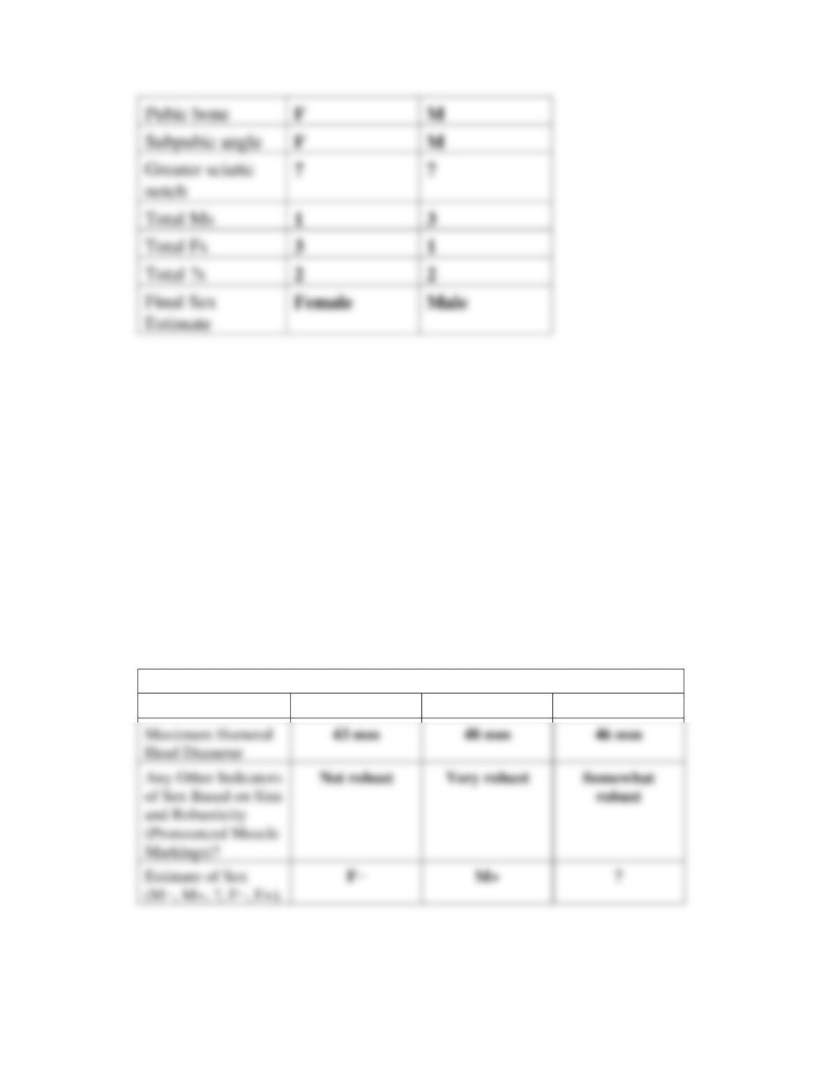
Estimate the sex of the three pelvises using the chart below. For each pelvic feature on the list,
score it as either M, F, or ? (if you are unsure or the feature is not visible). Compare the three
pelvises with one another, other pelvises you have in the classroom or the pelvises in Figure 7.7.
Once you have completed the table, tally the total number of Ms and Fs for each pelvis and
determine the estimated sex.
Pelvic Trait
Pelvis A
Pelvis B
IM 7-7
[Note: Answers may vary depending on specimens available. The answers here are based on the
Image Library specimens.]
PART C
Use the skeletal material (or measurements) provided by your instructor to complete the
following exercise.
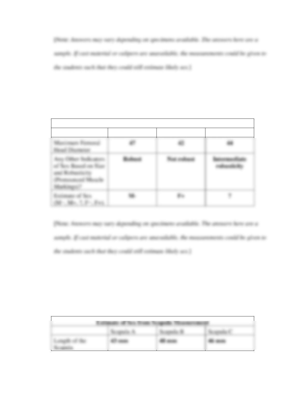
1. Measure the maximum humeral head diameter (assume the bone is of white European
ancestry). Record your measurements and use the table on p. 189 to give an estimated
sex (M−, M+, ?, F−, F+).
Estimate of Sex from Humeral Head Diameter
Humerus A
Humerus B
Humerus C
Head Diameter
Markings)?
IM 7-8
2. Measure the maximum femoral head diameter (assume the bone is of white European
ancestry). Record your measurements in the chart below and use the table on p. 189
to determine an estimated sex (M−, M+, ?, F−, F+).
Estimate of Sex from Femoral Head Diameter
Femur A
Femur B
Femur C
Head Diameter
Markings)?
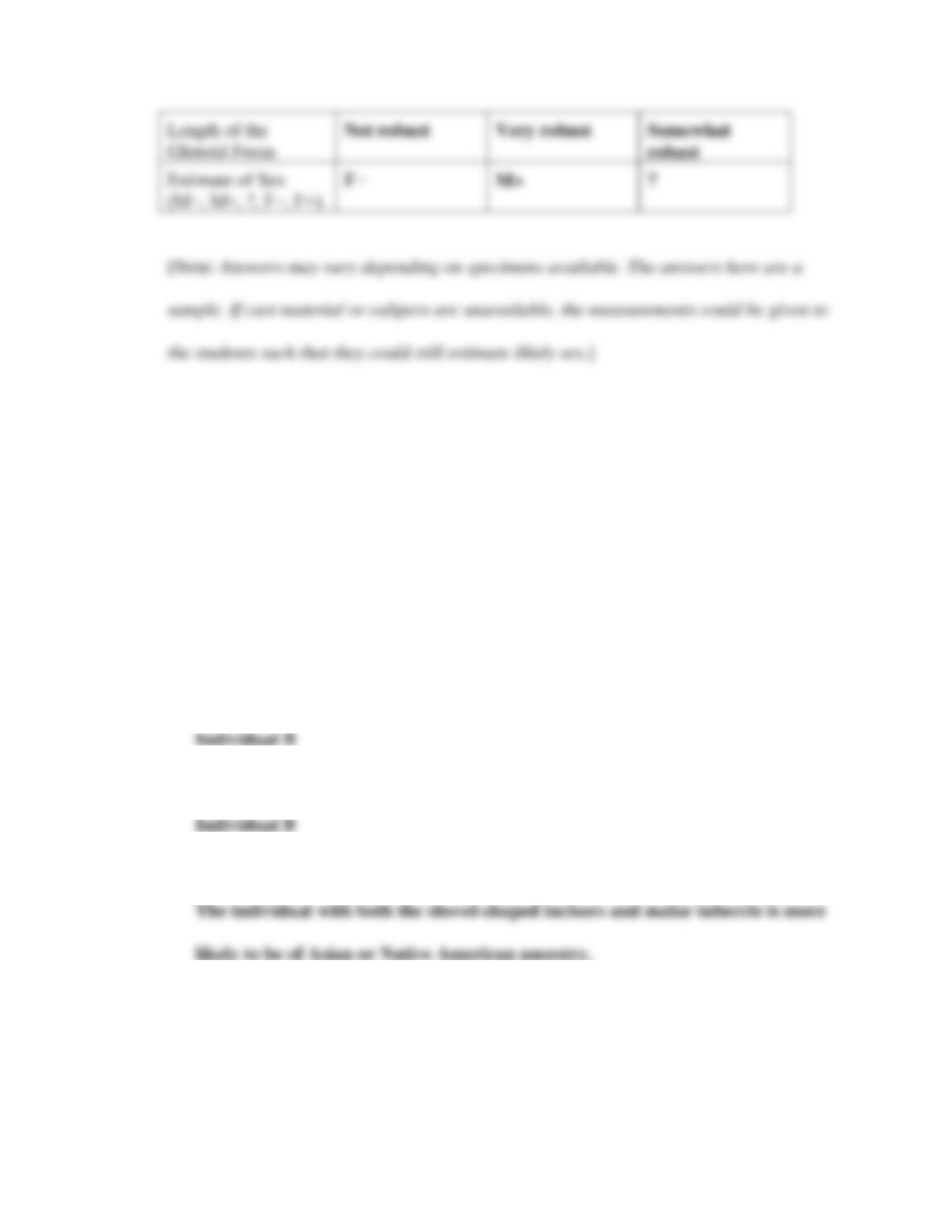
3. Measure the scapula length and length of the glenoid fossa (following the guidelines
provided in Figure 7.8 on p. 189). Record your measurements in the chart below and
use the numeric markers described in the text to determine an estimated sex (M−,
M+, ?, F−, F+).
Scapula A
Scapula B
Scapula C
Scapula
IM 7-9
Exercise 5: Ancestry (5 minutes)
For this exercise, you could use materials from your own collection to supplement or replace the
images provided of incisors and crania of two individuals (A without and B with shovel-shaped
incisors and malar tubercle).
Refer to the skeletal material provided by your instructor (or the images in the lab Image
Library) to answer the following questions.
1. Which individual has shovel-shaped incisors?
2. Which individual has the malar tubercle?
3. What might these two traits indicate about these individuals’ ancestries?
4. Are these two traits alone enough to make an ancestry determination? Why or why
not?
IM 7-10
Exercise 6: Stature (5 to 15 minutes)
Your students will probably need access to a calculator to complete the math in this exercise.
You may want to provide your students with the conversion information needed to convert
centimeters to inches (2.54 cm = 1 in.). Students should divide their centimeter stature estimate
by 2.54 to determine stature in inches.
As always, you could use materials from your own collection to supplement or replace the
material provided. If you do so, you could ask students to take their own measurements using an
osteometric board, calipers, or tape measure. If you ask students to take their own
measurements, be sure to allow for the extra class time this will require.
Review the skeletal material provided by your instructor (or the description below) and answer
the questions.
A forensic anthropologist has determined that a female victim has a maximum femur
length of 49.5 cm. The anthropologist also determined that the victim is of African
ancestry. Use the table on p. 197 to estimate this individual’s stature. (Be sure to give the
Exercise 7: Pathology (10 to 15 minutes)
For this exercise, you could use materials from your own collection to supplement or replace the
images provided.
Review the skeletal material provided by your instructor (or the images in the Lab 7 Exercise
Image Library on p. 222) and answer the following questions.
PART A
1. What bone is this?
2. What pathology is indicated on this bone?
3. Describe one trait you used to make this pathology identification.
4. Is this pathology antemortem or perimortem?
PART B
1. What bone is this?
2. What pathology is indicated on this bone?
3. Describe one trait you used to make this pathology identification.
4. Is this pathology antemortem or perimortem?
IM 7-12
Exercise 8: Fragmentary Remains (15 to 20 minutes)
For this exercise, you could use materials from your own collection to supplement or replace the
images provided.
Refer to the skeletal material provided by your instructor (or the images in the Lab 7 Exercise
Image Library on p. 223). These partial remains have been found in a shallow grave in a wooded
lot. Use this information to answer the following questions.
1. What bones are present?
2. What is the MNI?
3. What is (are) the age(s) of the individual(s), and how did you determine this?
Exercise 9: Tying It All Together (15 to 20 minutes)
For this exercise, you could use materials from your own collection to supplement or replace the
images provided.
A collection of skeletal remains has just been unearthed at a crime scene. You have been asked
to help law enforcement officials in their investigation of this skeletal material. Refer to the
IM 7-13
skeletal material provided by your instructor (or the mystery assemblage in the Lab 7 Exercise
Image Library on p. 224) to answer the following questions.
1. Is any of this skeletal material nonhuman? If so, which bone(s) is(are) nonhuman? Why
do you think this?
2. List all of the human skeletal elements, being as specific as possible. What is the
minimum number of individuals (MNI) represented here?
3. Does any of the skeletal material appear to be juvenile? If so, which bone(s)? Why do
you think this?
4. Can any of the skeletal material be used to determine the biological sex(es) of the
victim(s)? If so, which bone(s)? What is (are) the sex(es) you determined? What evidence
supports that conclusion?
5. Bone I in the assemblage has been measured with an osteometric board. It has a
IM 7-14
the estimated stature of this individual? (Be sure to give the estimation in feet and
inches.)
6. Based on the materials recovered, can you make any suggestions for additional analyses
you might use to further understand the circumstances surrounding the death of the
victim(s)?
ANSWERS TO LAB 7 CRITICAL THINKING QUESTIONS
1. The minimum number of individuals (MNI) represented by the bioarchaeological
IM 7-15
juvenile skeleton is undergoing these stages, we can pinpoint age. Adults are no longer
3. The estimated stature of the individual from Exercise 6 based on the new right humerus
(68.0 in.) and similar ranges.
4. Antemortem pathology occurs any time before death, while perimortem pathology occurs
around the time of death and may have contributed to death. The forensic anthropologist
5. Similar: Both bioarchaeology and forensic anthropology use similar methods of analysis;
they follow similar protocols, and seek to understand much of the same foundational
IM 7-16
6. Answers to the questions concerning the role forensic anthropologists in identifying the
remains of border migrants will vary depending on the materials and information used by
7. Answers regarding the steps taken in analyzing the remains of 200 people from a
medieval Italian cemetery for gender differences in health will vary but should include
these: The first analyses that should be conducted are biological sex determinations and
8. Answers to the question of whether Julia or Marco has correctly identified the pictured
bones as being from a female or male individual will vary. This question is designed to
help students realize that there may be uncertainty when conducting this kind of analysis
IM 7-17
and that our interpretations are the best we can do with the evidence provided. Possible
responses include these: The anthropologists disagree because a person may not have
9. The disagreement in ancestry determination may reflect differences in metric and
nonmetric traits. There is more within-population variation in metric traits than in
nonmetric traits. For example, there is variation in stature and robusticity within each