IP 6-1
LABORATORY MANUAL AND WORKBOOK FOR BIOLOGICAL
ANTHROPOLOGY INSTRUCTOR’S MANUAL
Lab 6: Bones of the Skeleton
ANSWERS TO LAB 6 CONCEPT REVIEW QUESTIONS
1. The occipital bone forms the posterior (back) of the cranium.
vertebra).
5. The ribs that articulate directly, indirectly, or not at all with the sternum are:
each arm.
ossa coxae.
side of the body.
GUIDE & ANSWERS TO LAB 6 EXERCISES
Exercise 1: Cranium (15 to 20 minutes)
For this exercise, you could substitute a cast with numbered stickers or labels instead of the
image provided in the lab Image Library.
Part A: Human Cranium (anterior and lateral views)
Refer to the skeletal material provided by your instructor (or the human cranium pictures in the
lab Image Library). Some of the major bones of the cranium have been assigned numbers. For
each number, provide the appropriate bone name in the space below:
Note that the zygomatic bone (7) fuses with the maxilla anteriorly and with the zygomatic
process of the occipital posteriorly, forming a bony zygomatic arch that creates a space for the
passage of cheek muscles.
IP 6-3
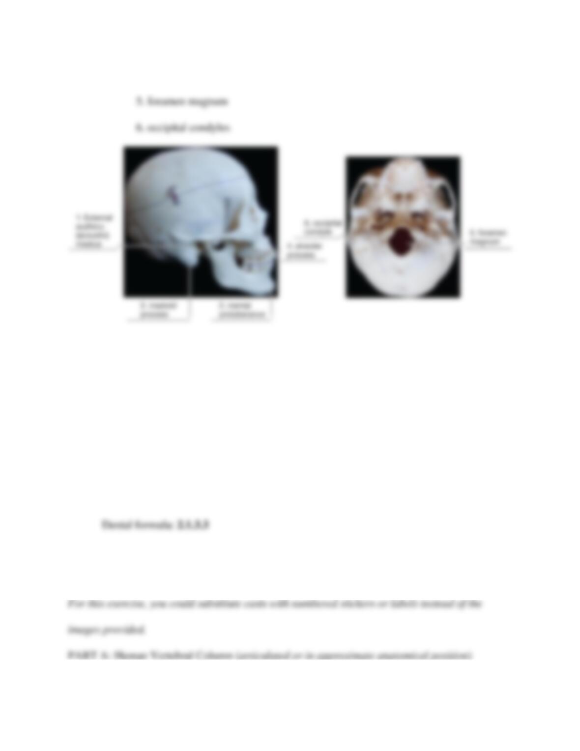
4. alveolar process
Exercise 2: Dentition (5 minutes)
For this exercise, you could substitute your own material for the image provided, but be sure to
adjust the dental formula answer as needed depending on the material you provide.
Refer to the skeletal material provided by your instructor (or the mystery animal dentition picture
in the lab Image Library) to calculate the dental formula for the animal and write it in the space
provided.
Exercise 3: Vertebral Column (10 to 15 minutes)
IP 6-4
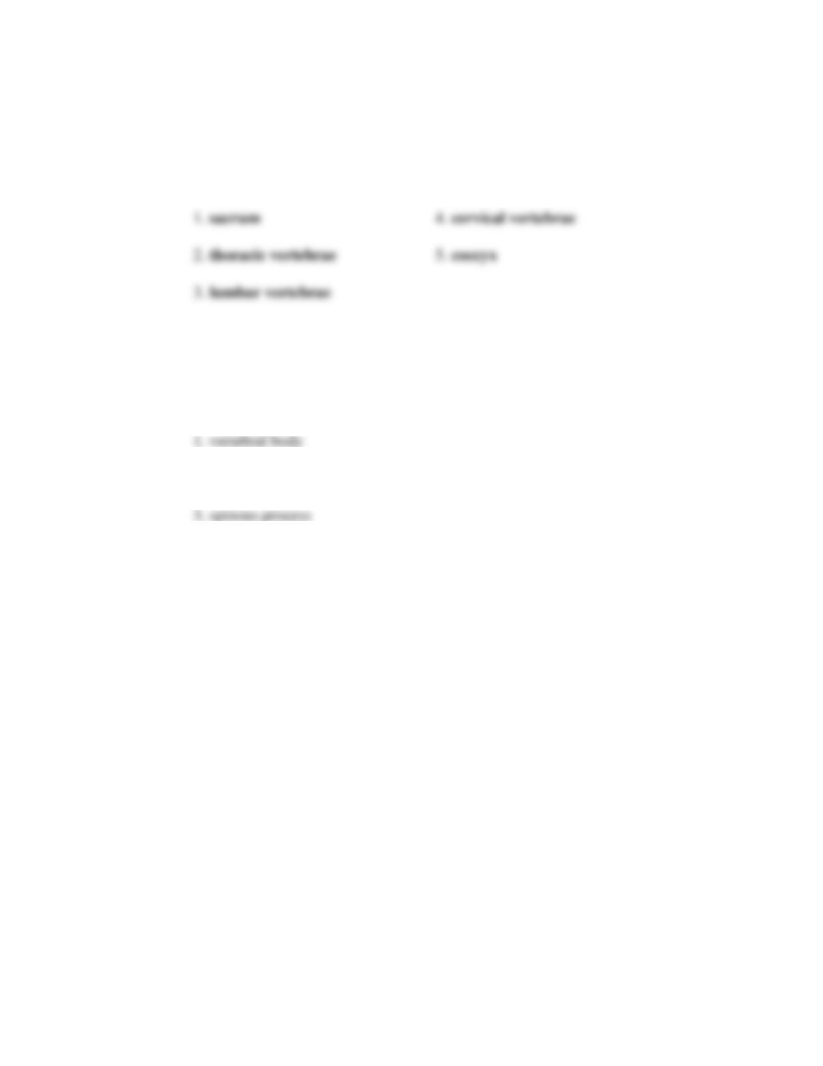
Refer to the skeletal material provided by your instructor (or the human vertebral column
diagram in the lab Image Library). The major bones of the vertebral column have been assigned
numbers. For each number, provide the appropriate name in the space below:
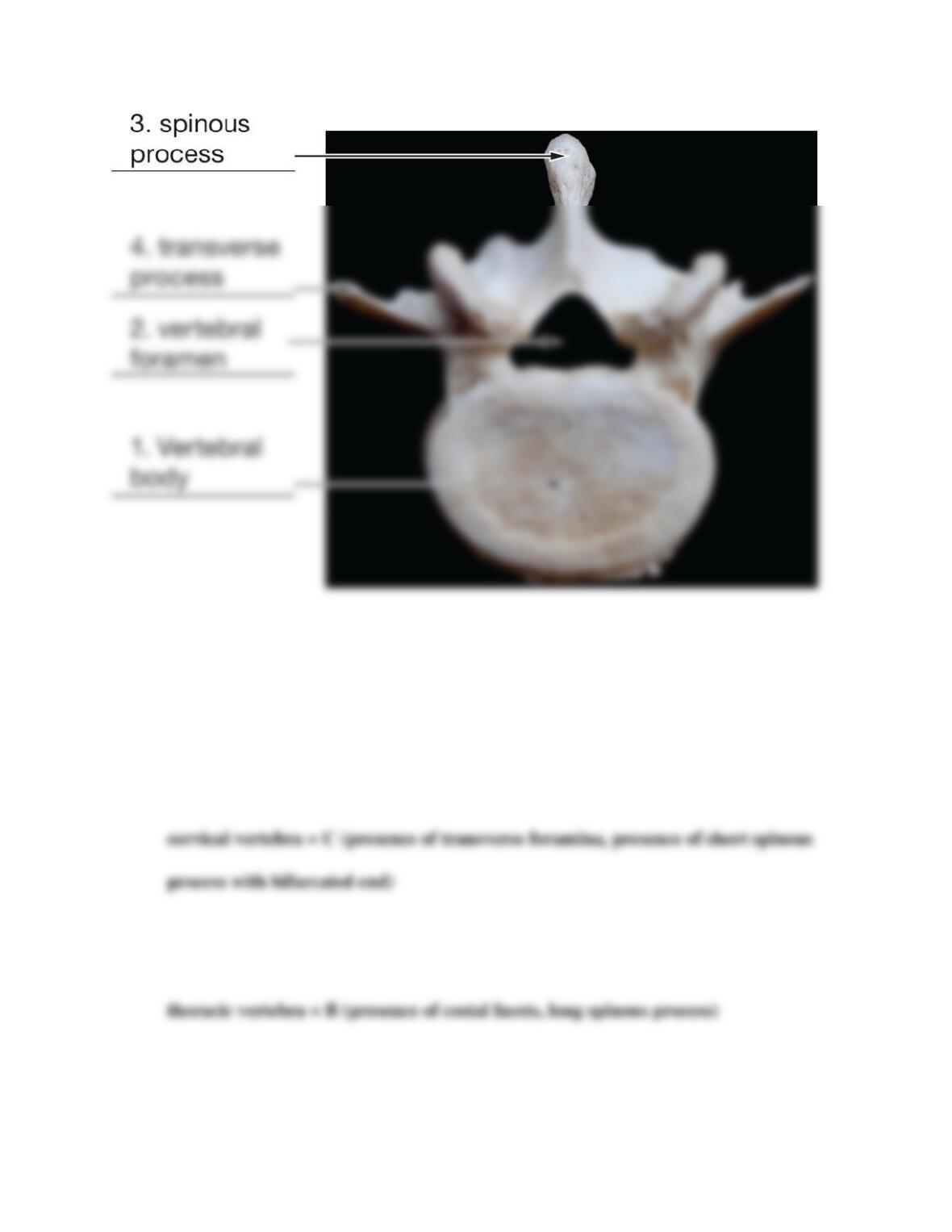
PART B: Typical Human Vertebra (such as L3)
Refer to the skeletal material provided by your instructor (or the typical human vertebra pictured
in the lab Image Library) and label the following key features on the vertebra:
2. vertebral foramen
4. transverse process
IP 6-5
PART C: Types of Vertebrae (one each of cervical, thoracic, and lumbar vertebrae)
Refer to the skeletal material provided by your instructor (or the photos of three mystery
vertebrae pictured in the lab Image Library) to answer the following questions.
1. Which mystery vertebra is a cervical vertebra? Describe two features that helped you
determine this.
2. Which mystery vertebra is a thoracic vertebra? Describe two features that helped you
determine this.
3. Which mystery vertebra is a lumbar vertebra? Describe two features that helped you
determine this.

Exercise 4: Thoracic Cage (Rib Cage) (5 minutes)
For this exercise, you could substitute a cast with numbered stickers or labels instead of the
image provided showing an articulated thoracic cage.
Refer to the skeletal material provided by your instructor (or the human rib cage pictured in the
lab Image Library) and label the following bones of the rib cage:
1. manubrium
2. sternal body
3. xiphoid process
4. true ribs
5. false ribs
6. floating ribs
Exercise 5: Upper and Lower Limb Comparison (10 minutes)
For this exercise, you could substitute a cast with numbered stickers or labels instead of the
images of limb bones provided.
PART A: Human Upper Limb
Refer to the skeletal material provided by your instructor (or the human upper limb picture in the
lab Image Library). The major bones have been numbered. For each number, provide the
appropriate name in the space provided.
IP 6-7
Note that some students may name the general bone groups or specific bones within the wrist,
hand, or fingers.
PART B: Human Lower Limb
Refer to the skeletal material provided by your instructor (or the human lower limb picture in the
lab Image Library). The major bones have been numbered. For each number, provide the
appropriate bone name in the space provided.
Exercise 6: Upper Limb (10 minutes)
For this exercise, you could substitute a cast with numbered stickers or labels instead of the
human humerus images provided.
IP 6-8
PART A: Human Humerus
Refer to the skeletal material provided by your instructor (or the human humerus pictured in the
lab Image Library) and label the following key features of the humerus:
1. head
2. lesser tubercle
IP 6-9
PART B: Human Hand
Review the bones of the hand provided by your instructor or depicted in Figure 6.37 Describe
two things that make the first finger (the thumb) different from the other fingers.
Exercise 7: Lower Limb (15 minutes)
For this exercise, you could substitute a cast with numbered stickers instead of the human pelvis
and femur images provided.
PART A: Human Pelvis (articulated with sacrum)
Review the skeletal material provided by your instructor (or the human pelvis pictured in the lab
Image Library). The major bones and features have been numbered. For each number, provide
the appropriate name in the space provided.
IP 6-10
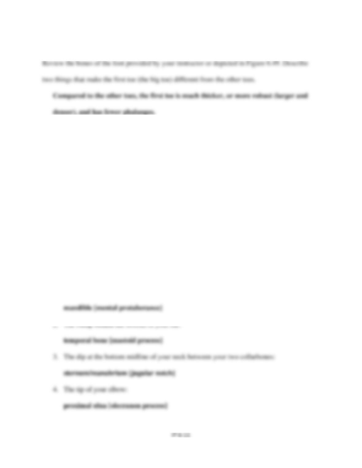
PART B: Human Femur
Review the skeletal material provided by your instructor (or the human femur pictured in the lab
Image Library) and label the following key features of the femur:
1. head
PART C: Human Foot
Exercise 8: Surface Anatomy of the Skeleton (20 to 25 minutes)
If available, you may want to provide students with access to an articulated full human skeleton
to help them visualize how their externally palpable features translate to internal features on the
skeleton. For this exercise, it is particularly important that students work in small groups.
Depending on individual variations in student body mass, some features may be harder for them
to locate on their own bodies. Having multiple students in the group will help alleviate some of
these potential problems.
Use your own body (in anatomical position) to identify the following bones and features from
the skeleton. Name the specific bone(s) [and feature(s) if possible] for each:
1. Your chin:
IP 6-12
5. The sides of your elbow:
7. The protrusion or bump you can feel on the corner of your wrist (palmar surface):
8. The protrusion or ridge at the front and top of your hips:
9. The protrusion or bump on your outer ankle:
10. The protrusion or bump on your inner ankle:
ANSWERS TO LAB 6 CRITICAL THINKING QUESTIONS
1. The cranium must form a round, protective shell for the brain. Many smaller bones must
2. Similar features of vertebrae: All have same basic parts (body, vertebral arch, vertebral
IP 6-13
specific shapes (rib attachment areas, orientation of spinous process, presence or absence
3. It is important that much of the anterior rib cage is made of cartilage because this
4. Similar features of shoulder and elbow: The humerus is part of both joints. Differences:
More bones are involved in the elbow than in the shoulder. The shoulder is more mobile
5. Similar types of bones and layout of upper and lower limbs: The same general types of
bones are involved (long, short), the same relative numbers of bones (one upper arm/leg
some similarities in overall function and mobility. For example, the limb overall is fairly
6. Differences in physical features of the hands and feet: Feet have arches and short toes.
Hands do not have an arch, and the fingers are long. Hands are for grasping and fine