IP 5-1
LABORATORY MANUAL AND WORKBOOK FOR BIOLOGICAL
ANTHROPOLOGY INSTRUCTOR’S MANUAL
Lab 5: Introduction to the Skeleton
ANSWERS TO LAB 5 CONCEPT REVIEW QUESTIONS
1. The main functions of the skeleton inside the body include:
bone forms the exterior and trabecular bone forms the interior).
osteodon, or osteocyte; “blasts build”).
6. Bone remodels for one reason, among the following: allow for growth; extract
IP 5-2
7. Students may name any bone in the appendicular skeleton (the arm or leg, including the
flat bone, irregular bone, or short bone).
GUIDE & ANSWERS TO LAB 5 EXERCISES
Exercise 1: Bone Remodeling (15 minutes)
1. Describe one hypothetical situation that illustrates the “use it” portion of the use it or lose
it principle.
Description of the situation/behavior:
Explanation for why more bone is present under these circumstances:
2. Describe one hypothetical situation of the “lose it” portion of the use it or lose it
principle.
IP 5-3
Description of the situation/behavior:
Explanation for why less bone is present under these circumstances:
Answers will vary but should address these issues: Decreased functional need/stress,
Exercise 2: Axial and Appendicular Skeleton (10 minutes)
Students are to use the diagram of the human skeleton provided to show the major skeletal
divisions.
1. On the diagram, draw circles around bones that are part of the axial skeleton.
2. On the diagram, draw boxes around bones that are part of the appendicular skeleton.
Exercise 3: Directional Terminology (15 to 20 minutes)
For this exercise, you could use your own materials to supplement or replace the diagram
provided in Figure 5.7.
1. Name three (3) bones that are proximal to the carpals.
IP 5-4
2. Name two (2) bones that are anterior to the occipital bone.
3. Name one (1) bone that is superior to the temporal bone.
4. Name four (4) bones that are distal to the femur.
5. Name one (1) bone that is posterior to the sternum.
6. Name one (1) bone that is inferior to the cervical vertebrae.
7. Draw a circle around the toe of each foot that is the most medial.
8. Draw a box around the toe of each foot that is most lateral.
Exercise 4: Bone Shapes (15 to 20 minutes)
For this exercise, you will need to provide students with colored pencils, markers, or crayons for
coloring four types of bones (long, short, flat, and irregular) on a diagram of the human skeleton
(the diagram also used for Exercise 2). Specific answers (color schemes) will vary, but the major
bone types should be similarly distinguished across students.
Step 1: For each of the four bone shapes, choose a different color.
IP 5-5
Step 2: Review the skeleton diagram on p. 126. Classify each of the bones on the skeleton
diagram as one of the four bone shapes.
Exercise 5: Bone Features (15 to 20 minutes)
For this exercise, you could use your own materials to supplement or replace the images
provided in the Image Library. If doing so, be sure to number the material accordingly. [Note:
This exercise is meant to help students apply what they have learned about general types of bone
features to real examples from the human skeleton. It is less important that they know the
specific name (such as transverse foramen) and more important that they recognize what
constitutes a foramen. In Lab 6, students are taught about the individual bones and their specific
features in greater detail.]
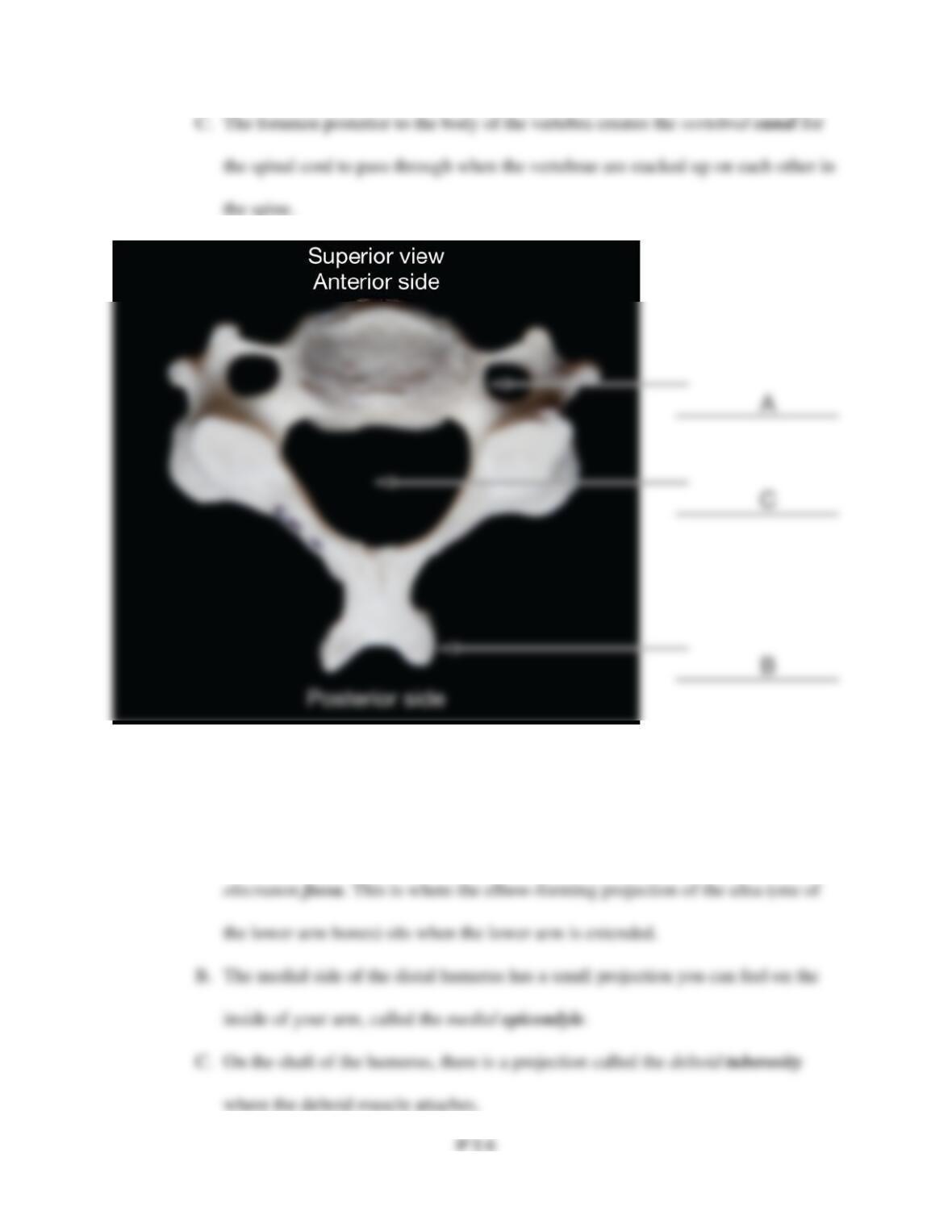
1. Examine the cervical vertebra (neck vertebra) depicted. Locate the features boldfaced and
described below, and write the corresponding letters in the blanks provided on the image on p.
131.
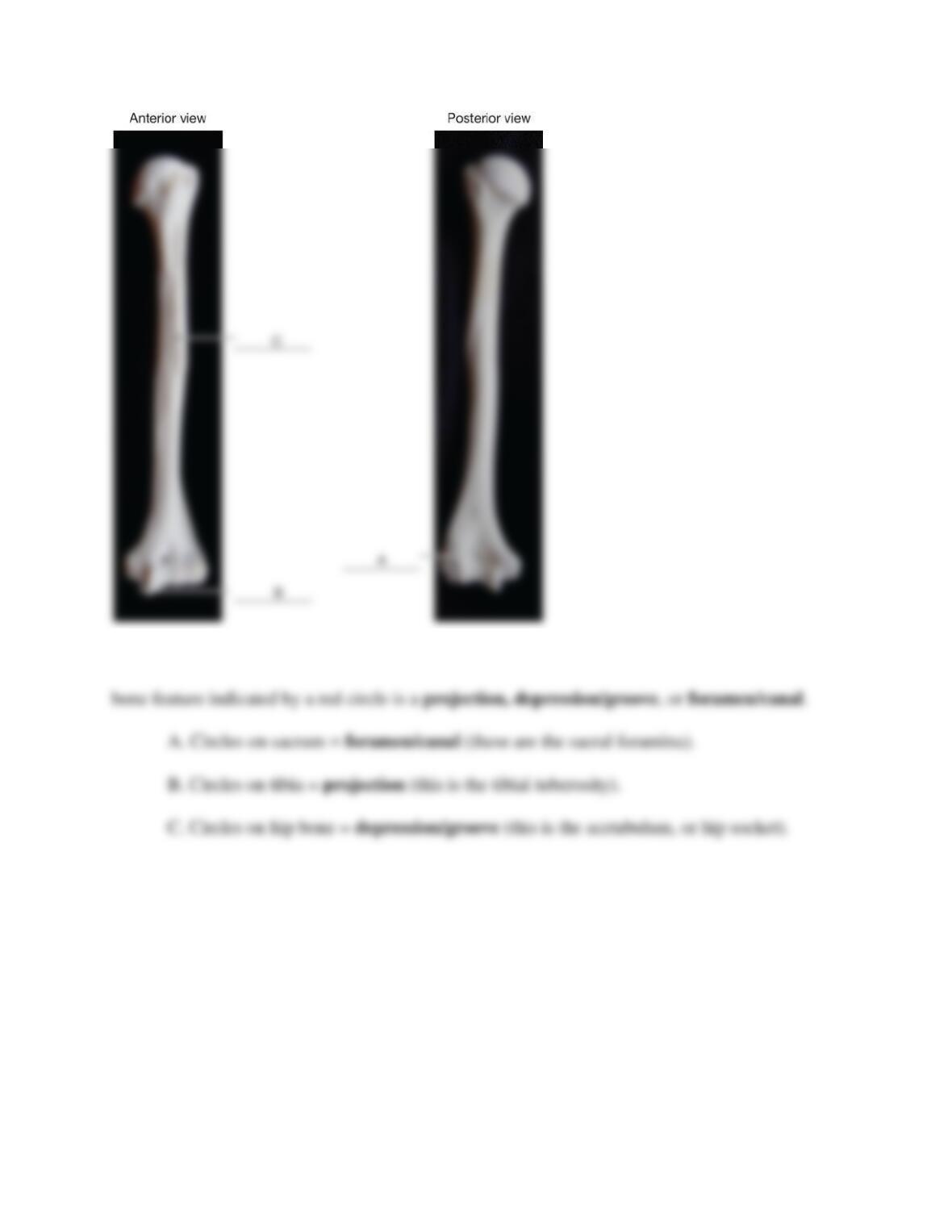
2. Examine the humerus (upper arm bone). Locate the features boldfaced and described
below, and write the corresponding letters in the blanks provided on the image on p. 131.
A. On the posterior side of the distal end of the humerus, there is a feature called the
IP 5-7
3. Examine the sacrum, shinbone (tibia), and hip bone (pelvis). For each bone, state whether the
ANSWERS TO LAB 5 CRITICAL THINKING QUESTIONS
1. Relation of the skeletal system to the muscular system: Bones provide the support and a
place of attachment for muscles; bones will have projections where muscles need extra
IP 5-8
attachment area; bones will have grooves where muscle tendons or ligaments insert on or
3. Examples of “use it” and “lose it” effects on bone will vary but should address similar
4. Bones of the appendicular skeleton include the bones that comprise the appendages. This
includes bones that may lie close to the midline or central axis but are more functionally
5. The cranium is superior to the pelvis (the pelvis is inferior to the cranium).
6. Short bones are found clustered together in areas with limited range of motion (e.g., wrist
and ankle). They are well suited for the wrist because they restrict movement. Long
7. The mandibular canal is an area for nerves, veins, and arteries in the mandible (lower