
562 Chapter 20 ■ SPELLING QUIZ
Chapter Twenty
SPELLING QUIZ
Name: ______________________________
A. Circle the term that is spelled correctly and write its meaning in the space provided:
1. en vivo in vivo _____________________________________________________
2. therapeutic therapreutic ________________________________________________
3. colangiography cholangiography ____________________________________________
4. radionuclide radioneuclide _______________________________________________
5. radiolucent radiolucant ________________________________________________
6. anterioposterior anteroposterior _____________________________________________
7. transducer transduser _________________________________________________
8. radiopharmaceutical radiopharmaseutical _________________________________________
9. traser studies tracer studies _______________________________________________
10. in vetro in vitro ____________________________________________________
B. Circle the term that is spelled correctly. The meaning of each term is given.
11. X-ray record of the spinal cord …………….. myleogram myelogram mielogram
12. Moving toward the midline ………………….. abduction adduckshun adduction
13. Lying down position ……………………………. rekumbent recumbant recumbent
14. Lying on the back ……………………………….. supine soupine suppine
15. Obstructing the passage of x-rays …………. radiopaquie radiopaque radioopaque
16. X-ray of the renal pelvis ………………………. pyleogram pyelogram pyilogram
17. X-ray record of vessels …………………………. anjiogram angeiogram angiogram
18. Radioactive form of a substance ……………. radioisotope radioiceotope radioisotop
19. Lying on the belly ……………………………….. prone proone pron

Chapter 20 ■ PRONUNCIATION QUIZ 563
Chapter Twenty
PRONUNCIATION QUIZ
Name: ______________________________
A. Underline the accented syllable in the following terms:
1. radioisotope 4. supine 7. ionization
2. ultrasonography 5. recumbent 8. scintigraphy
3. angiography 6. echocardiography 9. lateral decubitus
B. Match the term in Column I with its meaning in Column II:
Column I Column II
1. flexion ________ A. Turning outward
2. extension ________ B. Lying on one’s belly; face down
3. prone ________ C. Bending a part of the body
4. abduction ________ D. Lying on one’s back
5. supine ________ E. Radionuclides are used as tags attached to
chemicals and followed throughout the body
6. eversion ________ F. Test of blood flow to heart muscle
7. oblique ________ G. Lengthening or straightening a flexed limb
8. 99mtechnetium
sestamibi scan ________ H. Carrying a limb away from the body
9. tracer studies ________ I. Positioned at an angle
C. Complete the following medical terms from their definitions:
1. ____________________________ gram X-ray record of the urinary tract
2. ____________________________ gram X-ray record of the bile vessels
3. radio ___________________________ A radioactive drug used in diagnosis of disease
4. ____________________________ gram X-ray record of the uterus and the fallopian tubes
5. ____________________________ ology Study of x-rays
6. radio ___________________________ Permitting the passage of x-rays

564 Chapter 20 ■ REVIEW SHEET QUIZ
Chapter Twenty
REVIEW SHEET QUIZ
Name: ______________________________
Give meanings for the following combining forms, suffixes and prefixes:
1. therapeut/o _____________________ 8. echo- ___________________________________
2. is/o ____________________________ 9. -gram __________________________________
3. pharmaceut/o ___________________ 10. -graphy _________________________________
4. radi/o __________________________ 11. -lucent __________________________________
5. son/o __________________________ 12. -opaque _________________________________
6. viv/o ___________________________ 13. ultra- ___________________________________
7. vitr/o __________________________

Chapter 20 ■ MEDICAL SCRAMBLE 565
Chapter Twenty
MEDICAL SCRAMBLE
Name: ______________________________
Unscramble the letters to form radiology and nuclear medicine terms from the clues.
Use the letters in the squares to complete the bonus term.
1. Clue: Movement toward the midline of the body
____ ____ ____ ____ ____ ____ ____ C I D T N O U D A
2. Clue: Obstructing the passage of x-rays
____ ____ ____ ____ ____ ____ ____ ____ P U R D O Q E I A A
3. Clue: Lying on one’s back
____ ____ ____ ____ N E S I P U
4. Clue: X-ray image of blood vessels after injecting contrast material into the vessels
____ ____ ____ ____ ____ ____ ____ M A G A G O R N I
5. Clue: Permitting the passage of x-rays
____ ____ ____ ____ ____ ____ ____ ____ ____ C U T L E D A I R O N
BONUS TERM: Clue: Handheld device that sends and receives ultrasound signals
566 Chapter 20 ■ CROSSWORD PUZZLE
Chapter Twenty
CROSSWORD PUZZLE
Name: ______________________________
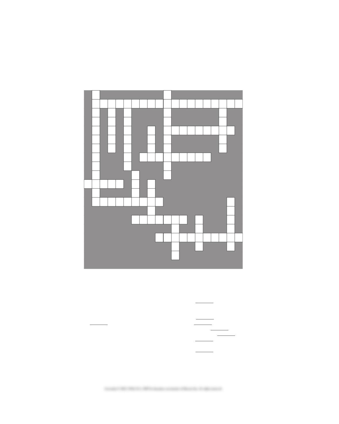
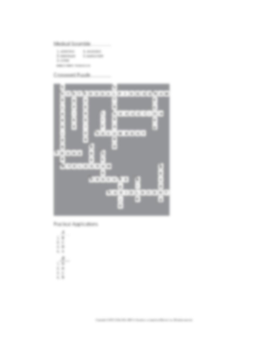
Fill in the crossword puzzle below using the clues listed underneath it.
12
3456
78
9
11
10 14
13 18
15 16
17
12
Across Clues Down Clues
3. An x-ray record of the uterus and fallopian tubes to
determine patency.
8. Moving a part of the body away from the midline.
9. Lying down; synonym of decubitus.
10. Lying on the belly.
13. An x-ray recording of the spinal cord.
15. -opaque means .
17. Permitting the passage of most x-rays.
1. An x-ray recording of the bile vessels.
2. Handheld device that projects high frequency sound
waves is a/an transducer.
4. Lying on the back.
5. Turning outward.
6. -gram means .
7. Viv/o means .
11. Pharmaceut/o means .
12. Cholangiography detects duct abnormality.
14. Vitr/o means .
16. The combining form meaning x-rays.
18. Ultra- means .

Chapter 20 ■ PRACTICAL APPLICATIONS 567
Chapter Twenty
PRACTICAL APPLICATIONS
Name: ______________________________
A. Radiology Report
Mass Adjacent to Thyroid—Lt side on US done 5/5/10
EXAM: NECK W/WOUT CONTRAST
5/9/10
Initially scans were obtained throughout the neck without IV contrast. Following this, a bolus of
IV contrast was given and rapid scans obtained through the region of interest in the neck, related
to the left lobe of the thyroid. As demonstrated on the recent ultrasound, there is a well-defined
oblong mass in the left neck measuring 2.2 × 3 × 1.8 cm in size. This lies posterolateral to the
left thyroid lobe and is predominantly between the left common carotid artery, which is displaced
posteromedially, and the left internal jugular vein, which is displaced laterally. No other adenopathy,
and the remainder of the thyroid appears unremarkable.
IMPRESSION: The mass appears to be extrinsic to the thyroid, possible even within the carotid
sheath. The lesion appears to be relatively avascular. The differential diagnosis would include an
unusually enlarged lymph node, a very atypical thyroid nodule, or a soft tissue tumor. The lesion is
very easily accessible to needle biopsy and this would certainly be the easiest method of obtaining
positive confirmation. This could be done using US guidance.
1. What type of radiologic test is
described here? ……………………………..
A. Ultrasound
B. CT scans
C. Pyelogram
D. PET scan
2. Where is the mass located? ……………..
A. Within the left lobe of the thyroid gland
B. Between the left and right carotid arteries
C. Behind and to the side of the left lobe of
the thyroid gland
D. In front and to the side of the thyroid
gland
3. Which of the following is not a
possible diagnosis? ………………………..
A. An unusual mass on the thyroid gland
B. Lymphadenopathy
C. Tumor mass adjacent to the thyroid gland
D. Lesion composed of many blood vessels
4. What procedure will help determine
the diagnosis? ………………………………
A. Aspiration of tissue and pathologic
examination
B. Removal of the thyroid gland
C. Removal of thyroid tissue for biopsy
D. Further scans with ultrasound guidance
568 Chapter 20 ■ PRACTICAL APPLICATIONS
B. Chart Note
The pt underwent a bone scan, which revealed irregular foci of tracer in lower T spine (T11-T12)
consistent with compression fracture. There was also increased tracer in the posterior/lateral right
ribs. On physical exam she was diffusely tender and in pain throughout her chest/ribs and spine.
The pt also underwent chest CT, which demonstrated extensive parenchymal and pleural disease
encasing the entire chest and involving vessels and bronchi. Her adrenals and liver, vertebral bodies,
and R scapula were also involved with metastatic disease.
1. What type of test is a bone scan? ………
A. Chest x-ray of ribs and bones of back
B. MRI of the skeleton
C. Dye is injected and traced in blood vessels
D. Radioisotope is injected and traced in
bones
2. What type of doctor administers
this test? ……………………………………..
A. Diagnostic radiologist
B. Nuclear medicine specialist
C. Radiation oncologist
D. Medical oncologist
3. Extensive parenchymal and pleural
disease means: ……………………………..
A. Tumor is in the spinal cord
B. Tumor is in the backbones
C. Tumor is in the lungs and membranes
around the lungs
D. Tumor is in the abdominal, pelvic, and
chest regions
4. The bone scan revealed: ………………….
A. Disease in the lungs
B. Areas of tumor in the lower chest region
and ribs
C. Disease in the adrenals and liver
D. Metastatic disease in the right shoulder
bone
Chapter 20 ■ ANSWERS TO THE QUIZZES 569
Chapter Twenty
ANSWERS TO THE QUIZZES
570 Chapter 20 ■ ANSWERS TO THE QUIZZES
Chapter 20 ■ ANSWERS TO THE QUIZZES 571
572 Chapter 20 ■ ANSWERS TO TERMINOLOGY SECTION