
Chapter 12 ■ DICTATION AND COMPREHENSION QUIZ: VOCABULARY 297
Chapter Twelve
DICTATION AND COMPREHENSION QUIZ:
VOCABULARY AND TERMINOLOGY
Name: _____________________
A. Dictation of Terms
1. ____________________________________ 11. ______________________________________
2. ____________________________________ 12. ______________________________________
3. ____________________________________ 13. ______________________________________
4. ____________________________________ 14. ______________________________________
5. ____________________________________ 15. ______________________________________
6. ____________________________________ 16. ______________________________________
7. ____________________________________ 17. ______________________________________
8. ____________________________________ 18. ______________________________________
9. ____________________________________ 19. ______________________________________
10. ____________________________________ 20. ______________________________________
B. Comprehension of Terms: Match number of the above term with its meaning below.
Condition of increased carbon dioxide in the blood
Space in the chest between the lungs
Essential tissue of the lung
Surgical repair of the nose
Drug that opens up (widens) the bronchial tubes
Spitting up blood
Instrument to measure breathing
Incision of the chest
Inflammation of the flap of cartilage over the windpipe
Pertaining to the throat
Resection of a lung
Inflammation of the small bronchial tubes
Pertaining to the voice box
Inflammation of the membrane lining the lungs
Widening of bronchial tubes
Difficult, painful breathing
Absence of a sense of smell
Pus in the chest (between the membranes around the lung)
Incision of the windpipe
Muscle that aids in breathing and is located between the chest and the abdomen

298 Chapter 12 ■ DICTATION AND COMPREHENSION QUIZ: PATHOLOGY
Chapter Twelve
DICTATION AND COMPREHENSION
QUIZ: PATHOLOGY
Name: ______________________________
A. Dictation of Terms
1. ____________________________________ 11. ______________________________________
2. ____________________________________ 12. ______________________________________
3. ____________________________________ 13. ______________________________________
4. ____________________________________ 14. ______________________________________
5. ____________________________________ 15. ______________________________________
6. ____________________________________ 16. ______________________________________
7. ____________________________________ 17. ______________________________________
8. ____________________________________ 18. ______________________________________
9. ____________________________________ 19. ______________________________________
10. ____________________________________ 20. ______________________________________
B. Comprehension of Terms: Match the number of the above term with its meaning below.
Pertaining to containing pus
Escape of fluid into the pleural cavity
Visual examination of the voice box
Adenocarcinoma and small cell carcinoma are examples
Musical sounds heard during expiration
Whooping cough
Swelling and fluid in alveoli and bronchioles
Spasm and narrowing of bronchi leading to airway obstruction
Creation of an opening into the windpipe
Coal dust accumulation in the lungs
Malignant tumor arising in the pleura
Collapsed lung
Infectious disease of the lungs; caused by bacilli
Nosebleed
Strained, high-pitched noisy breathing
Listening to sounds within the body
Surgical puncture to remove fluid from the chest (pleural cavity)
Hyperinflation of alveoli with damage to alveolar walls; type of COPD
Pertaining to a sudden occurrence
Injecting and retrieving fluid from the bronchial tubes

Chapter 12 ■ SPELLING QUIZ 299
Chapter Twelve
SPELLING QUIZ
Name: ______________________________
A. Circle the term that is spelled correctly and write its meaning in the space provided.
1. epiglottis epiglottus __________________________________________________
2. diaphrame diaphragm _________________________________________________
3. ascultation auscultation ________________________________________________
4. astmah asthma ____________________________________________________
5. emphysema emphyzema ________________________________________________
6. cilia cili ________________________________________________________
7. traychea trachea ____________________________________________________
8. plural pleural ____________________________________________________
9. pnuemonia pneumonia _________________________________________________
10. alveoli alveroli ____________________________________________________
B. Circle the term that is spelled correctly. The meaning of each term is given.
11. Incision of the chest ……………………………. thorocotomy thorecotomy thoracotomy
12. Collapsed lung ……………………………………. atelactasis atelectasis atelelectisis
13. Rod-shaped bacteria ……………………………. bacilli basilli basceilli
14. Collection of pus …………………………………. absess absecess abscess
15. Surgical repair of the nose …………………… rhinoplasty rrhinoplasty rinoplasty
16. Removal of the tonsils …………………………. tonsilectomy tonselectomy tonsillectomy
17. Whooping cough ………………………………… pertusis pertussis partussus
18. Visual examination of the voice box………. larnygoscopy larnygoscipe laryngoscopy
19. Pain of the pleura (chest wall) ……………… phrenodynia frenodynia phrenodinia
20. Incision of the windpipe ………………………. trachiotomy tracheotomy traycheotomy

300 Chapter 12 ■ PRONUNCIATION QUIZ
Chapter Twelve
PRONUNCIATION QUIZ
Name: ______________________________
A. Underline the accented syllable in the following terms:
1. dyspnea 4. rhinoplasty 7. bronchoscopy 10. tonsillectomy
2. bacilli 5. pleural effusion 8. expectoration
3. larynx 6. adenoids 9. hypoxia
B. Match the term in Column I with its meaning in Column II:
Column I Column II
1. mediastinum _______ A. Throat
2. empyema _______ B. Collection of fluid in tissues
3. auscultation _______ C. Membranes surrounding the lungs
4. edema _______ D. Central cavity between the lungs in the chest
5. atelectasis _______ E. The windpipe
6. pleura _______ F. Condition of imperfect lung expansion; collapsed lung
7. pharynx _______ G. Thin hairs attached to the lining of the respiratory tract
8. trachea _______ H. Pus in the pleural cavity
9. cilia _______ I. Listening to the sounds in the chest
10. diphtheria _______ J. Infectious disease of the throat and upper respiratory
tract; caused by bacteria
C. Complete the following terms using the definitions given:
1. dys _____________________________ Difficult breathing
2. hemo ___________________________ Spitting up blood
3. ______________________________ itis Inflammation of a small bronchial tube
4. _____________________________ osis Abnormal condition of dust in the lung
5. ___________________________ otomy Incision of the windpipe
6. par _____________________________ Essential cells of an organ
7. pleuro __________________________ Pain of the pleura (chest wall)
8. ______________________________ itis Inflammation of the nose and throat
9. em _____________________________ Lung disease marked by distention or swelling
of the alveoli
10. ____________________________ pnea Breathing is easier in an upright position
Chapter 12 ■ DIAGRAM QUIZ 301
Chapter Twelve
DIAGRAM QUIZ
Name: ______________________________
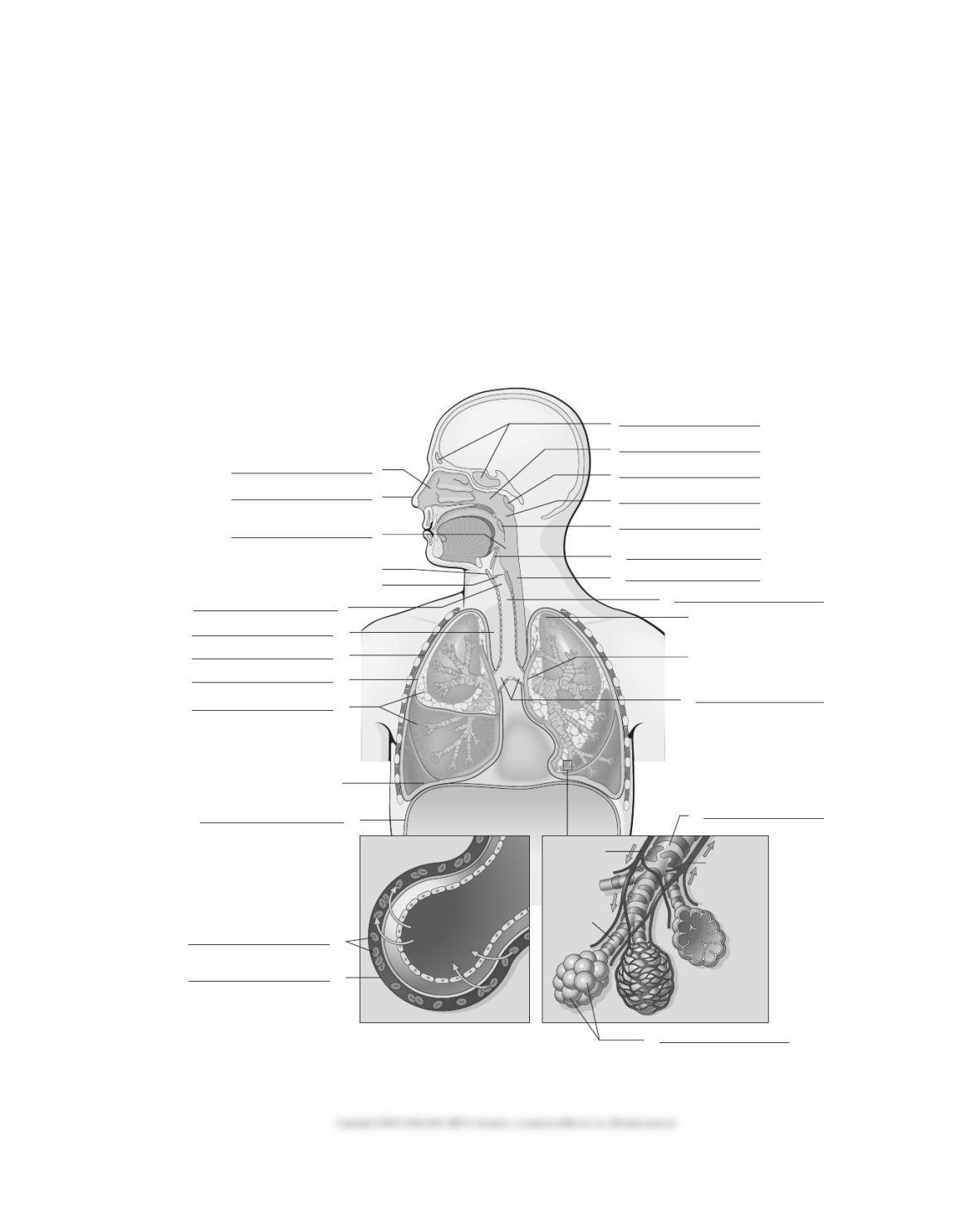
Label the diagram below using the following terms:
Adenoids
Alveoli
Bronchi
Capillary
Diaphragm
Epiglottis
Erythrocytes
Esophagus
Laryngopharynx
Larynx
Lung
Mediastinum
Nasal cavity
Nasopharynx
Nose
Oropharynx
Palatine tonsils
Paranasal sinuses
Parietal pleura
Terminal bronchiole
Trachea
Visceral pleura
3
2
4
5
6
7
11
10
12
14
Apex of the lung
Hilum of the lung
17
16
1
8
9
13
20
21
22
19
18
15
Thyroid cartilage
Glottis and vocal cords
Base of the lung
Exchange of gases between an
alveolus and a lung capillary
Heart
Alveolus
CO2
O2
Right Left
Alveolar
duct
Pulmonary
arteriole Pulmonary
venule
302 Chapter 12 ■ FLOW CHART QUIZ
1
2
3
4
5
6
7
8
9
Adenoids
Tonsils
Epiglottis
Chapter Twelve
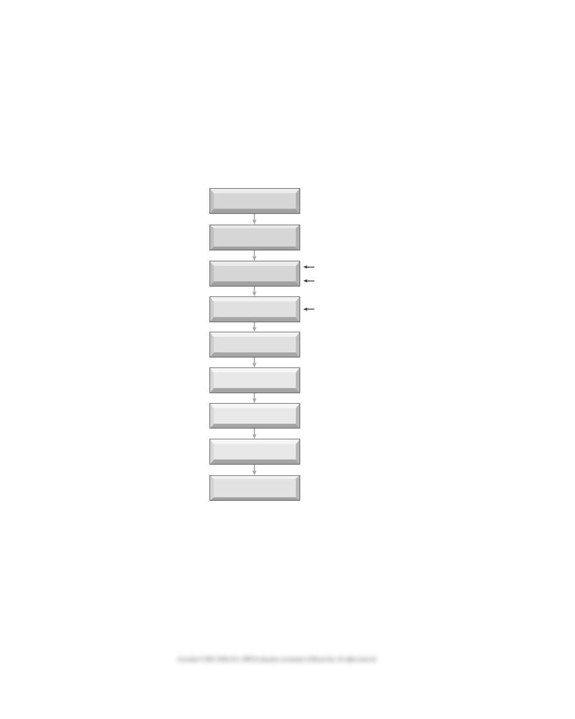
FLOW CHART QUIZ
Name: ______________________________
Alveoli Nasal cavities and paranasal sinuses
Bronchi Nose
Bronchioles Pharynx
Larynx Trachea
Lung capillaries (bloodstream)

Chapter 12 ■ REVIEW SHEET QUIZ 303
Chapter Twelve
REVIEW SHEET QUIZ
Name: ______________________________
A. Give meanings for the following combining forms:
1. adenoid/o _______________________ 6. nas/o ___________________________________
2. alveol/o ________________________ 7. or/o ____________________________________
3. bronch/o _______________________ 8. ox/o ____________________________________
4. bronchiol/o _____________________ 9. pector/o _________________________________
5. pulmon/o _______________________ 10. orth/o __________________________________
B. Give combining forms for the following meanings:
1. voice box _______________________ 6. dust ____________________________________
2. throat __________________________ 7. pus _____________________________________
3. blue ___________________________ 8. complete ________________________________
4. voice ___________________________ 9. windpipe ________________________________
5. diaphragm ______________________ 10. tonsils __________________________________

304 Chapter 12 ■ REVIEW SHEET QUIZ
C. Give meanings for the following suffixes and prefixes:
1. -centesis ________________________ 6. -ptysis __________________________________
2. -osmia _________________________ 7. brady- __________________________________
3. -pnea __________________________ 8. per– ____________________________________
4. -stenosis ________________________ 9. hypo- ___________________________________
5. -ectasis _________________________ 10. para- ___________________________________

Chapter 12 ■ MEDICAL SCRAMBLE 305
Chapter Twelve
MEDICAL SCRAMBLE
Name: ______________________________
Unscramble the letters to form respiratory system–related terms from the clues. Use the letters in
the squares to complete the bonus term.
1. Clue: Flap of cartilage above the trachea
____ ____ ____ ____ ____ ____ ____ I O P T L G E T S I
2. Clue: Bacteria causing tuberculosis
____ ____ ____ ____ L A L C B I I
3. Clue: Small airway leading to air sacs
____ ____ ____ ____ ____ ____ ____ ____ ____ O N E B H L R I C O
4. Clue: Chronic allergic condition marked by airway obstruction
____ ____ S M T A H A
BONUS TERM: Clue: Collapsed lung
306 Chapter 12 ■ CROSSWORD PUZZLE
Chapter Twelve
CROSSWORD PUZZLE
Name: ______________________________
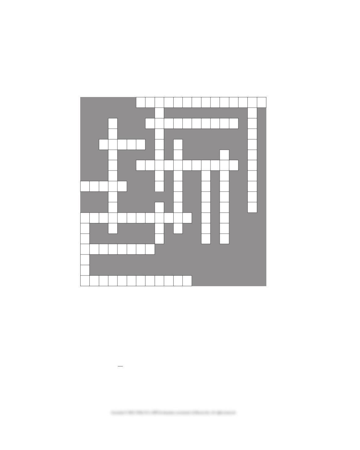
Fill in the crossword puzzle below using the clues listed underneath it.
Across Clues Down Clues
1. Abnormal condition caused by dust in 2. Air is trapped in lungs, and bronchioles are plugged
the lungs. with mucus; a type of COPD.
5. Lid-like piece of cartilage that covers the larynx. 3. Breathing in.
6. Phon/o means sound or _____. 4. Smallest branches of the bronchi.
9. Collapsed lung; or incomplete expansion of lung. 7. Acute inflammation and infection of the lung caused
11. Thin hairs attached to the mucous membranes. by bacteria (pneumococci), viruses, or fungi.
13. An infectious disease caused by bacilli and 8. Muscle separating the chest and abdomen.
treated with INH (isoniazid). 10. Collection of lymph tissue in the nasopharynx.
14. Tel/o means _____, as in atelectasis. 12. Rhin/o means ________.
15. Listening to sounds within the body. 13. Windpipe.
1
910
2
5
7
8
4
6
11
12
13
14
15
3

Chapter 12 ■ PRACTICAL APPLICATIONS 307
Chapter Twelve
PRACTICAL APPLICATIONS
Name: ______________________________
Questions for the Case Report on page 485 of the text
1. What did the initial chest x-ray
show? ………………………………………….
A. Collapsed lung
B. Shallow respirations
C. Pleurodynia
D. Collection of fluid between the pleura
2. What term indicates that the
condition was caused by fractured
ribs? ……………………………………………
A. Secondary
B. Comatose
C. Heroin
D. Effusion
3. What procedure was used to
relieve the condition? ……………………..
A. Chest x-ray
B. Thoracotomy and tube insertion
C. Paracentesis
D. Pericardiocentesis
4. What is the lesson from this case
report? ………………………………………..
A. Get a chest x-ray immediately upon
entering the ER
B. Removal of fluid from the pleural space
showed no blood was present
C. Be sure that a chest x-ray is read correctly
D. Be careful when injecting heroin

308 Chapter 12 ■ PRACTICAL APPLICATIONS
PATHOLOGY REPORT
Date: November 16, 2014 Pathology No. 450231
Patient: Carolyn Jones Room No. 422
Physician: Howard T. Waxman, MD Hospital No. 550330
Specimen: Biopsy of left bronchus
GROSS DESCRIPTION: The specimen consisted of a very tiny, wispy portion of soft
whitish pink tissue measuring 3 × 2 × 1 mm in toto. The entire
specimen is submitted.
MICROSCOPIC DESCRIPTION: The sections of the bronchial biopsy show approximately
half of the mucosa to be composed of pseudostratified,
ciliated, respiratory-type epithelium, and the second half to
be composed of respiratory epithelium that has undergone
squamous metaplasia. There is one small area of cells that
has become detached from the mucosa, which is composed
of rather pleomorphic and hyperchromatic cells with loss of
polarity. This small area of tissue would be classified as the
squamous carcinoma. However, I see no evidence of (the)
infiltration through the basement membrane in this section.
Additional tissue may show more extensive involvement with
the carcinoma.
DIAGNOSIS: Small fragment of squamous carcinoma without evidence
of infiltration into the underlying submucosa, left bronchus,
biopsy (see description).
Pathologist
Mark M. Mosley, MD
New Terms:
hyperchromatic Pertaining to cells that stain intensely (chrom/o = color)
loss of polarity Cells lose normal sense of organization (characteristic of
malignancy)
pleomorphic Pertaining to cells with many (ple/o = more) different shapes
and form characteristic of malignancy
pseudostratified Type of layered epithelium in which nuclei of adjacent cells are
at different levels.
squamous metaplasia Reversible conversion of normal cells into another, less
specialized cell type. Often, these cells can transform into
cancerous cells.
Chapter 12 ■ ANSWERS TO THE QUIZZES 309
Chapter Twelve
ANSWERS TO THE QUIZZES
310 Chapter 12 ■ ANSWERS TO THE QUIZZES
Chapter 12 ■ ANSWERS TO THE QUIZZES 311
312 Chapter 12 ■ ANSWERS TO THE QUIZZES
Chapter 12 ■ ANSWERS TO TERMINOLOGY SECTION 313
Chapter Twelve
Answers to Terminology Section
(textbook pages 465–470)
314 Chapter 12 ■ ANSWERS TO TERMINOLOGY SECTION