Chapter 34: Interpretation of Periodontal Disease
Iannucci: Dental Radiography, 5th Edition
MULTIPLE CHOICE
1. In health, the lamina dura around the roots of the teeth appears as a(n) _________ line.
a.
dense radiolucent
b.
dense radiopaque
c.
intermittent radiolucent
d.
intermittent radiopaque
2. Your patient has been diagnosed as having a periodontal classification of ADA case type I.
How far below the cement-enamel junction would you expect to see the crestal bone on the
dental images?
a.
0.5 to 1.0 mm
b.
1.0 to 1.5 mm
c.
1.5 to 2.0 mm
d.
2.0 to 2.5 mm
3. The normal periodontal ligament space appears as a ______________ line.
a.
thin radiopaque
b.
thin radiolucent
c.
thick radiopaque
d.
thick radiolucent
4. The periodontal ligament space is located between the root of the tooth and the
a.
crest of alveolar bone.
b.
lamina dura.
c.
cementum.
d.
spongy bone.
5. Dental images permit the evaluation of ______ in the detection of periodontal disease.
a.
pocket depth
b.
soft tissue
c.
inflammation
d.
bone
6. You suspect your patient has periodontal disease in the molar regions. Which of the following
dental images should you take?
a.
Horizontal bite-wing
b.
Vertical bite-wing
c.
Panoramic
d.
Periapical
7. The _________________ of adjacent teeth can be used as a plane of reference in determining
the pattern of bone loss present.
a.
marginal ridge height
b.
cemento-enamel junctions (CEJs)
c.
pulp chambers
d.
cementum thickness
8. A patient with localized bone loss will exhibit bone loss in less than ______ of the areas.
a.
10%
b.
20%
c.
30%
d.
50%
9. Which of the following should you use to determine the extent of your patient’s periodontal
disease?
a.
Pocket depth
b.
Recession
c.
Amount of inflammation
d.
Clinical attachment loss
10. Severe bone loss is defined as a loss of __________.
a.
1 to 2 mm
b.
2 to 3 mm
c.
3 to 4 mm
d.
5 mm or greater
11. ADA Case Type I is
a.
gingivitis.
b.
mild or slight periodontitis.
c.
moderate periodontitis.
d.
advanced or severe periodontitis.
12. Your patient is viewing her bite-wing images and has noticed the small radiopaque pieces on
the interproximal surfaces below the contacts of some of her teeth. What is she looking at?
a.
Crestal bone
b.
Cemento-enamel junction
c.
Composite restorations
d.
Calculus
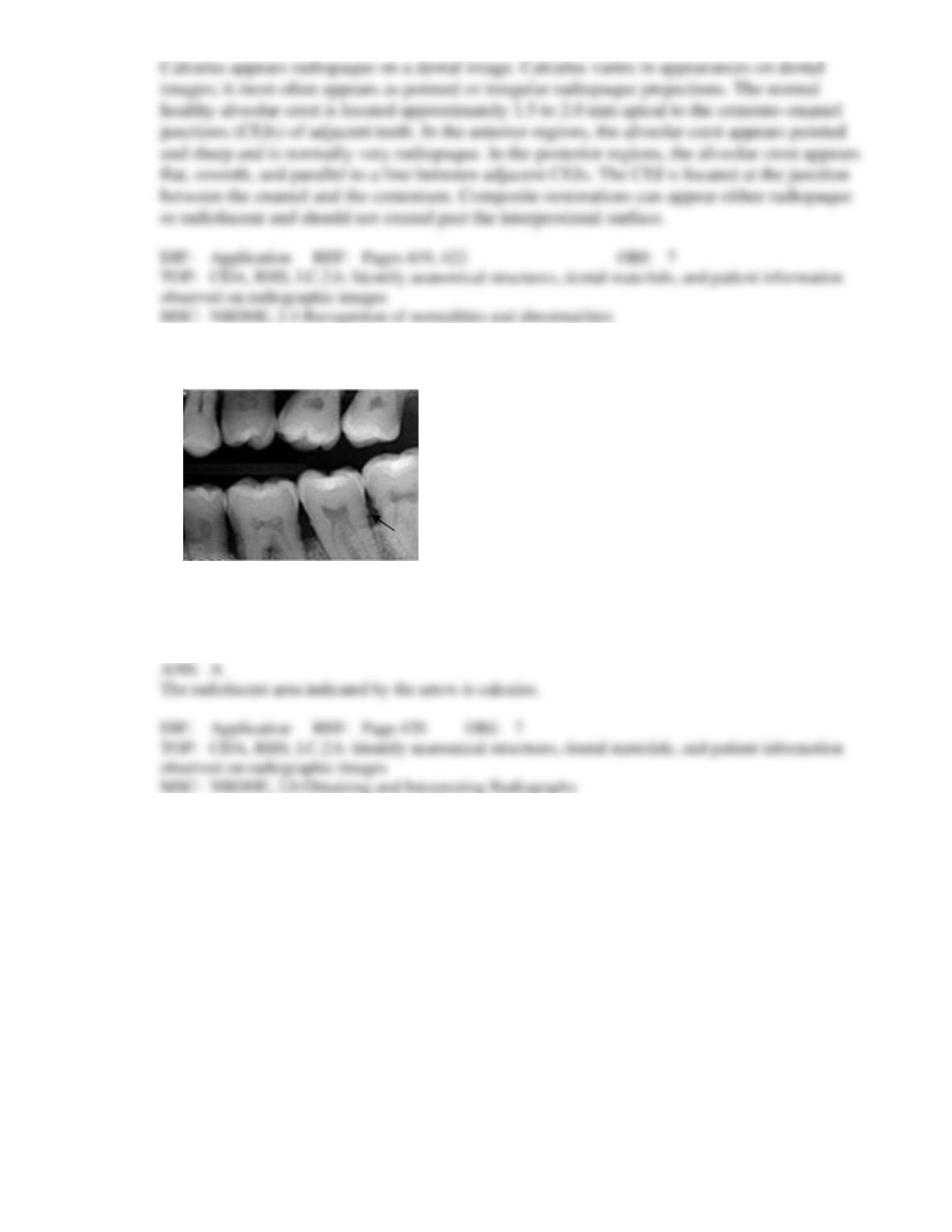
13. Identify the radiopaque area the arrow is pointing to on your patient’s dental image.
a.
Calculus
b.
Caries
c.
Cemento-enamel junction
d.
Cementum