
Chapter 33: Interpretation of Dental Caries
Iannucci: Dental Radiography, 5th Edition
MULTIPLE CHOICE
1. In the practice of dentistry, ___________ is probably the most frequent reason for taking
dental images.
a.
periodontitis
b.
caries
c.
impacted teeth
d.
orthodontia
2. A dental image is most useful for detection of _________ caries.
a.
buccal
b.
lingual
c.
occlusal
d.
interproximal
3. A carious lesion appears _________ on a dental image.
a.
radiopaque
b.
radiolucent
c.
white
d.
clear
4. You suspect your patient has caries in the molar regions. Which of the following dental
images should you take?
a.
Periapical
b.
Bite-wing
c.
Panoramic
d.
Occlusal
5. ____________ caries are found between two teeth.
a.
Occlusal
b.
Buccal
c.
Lingual
d.
Interproximal
6. On a dental image, interproximal caries are typically seen at or just below the
a.
marginal ridge.
b.
contact point.
c.
cementoenamel junction.
d.
crest of the alveolar bone.
7. Which of the following interproximal lesions does not involve the dentin, but is does extend
more than half way through the enamel?
a.
Incipient
b.
Moderate
c.
Advanced
d.
Severe
8. You are viewing your patient’s bite-wing images. He has an interproximal carious lesion that
is almost to the pulp. What is the classification of the lesion?
a.
I
b.
II
c.
III
d.
IV
9. Which of the following carious lesions cannot be found on a dental image and must be
identified clinically?
1. Incipient
2. Moderate
3. Severe
a.
1, 2, 3
b.
1, 3
c.
2, 3
d.
1 only

10. Buccal or lingual caries appear as a _____________________ on dental images.
a.
radiolucent triangle
b.
radiopaque triangle
c.
radiolucent small circle
d.
radiopaque small circle
11. Which of the following statements is true of root surface caries?
a.
Root surface caries involve the loss of enamel.
b.
Root surface caries are clinically difficult to detect on exposed root surfaces.
c.
Root surface caries appear as a cupped-out or crater-shaped radiolucency just
below the cement-enamel junction (CEJ).
d.
Root surface caries appear as a cupped-out or crater-shaped radiolucency just
above the CEJ.
12. Rampant caries are typically seen in children with ________ dietary habits or in adults with
__________ salivary flow.
a.
poor; increased
b.
poor; decreased
c.
good; increased
d.
good; decreased
13. Which type of caries occurs under an existing restoration?
a.
Buccal
b.
Recurrent
c.
Rampant
d.
Root
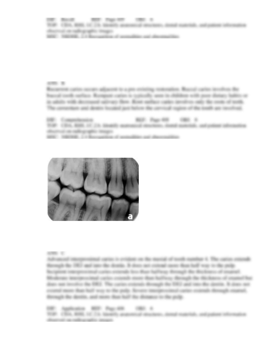
14. Identify the type of decay present that your patient has on the mesial of tooth number 4.
a.
Incipient interproximal caries
b.
Moderate interproximal caries
c.
Advanced interproximal caries
d.
Severe interproximal caries