Chapter 32: Identification of Restorations, Dental Materials, and Foreign Objects
Iannucci: Dental Radiography, 5th Edition
MULTIPLE CHOICE
1. You are having difficulty determining whether the radiolucent area on tooth number 7 on the
dental image is decay or a composite restoration. Which of the following should you do?
a.
Ask your patient.
b.
Perform a clinical examination.
c.
Search on the Internet.
d.
Look in a textbook.
2. Because metallic restorations absorb x-rays, the area of the image that corresponds to their
location remains unexposed, and the metallic restorations appear completely ___________ on
a dental image.
a.
radiosensitive
b.
radioresistant
c.
radiopaque
d.
radiolucent
3. Of the nonmetallic restorations, ____________ is the most dense and least radiolucent.
a.
unfilled acrylic
b.
filled acrylic
c.
composite
d.
porcelain
4. Which of the following is the most common restorative material used in dentistry?
a.
Porcelain
b.
Cast gold
c.
Amalgam
d.
Acrylic
5. Amalgam overhangs, excess amalgam material that extends beyond the cavity margin, can be
seen on dental images in the ____________ region of the crown of the tooth.
a.
Buccal
b.
Lingual
c.
Interproximal
d.
Occlusal
6. The greatest potential negative consequence of an amalgam overhang is
a.
unesthetic contour.
b.
destruction of interproximal bone.
c.
impingement of the interdental papilla.
d.
stretching of periodontal ligament fibers.
7. You are looking at your patient’s dental images. On tooth number 30, you see a large
restoration that has smooth margins, doesn’t conform to the tooth, and has a ghostlike
radiopaque appearance. Which type of restoration does your patient have on tooth number 30?
a.
Silver amalgam
b.
Gold crown
c.
Porcelain crown
d.
Stainless steel crown
8. Post and core restorations can be seen in ____________ treated teeth.
a.
periodontally
b.
orthodontically
c.
endodontically
d.
nonendodontically
9. Porcelain restorations are
a.
radiolucent.
b.
slightly radiolucent.
c.
slightly radiopaque.
d.
radiopaque.
10. A thin radiopaque line outlining the prepared tooth may be evident on a dental image of an
all-porcelain crown. This thin line represents
a.
die spacer.
b.
cement.
c.
an air pocket.
d.
a metal substructure.
11. A porcelain-fused-to-metal crown appears
a.
uniformly radiopaque.
b.
uniformly radiolucent.
c.
to have two components. The metal component appears completely radiopaque,
and the porcelain component appears slightly radiopaque.
d.
to have two components. The metal component appears completely radiolucent,
and the porcelain component appears slightly radiolucent.
12. Some manufacturers of composite materials add radiopaque particles to their products to help
the viewer differentiate a composite restoration from
a.
an amalgam restoration.
b.
dental caries.
c.
a cast gold restoration.
d.
a porcelain-fused-to-metal crown.
13. Gutta percha appears __________. If compared with amalgam, gutta percha appears
________ radiodense.
a.
radiolucent; more
b.
radiolucent; less
c.
radiopaque; more
d.
radiopaque; less
14. Diatorics are found in
a.
silver point endodontic fillings.
b.
gutta percha endodontic fillings.
c.
anterior porcelain denture teeth.
d.
anterior acrylic denture teeth.
15. (1) The metal framework on a removable partial denture appears radiopaque on dental images.
(2) The acrylic portion of a removable partial denture appears radiolucent on dental images.
a.
Both statements are true.
b.
Both statements are false.
c.
The first statement is true; the second statement is false.
d.
The first statement is false; the second statement is true.
16. You are going to take intraoral images on your patient. Which of the following should you ask
her to remove?
1. Necklace
2. Eyeglasses
3. Nose ring
4. Hearing aids
a.
1, 2, 3, 4
b.
1, 2, 3
c.
2, 3, 4
d.
2, 3
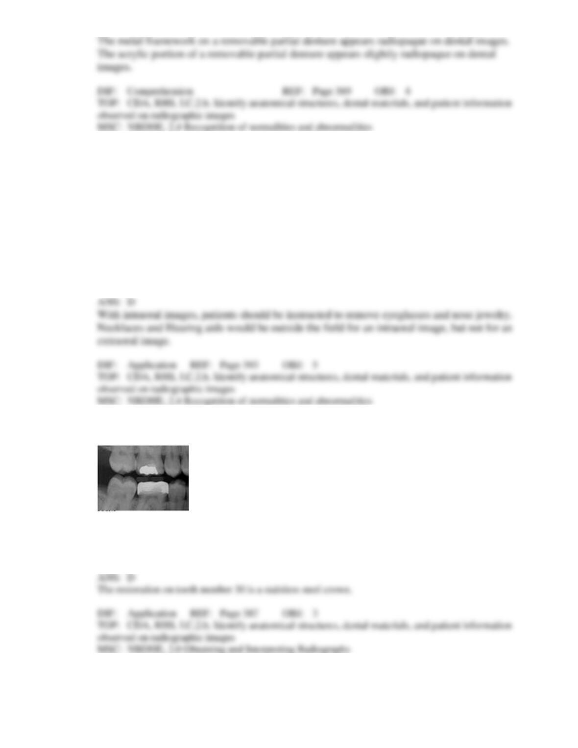
17. Identify the restoration your patient has on tooth number 30.
a.
Amalgam
b.
Stainless steel crown
c.
Gold crown
d.
Porcelain fused to metal crown