43. Your patient is pointing to the mental ridge on his dental image. He is pointing to the
____________________ portion of the mandible.
a.
external surface of the anterior
b.
internal surface of the anterior
c.
external surface of the posterior
d.
internal surface of the posterior
44. On a dental image, the mental ridge often appears superimposed over the ___________ teeth.
a.
maxillary anterior
b.
maxillary posterior
c.
mandibular anterior
d.
mandibular posterior
45. The mental fossa is located ________ the mental ridge in the mandibular __________ region.
a.
above; incisor
b.
below; incisor
c.
above; premolar
d.
below; premolar
46. The ____________ is an opening or hole in bone located on the external surface of the
mandible in the region of the mandibular premolars.
a.
nutrient canal
b.
mental fossa
c.
lingual foramen
d.
mental foramen
47. The mental foramen is frequently misdiagnosed as a _____________ because of its apical
location.
a.
dentigerous cyst
b.
radicular cyst
c.
periapical granuloma
d.
odontoma
48. The conduit that extends from just below the ramus to the premolar region of the mandible is
the
a.
external oblique ridge.
b.
mylohyoid ridge.
c.
submandibular fossa.
d.
mandibular canal.
49. On a dental image, the mylohyoid ridge is a ___________ band that may appear to be
continuous with the ______________.
a.
radiolucent; internal oblique ridge
b.
radiolucent; external oblique ridge
c.
radiopaque; internal oblique ridge
d.
radiopaque; external oblique ridge
50. On a mandibular periapical image, the mandibular canal is a ___________ band that appears
below or superimposed over the apices of the mandibular _________ teeth.
a.
radiolucent; incisor
b.
radiolucent; molar
c.
radiopaque; incisor
d.
radiopaque; molar
51. The internal oblique ridge extends from the third molar region to the ____________ area.
a.
canine
b.
first molar
c.
incisor area
d.
second premolar
52. On a mandibular molar periapical image, the external oblique ridge appears as a
_______________ band extending _______________________ from the anterior border of
the ramus of the mandible.
a.
radiopaque; upward and backward
b.
radiolucent; upward and backward
c.
radiopaque: downward and forward
d.
radiolucent: downward and forward
53. The external oblique ridge typically ends in the _____________ molar region.
a.
maxillary first
b.
mandibular first
c.
maxillary third
d.
mandibular third
54. The ______________ is found in the submandibular fossa.
a.
inferior alveolar nerve
b.
submandibular salivary gland
c.
mylohyoid muscle
d.
lingual nerve
55. On a periapical image, the submandibular fossa appears as a ___________ area in the molar
region _______ the mylohyoid ridge.
a.
radiopaque; above
b.
radiopaque; below
c.
radiolucent; above
d.
radiolucent; below
56. The coronoid process may be seen on a ______________ periapical image.
a.
maxillary incisor
b.
maxillary molar
c.
mandibular incisor
d.
mandibular molar
57. Which of the following tooth structures is most radiopaque?
a.
Enamel
b.
Dentin
c.
Cementum
d.
Pulp
58. Which of the following tooth structures is most radiolucent?
a.
Enamel
b.
Dentin
c.
Cementum
d.
Pulp
59. The ______________ is the space between the alveolar bone and the tooth, it contains the
attachment fibers.
a.
alveolar crest
b.
residual ridge
c.
periodontal ligament space
d.
lamina dura
60. On a dental image, the alveolar crest is typically located _________ mm below the junction of
the crown and the root surfaces.
a.
0.5 to 1.0
b.
1.5 to 2.0
c.
2.0 to 3.0
d.
1.5 to 3.0
61. On a dental image, the periodontal ligament space (PDL) space appears as a _____________
line around the root of a tooth.
a.
thin, radiopaque
b.
thin, radiolucent
c.
wide, radiopaque
d.
wide, radiolucent
62. The alveolar crest in the posterior region tends to appear ______ dense and ______
radiopaque than the alveolar crest seen in the anterior region.
a.
less; less
b.
more; more
c.
less; more
d.
more; less
63. Your patient’s mother is concerned because her two-year-old son doesn’t have all of his teeth.
What should you tell her?
a.
Your son is late in his eruption pattern. They should have all erupted a year ago.
b.
If your son’s teeth don’t all erupt in the next month, we will refer him.
c.
We need to take dental images to see if he has his teeth under the tissue.
d.
Don’t worry, his teeth should be in by the time he is three.
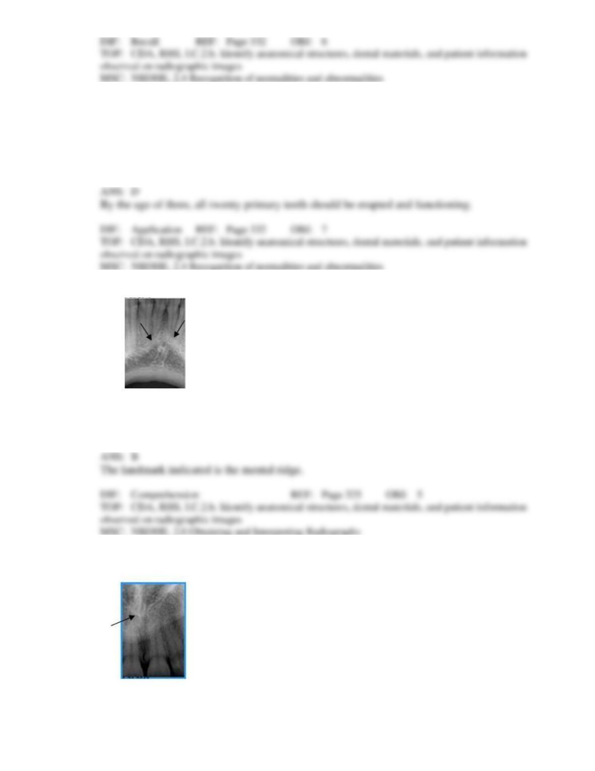
64. Identify this landmark.
a.
Genial tubercles
b.
Mental ridge
c.
Mental fossa
d.
Mylohyoid ridge
65. Identify this landmark.
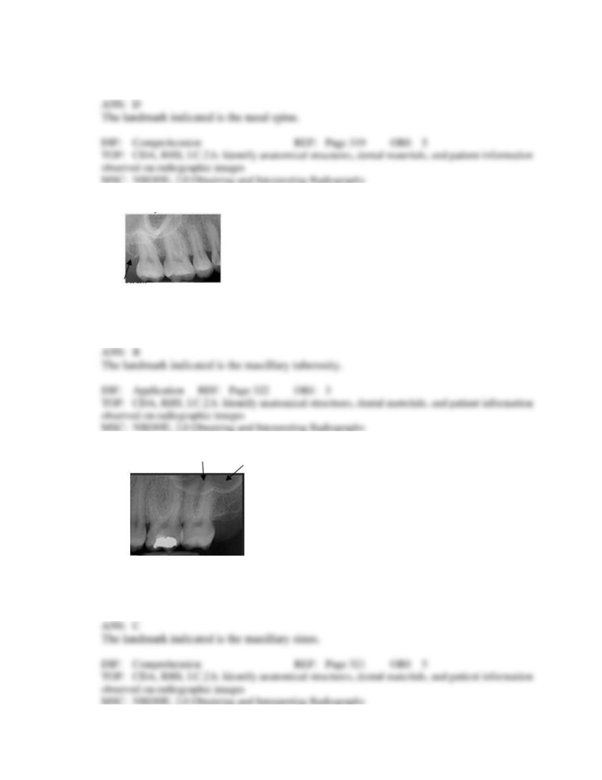
a.
Incisive foramen
b.
Nasal fossa
c.
Nasal septum
d.
Nasal spine
66. Identify this landmark on your patient’s dental image.
a.
Lateral pterygoid plate
b.
Maxillary tuberosity
c.
Zygoma
d.
Maxillary sinus
67. Identify this landmark.
a.
Maxillary tuberosity
b.
Zygoma
c.
Maxillary sinus
d.
Mylohyoid bone
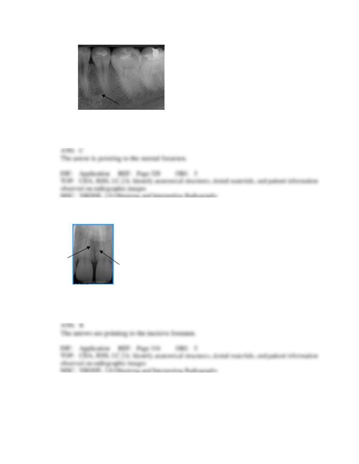
68. What is the arrow pointing to on your patient’s dental image?
a.
Periapical abscess
b.
Trabecular bone
c.
Mental foramen
d.
Periodontal abscess
69. Identify the landmark the arrows are pointing to on your patient’s dental image.
a.
Nose
b.
Incisive foramen
c.
Midpalatine suture
d.
Lateral fossa
70. Identify this landmark.
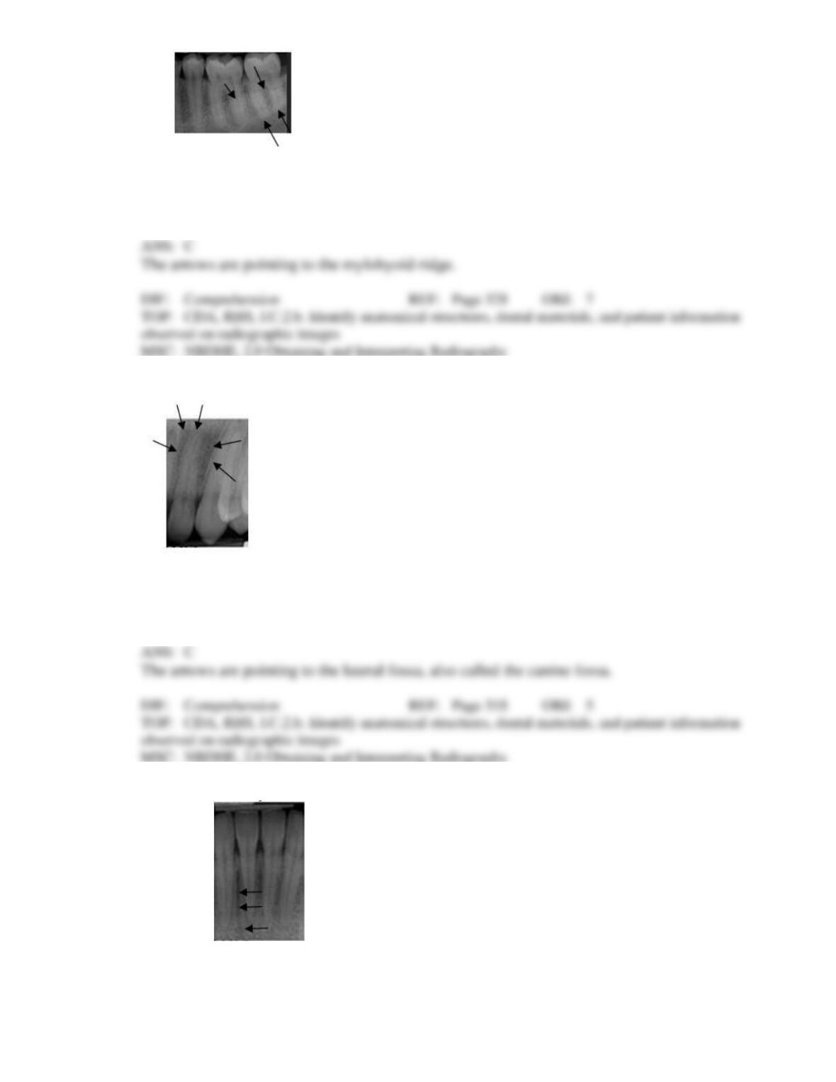
a.
External oblique ridge
b.
Submandibular fossa
c.
Mylohyoid ridge
d.
Mandibular canal
71. What are the arrows pointing to?
a.
Periodontal abscess
b.
Incisive foramen
c.
Lateral fossa
d.
Periapical abscess
72. Identify the radiolucent area the arrows are pointing to.
a.
Mental fossa
b.
Periodontal pocket
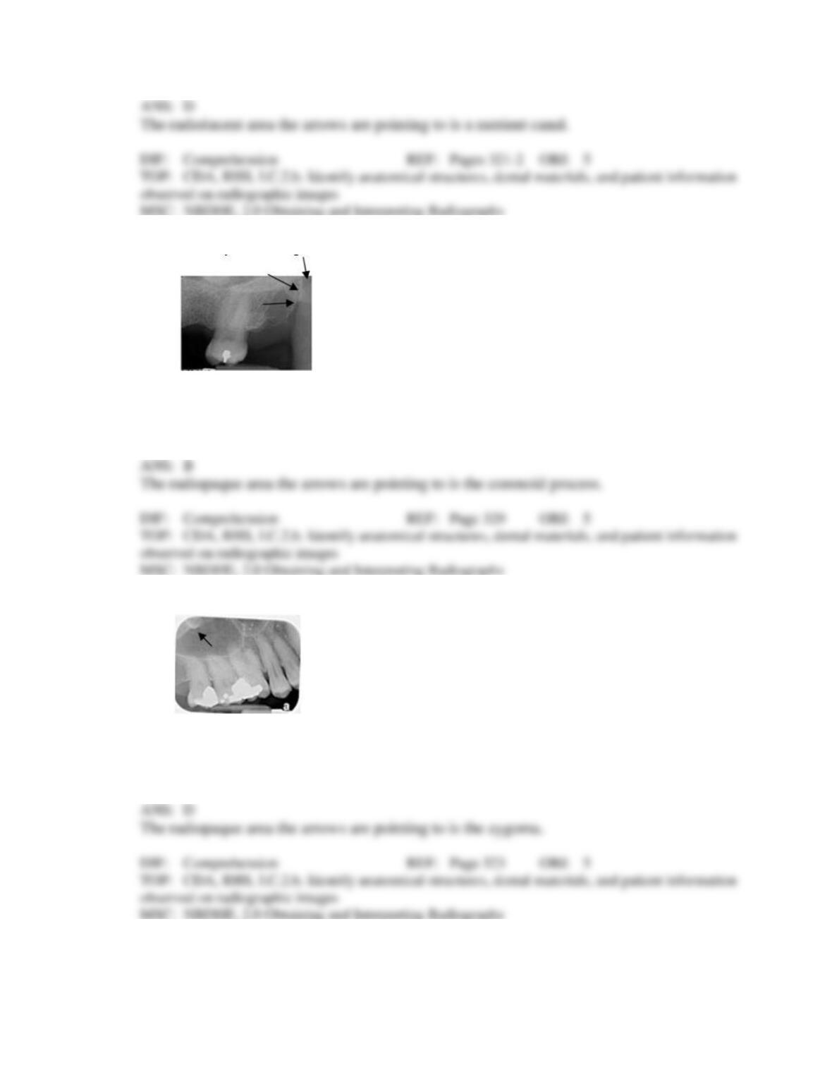
c.
Mental ridge
d.
Nutrient canal
73. Identify the radiopaque area the arrows are pointing to.
a.
Maxillary tuberosity
b.
Coronoid process
c.
Condyle
d.
Spine
74. Identify the radiopaque area the arrows are pointing to.
a.
Maxillary sinus
b.
Lateral pterygoid plate
c.
Orbit
d.
Zygoma
75. Identify the area the arrows are pointing to on your patient’s dental image.
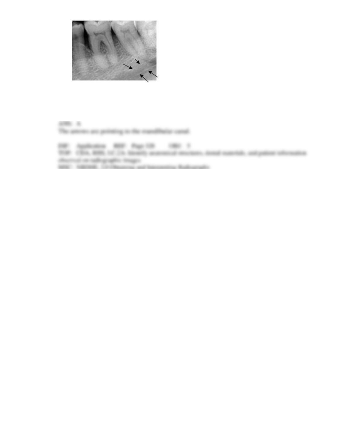
a.
Mandibular canal
b.
Submandibular fossa
c.
Mylohyoid ridge
d.
Border of the mandible