Chapter 20: Exposure and Technique Errors
Iannucci: Dental Radiography, 5th Edition
MULTIPLE CHOICE
1. An unexposed film appears
a.
clear.
b.
black.
c.
dark.
d.
light.
2. Which of the following choices may be the cause of an unexposed film or receptor?
a.
Excessive exposure time
b.
Failure to turn on the x-ray machine
c.
Excessive kilovoltage
d.
Excessive milliamperage
3. Which of the following films would most likely appear black?
a.
An unexposed film
b.
A film exposed to light
c.
An overexposed film
d.
An underexposed film
4. The possibility of exposure of radiographic film to white light can be reduced by
a.
turning off the safelight and unwrapping the films in a pitch-black room.
b.
pre-exposing the entire package of film to radiation before opening the package.
c.
not unwrapping the film in a room with white light.
d.
turning off the fluorescent lighting in the treatment room before exposing
radiographic films.
5. An underexposed receptor results from
a.
insufficient exposure time.
b.
inadequate tissue density.
c.
excessive milliamperage.
d.
excessive kilovoltage.
6. To prevent underexposure, check and increase ___________ as needed.
1. exposure time
2. kilovoltage
3. milliamperage
a.
1, 2, 3
b.
1, 2
c.
2, 3
d.
1, 3
7. The cause of an excessive margin of receptor edge (which appears as a black band) on a
nondiagnostic periapical image is
a.
the edge of the receptor was not placed parallel to the incisal-occlusal surfaces of
the teeth.
b.
the receptor was not positioned in the patient’s mouth to cover the apical regions
of the teeth.
c.
the receptor was underexposed.
d.
the central ray was not directed through the interproximal spaces.
8. To correct a dental image where apices do not appear,
a.
make certain that no more than 3/4 inch of the receptor edge extends beyond the
incisal-occlusal surfaces of the teeth.
b.
the teeth being imaged must be firmly in contact with the bite-block.
c.
increase the exposure time.
d.
direct the x-ray beam through the interproximal spaces.
9. When the occlusal plane appears tipped or tilted, the error is a(n)
a.
incorrect horizontal angulation.
b.
incorrect vertical angulation.
c.
dropped receptor corner.
d.
elongated image.
10. To prevent a dropped receptor corner,
a.
change the horizontal angulation.
b.
increase the vertical angulation.
c.
decrease the vertical angulation.
d.
make certain the edge of the receptor is placed parallel to the incisal-occlusal
surfaces of the teeth.
11. When overlapped contacts appear on a dental image, the cause is
a.
incorrect vertical angulation.
b.
incorrect horizontal angulation.
c.
a dropped receptor corner.
d.
an underexposed film.
12. To avoid overlapped contacts on a periapical image,
a.
increase vertical angulation.
b.
decrease vertical angulation.
c.
direct the x-ray beam through the interproximal regions.
d.
make certain that no more than 1/8 inch of the receptor edge extends beyond the
incisal-occlusal surfaces of the teeth.
13. Short teeth with blunted roots appear on the image when
a.
the vertical angulation is excessive.
b.
the vertical angulation is insufficient.
c.
the horizontal angulation is incorrect.
d.
there is a cone-cut.
14. Long, distorted teeth appear on the image when
a.
the vertical angulation is excessive.
b.
the vertical angulation is insufficient.
c.
the horizontal angulation is incorrect.
d.
there is a cone-cut.
15. A cone-cut occurs when
a.
the receptor is underexposed.
b.
the receptor is overexposed.
c.
the PID was not properly aligned with the periapical receptor holder.
d.
the exposure button was not depressed for a sufficient amount of time.
16. When the distal surfaces of the canines are not visible on a premolar bite-wing image, the
solution is to
a.
change the horizontal angulation of the tubehead.
b.
increase the vertical angulation of the tubehead.
c.
decrease the vertical angulation of the tubehead.
d.
position the anterior edge of the receptor at the midline of the mandibular canine.
17. When the third molar regions are not visible on a molar bite-wing image, the solution is to
a.
decrease the vertical angulation of the tubehead.
b.
increase the vertical angulation of the tubehead.
c.
position the anterior edge of the receptor at the midline of the mandibular first
premolar.
d.
position the anterior edge of the receptor at the midline of the mandibular second
premolar.
18. With ____________, images appear stretched and distorted.
a.
film creasing
b.
film bending
c.
double exposure
d.
patient movement
19. A thin radiolucent line appears on the film with
a.
film creasing.
b.
film bending.
c.
double exposure.
d.
patient movement.
20. The appearance of a patient’s finger on the image is called a
a.
hemangioma
b.
phalangioma
c.
myxoma
d.
pericytoma
21. What is the cause of a double exposure?
a.
The patient moved during receptor exposure.
b.
Two radiographs were stuck together during film exposure.
c.
A double packet of film was used.
d.
The film was exposed in the patient’s mouth twice.
22. To avoid a double exposure,
a.
only expose and process one half of the patient’s mouth at one time.
b.
mark exposed films with indelible ink.
c.
always separate exposed and unexposed film and PSP receptors.
d.
ask the patient to hold the unexposed film and PSP receptors.
23. Blurred images appear on the dental image when
a.
there is patient movement.
b.
the film is reversed.
c.
there is a double exposure.
d.
the film is creased.
24. Your left premolar bite-wing image is lighter than the rest of the images and has a
herringbone pattern. What can you do to prevent this problem from occurring? When the film
is reversed,
a.
change vertical angulation.
b.
make sure you do not double expose your receptor.
c.
make sure the film side is facing the PID.
d.
make sure the patient doesn’t move.
25. An unexposed sensor appears
a.
clear.
b.
black.
c.
gray.
d.
clear or black.
26. You have noticed that your mandibular incisors are foreshortened; how would you correct this
problem?
a.
Adjust horizontal angulation.
b.
Decrease vertical angulation.
c.
Increase vertical angulation.
d.
Place receptor parallel to the long axis of the teeth.
27. You have noticed that your maxillary premolars are elongated, how would you correct this
problem?
a.
Adjust horizontal angulation.
b.
Decrease vertical angulation.
c.
Increase vertical angulation.
d.
Place receptor parallel to the long axis of the teeth.
28. The mesial half of your right premolar bite-wing image is clear, what will you need to correct
when taking your retake to prevent this from reoccurring?
a.
Adjust receptor placement.
b.
Adjust horizontal angulation.
c.
Adjust vertical angulation.
d.
Adjust receptor exposure.
29. You will need to take a retake on your patient’s mandibular right premolars because of
overlapping. The overlapping is more excessive toward the mesial of the film than the distal
of the film. What do you need to do to prevent this error on the retake?
a.
Correct horizontal angulation, direct the PID more toward the distal.
b.
Correct horizontal angulation, direct the PID more toward the mesial.
c.
Correct vertical angulation, decrease vertical angulation.
d.
Correct vertical angulation, increase vertical angulation.
30. Radiopaque artifacts or radiolucent scratch marks on the image indicate which of the
following?
a.
Debris accumulation on the sensor
b.
Creasing of the film or PSP receptor
c.
Bending of the film or PSP receptor
d.
Double image
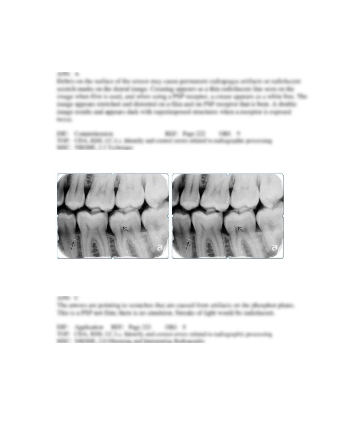
31. Identify the cause of the lines the arrows are pointing to on your patient’s dental image.
a.
Fractures in the root and crown
b.
Scratched film emulsion
c.
Artifacts on the phosphor plates
d.
Streaks of light
32. Identify the major technique error on your patient’s bitewing.
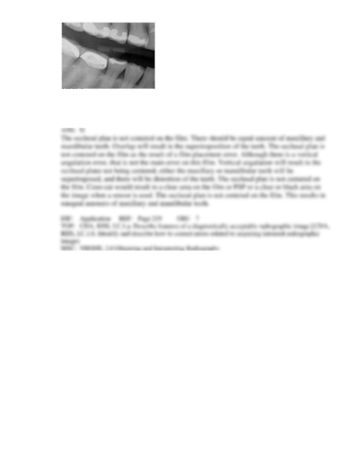
a.
Overlap
b.
Vertical angulation
c.
Cone-cut
d.
Occlusal plane not centered