Chapter 17: Paralleling Technique
Iannucci: Dental Radiography, 5th Edition
MULTIPLE CHOICE
1. Parallel is defined as
a.
cutting across or through.
b.
intersecting at or forming a right angle.
c.
moving or lying in the same plane, always separated by the same distance and not
intersecting.
d.
an angle of 90 degrees formed by two lines perpendicular to each other.
2. According to the basic principles of the paralleling technique, the receptor is placed in the
mouth _________ to the long axis of the tooth being x-rayed, and the central ray of the x-ray
beam is directed ____________ to the receptor and long axis of the tooth.
a.
parallel; perpendicular
b.
parallel; parallel
c.
perpendicular; parallel
d.
perpendicular; perpendicular
3. To achieve parallelism between the receptor and the tooth, the ____________ distance must
be increased to keep the receptor parallel with the long axis of the tooth.
a.
target-object
b.
object-receptor
c.
target-receptor
d.
operator-patient
4. Image magnification occurs when the ______________ distance is increased.
a.
target-object
b.
object-receptor
c.
target-receptor
d.
operator-patient
5. Image magnification can be decreased when the _______________ is increased.
a.
target-object
b.
object-receptor
c.
target-receptor
d.
operator-patient
6. Which of the following is a disposable receptor holder designed for one-time use only?
a.
Rinn XCP instruments
b.
Precision receptor holders
c.
Stabe bite-block
d.
Snap-a-ray receptor holder
7. Reusable receptor holders must be _________________ after each use.
a.
rinsed with tap water
b.
wiped with disinfectant
c.
placed in an immersion disinfectant
d.
sterilized
8. Which of the following receptor holders include aiming rings that aid in the alignment of the
position-indicating device (PID) with the receptor?
a.
Hemostat with bite-block
b.
Stabe bite-block
c.
Snap-a-ray receptor holder
d.
Rinn XCP instruments
9. The authors recommend using a size ____ receptor for anterior teeth and a size ____ receptor
for posterior teeth.
a.
1; 1
b.
2; 2
c.
1; 2
d.
2; 1
10. According to the text, a size 1 receptor is always positioned with the long portion of the
receptor in a ____________ direction, and a size 2 receptor is always positioned with the long
portion of the receptor in a ______________ direction.
a.
vertical; vertical
b.
horizontal; horizontal
c.
vertical; horizontal
d.
horizontal; vertical
11. (1) The dental radiographer should be careful not to stimulate the patient’s gag reflex. (2)
Once the gag reflex is stimulated, the patient may gag on receptors that could normally be
tolerated.
a.
Both statements are true.
b.
Both statements are false.
c.
The first statement is true; the second statement is false.
d.
The first statement is false; the second statement is true.
12. If you are using size 1 receptors, how many exposures will you need to take of the anterior
teeth?
a.
Four
b.
Six
c.
Seven
d.
Eight
13. If you are using size 2 receptors, how many exposures will you need to take of the anterior
teeth?
a.
Four
b.
Six
c.
Seven
d.
Eight
14. If you are taking an anterior image sequence, which tooth do the authors recommend you
begin with?
a.
6
b.
8
c.
9
d.
11
15. How many exposures of the posterior teeth will you need to take when using the paralleling
technique?
a.
Four
b.
Six
c.
Seven
d.
Eight
16. Which posterior quadrant would be the best for you to begin with when using the paralleling
technique?
a.
Maxillary left quadrant
b.
Maxillary right quadrant
c.
Mandibular left quadrant
d.
Mandibular right quadrant
17. Which posterior quadrant should you move to next (second) when using the paralleling
technique?
a.
Maxillary left quadrant
b.
Maxillary right quadrant
c.
Mandibular left quadrant
d.
Mandibular right quadrant
18. Your patient has a shallow palate; you should modify your technique when there is a lack of
parallelism ________________.
a.
greater than 10 degrees
b.
greater than 20 degrees
c.
greater than 30 degrees
d.
less than 30 degrees
19. (1) When taking maxillary periapical images on a patient with a shallow palate, there is only
one technique that can be used to compensate for lack of parallelism. (2) To compensate for
lack of parallelism, the vertical angulation can be increased by 5 to 15 degrees more than the
XCP instrument indicates.
a.
Both statements are true.
b.
Both statements are false.
c.
The first statement is true; the second statement is false.
d.
The first statement is false; the second statement is true.
20. Your patient has a palatal torus, where should you place the receptor?
a.
You cannot take a periapical, the patient will require a panoramic receptor.
b.
Place the receptor on the torus.
c.
Place the receptor in front of the torus next to the teeth you are exposing.
d.
Place the receptor on the far side of the torus.
21. When placing the receptor in the mandibular premolar region, the receptor must be placed
________ the tongue. When inserting the receptor holder into the mouth, the receptor is
tipped __________ the tongue.
a.
over; toward
b.
over; away from
c.
under; toward
d.
under; away from
22. (1) When using the paralleling technique, you must be conscious of fixed vertical angulations.
(2) The paralleling technique creates dental images free from dimensional distortion.
a.
Both statements are true.
b.
Both statements are false.
c.
The first statement is true; the second statement is false.
d.
The first statement is false; the second statement is true.
23. When using film, the dot should always be placed ________ in relation to the bite-block.
a.
down
b.
up
c.
distal
d.
in the slot
24. The lingual cusp of the first premolar usually obscures the distal contact of the canine on the
________________ exposure.
a.
maxillary canine
b.
maxillary incisor
c.
mandibular canine
d.
mandibular incisor
25. Which of the following images must include the crowns, roots, and surrounding bone of the
right or left central and lateral incisors?
a.
Maxillary canine
b.
Maxillary incisor
c.
Mandibular canine
d.
Mandibular incisor
26. Which of the following images must include the crowns, roots, and surrounding bone, sinus
and tuberosity region of the first, second, and third molars?
a.
Maxillary premolar
b.
Maxillary molar
c.
Mandibular premolar
d.
Mandibular molar
27. Which target-receptor distance should be used in the paralleling technique to compensate for
image magnification and loss of definition?
a.
4 inches
b.
8 inches
c.
12 inches
d.
16 inches
28. (1) The American Dental Association (ADA) and the American Academy of Oral and
Maxillofacial Radiology both recommend the use of a beam alignment device to reduce the
amount of radiation the patient receives. (2) Limiting the size of the x-ray beam not only
reduces the amount of skin that is exposed but also results in a significant reduction of
radiation to the patient, by as much as 20%.
a.
Both statements are true.
b.
Both statements are false.
c.
The first statement is true; the second statement is false.
d.
The first statement is false; the second statement is true.
29. Which of the Rinn beam alignment device includes one ring and one arm that can be used for
anterior, posterior, and bite-wing receptor positioning?
a.
Rinn XCP Extension Cone Paralleling System
b.
Rinn XCP-ORA System
c.
Rinn SCP-DS FIT Universal Sensor Holder
d.
Rinn Flip-Ray System
30. Which basic rule of paralleling states that the receptor must be parallel to the long axis of the
tooth?
a.
Receptor placement
b.
Receptor position
c.
Vertical angulation
d.
Receptor exposure
31. According to the basic rules of paralleling, the central ray should be directed
______________ to the receptor and the long axis of the tooth.
a.
parallel
b.
perpendicular
c.
horizontal
d.
adjacent
32. Which basic rule of paralleling indicates that the central ray should be directed through the
contact area of the teeth?
a.
Receptor placement
b.
Vertical angulation
c.
Horizontal angulation
d.
Receptor position
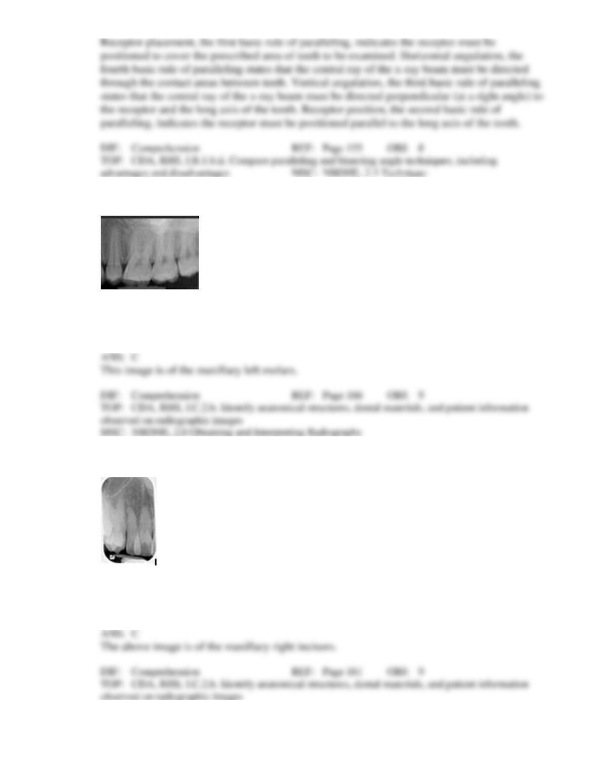
33. Identify this image; it is mounted using the labial view.
a.
Maxillary right molars
b.
Maxillary left premolars
c.
Maxillary left molars
d.
Mandibular left molars
34. Identify this image; it is mounted using the labial view.
a.
Maxillary right canine
b.
Maxillary left canine
c.
Maxillary right incisors
d.
Maxillary left incisors
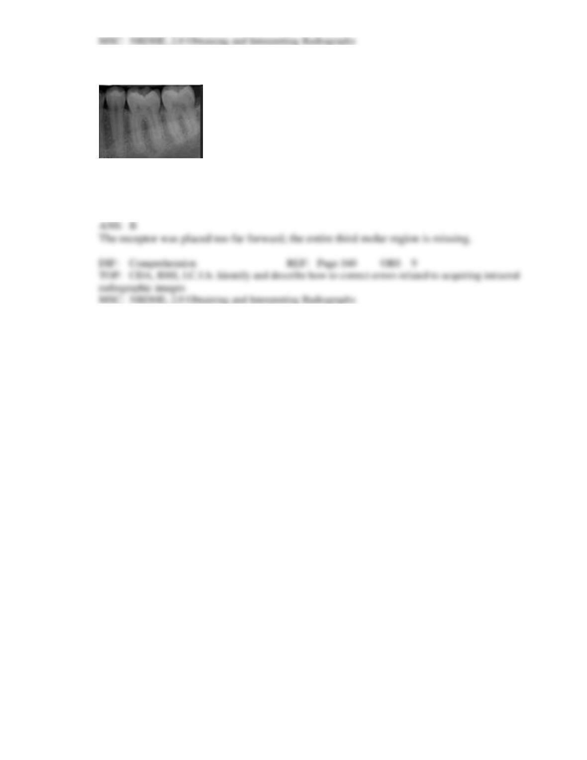
35. Identify the major placement error in this molar radiograph.
a.
Too far mesial
b.
Too far distal
c.
Vertical angulation
d.
Horizontal angulation