
50) Familial hypercholesterolemia is characterized by which of the following?
A) defective LDL receptors on the cell membranes
B) poor attachment of the cholesterol to the extracellular matrix of cells
C) a poorly formed lipid bilayer that cannot incorporate cholesterol into cell membranes
D) inhibition of the cholesterol active transport system in red blood cells
E) a general lack of glycolipids in the blood cell membranes
51) The difference between pinocytosis and receptor-mediated endocytosis is that
A) pinocytosis brings only water molecules into the cell, but receptor-mediated endocytosis brings in
other molecules as well.
B) pinocytosis increases the surface area of the plasma membrane whereas receptor-mediated
endocytosis decreases the plasma membrane surface area.
C) pinocytosis is nonselective in the molecules it brings into the cell, whereas receptor-mediated
endocytosis offers more selectivity.
D) pinocytosis requires cellular energy, but receptor-mediated endocytosis does not.
E) pinocytosis can concentrate substances from the extracellular fluid, but receptor-mediated
endocytosis cannot.
52) In receptor-mediated endocytosis, receptor molecules initially project to the outside of the cell.
Where do they end up after endocytosis?
A) on the outside of vesicles
B) on the inside surface of the cell membrane
C) on the inside surface of the vesicle
D) on the outer surface of the nucleus
E) on the ER
53) A bacterium engulfed by a white blood cell through phagocytosis will be digested by enzymes
contained in
A) peroxisomes.
B) lysosomes.
C) Golgi vesicles.
D) vacuoles.
E) secretory vesicles.
Art Questions
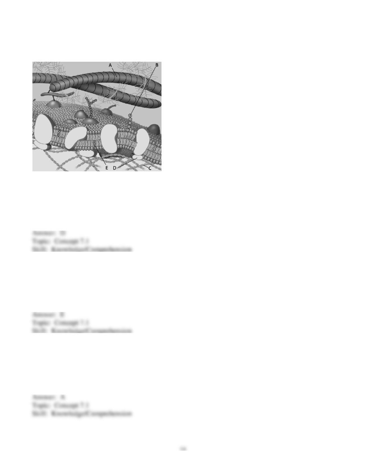
For the following questions, match the labeled component of the cell membrane in the figure with its
description.
54) Which component is the peripheral protein?
A) A
B) B
C) C
D) D
E) E
55) Which component is cholesterol?
A) A
B) B
C) C
D) D
E) E
56) Which component is the fiber of the extracellular matrix?
A) A
B) B
C) C
D) D
E) E
57) Which component is a microfilament of the cytoskeleton?
A) A
B) B
C) C
D) D
E) E
58) Which component is a glycolipid?
A) A
B) B
C) C
D) D
E) E
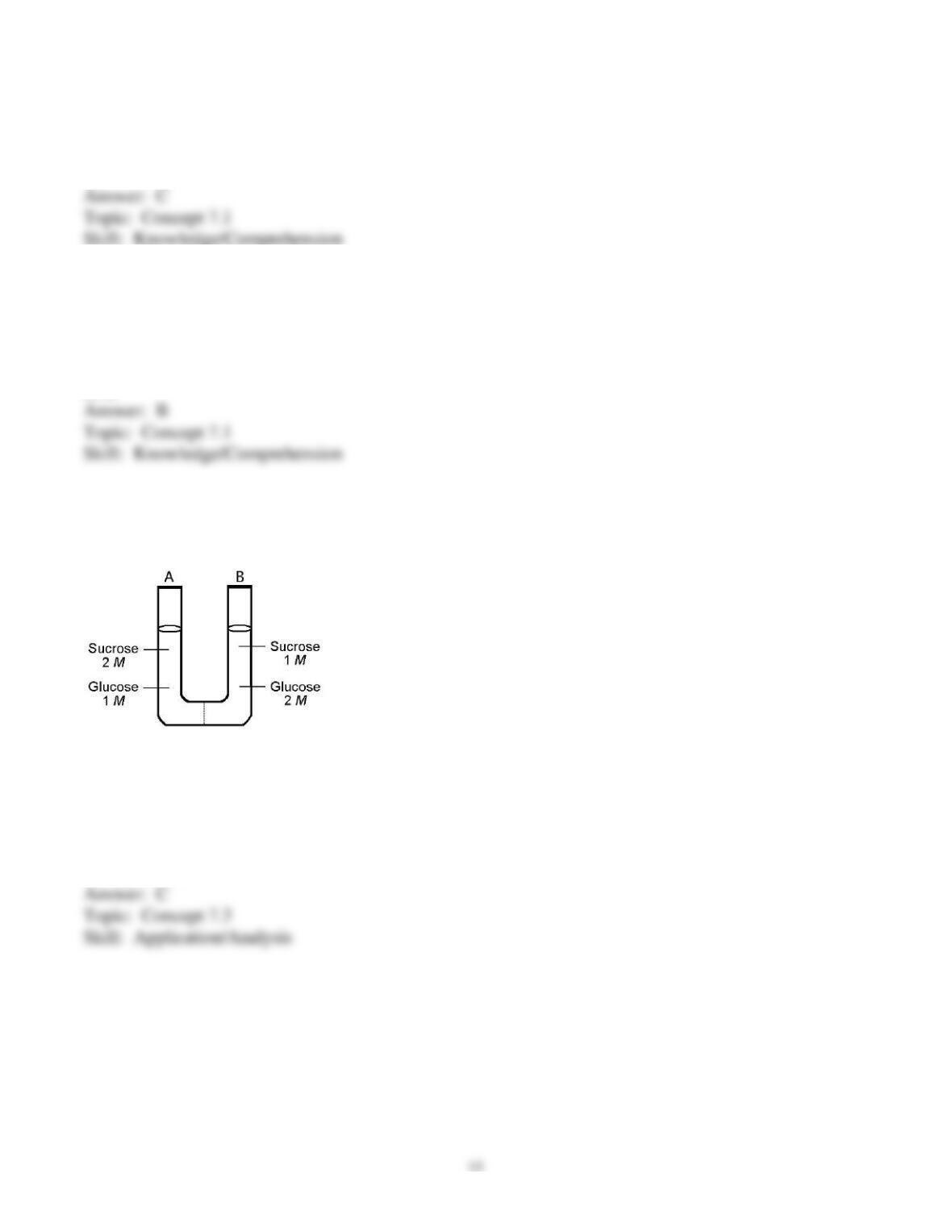
The solutions in the two arms of this U-tube are separated by a membrane that is permeable to water and
glucose but not to sucrose. Side A is half-filled with a solution of 2 M sucrose and 1 M glucose. Side B
is half-filled with 1 M sucrose and 2 M glucose. Initially, the liquid levels on both sides are equal.
59) Initially, in terms of tonicity, the solution in side A with respect to that in side B is
A) hypotonic.
B) plasmolyzed.
C) isotonic.
D) saturated.
E) hypertonic.
60) After the system reaches equilibrium, what changes are observed?
A) The molarity of sucrose and glucose are equal on both sides.
B) The molarity of glucose is higher in side A than in side B.
C) The water level is higher in side A than in side B.
D) The water level is unchanged.
E) The water level is higher in side B than in side A.
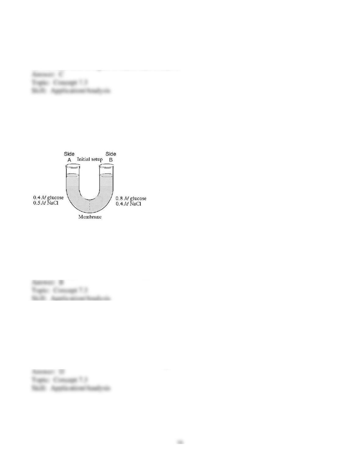
The solutions in the arms of a U-tube are separated at the bottom of the tube by a selectively permeable
membrane. The membrane is permeable to sodium chloride but not to glucose. Side A is filled with a
solution of 0.4 M glucose and 0.5 M sodium chloride (NaCl), and side B is filled with a solution
containing 0.8 M glucose and 0.4 M sodium chloride. Initially, the volume in both arms is the same.
Refer to the figure to answer the following questions.
61) At the beginning of the experiment,
A) side A is hypertonic to side B.
B) side A is hypotonic to side B.
C) side A is isotonic to side B.
D) side A is hypertonic to side B with respect to glucose.
E) side A is hypotonic to side B with respect to sodium chloride.
62) If you examine side A after three days, you should find
A) a decrease in the concentration of NaCl and glucose and an increase in the water level.
B) a decrease in the concentration of NaCl, an increase in water level, and no change in the
concentration of glucose.
C) no net change in the system.
D) a decrease in the concentration of NaCl and a decrease in the water level.
E) no change in the concentration of NaCl and glucose and an increase in the water level.
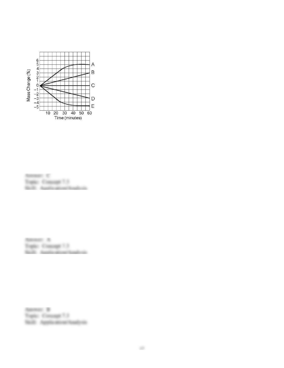
Five dialysis bags, constructed from a semipermeable membrane that is impermeable to sucrose, were
filled with various concentrations of sucrose and then placed in separate beakers containing an initial
concentration of 0.6 M sucrose solution. At 10-minute intervals, the bags were massed (weighed) and
the percent change in mass of each bag was graphed.
63) Which line in the graph represents the bag that contained a solution isotonic to the 0.6 M solution at
the beginning of the experiment?
A) A
B) B
C) C
D) D
E) E
64) Which line in the graph represents the bag with the highest initial concentration of sucrose?
A) A
B) B
C) C
D) D
E) E
65) Which line or lines in the graph represent(s) bags that contain a solution that is hypertonic at 50
minutes?
A) A and B
B) B
C) C
D) D
E) D and E
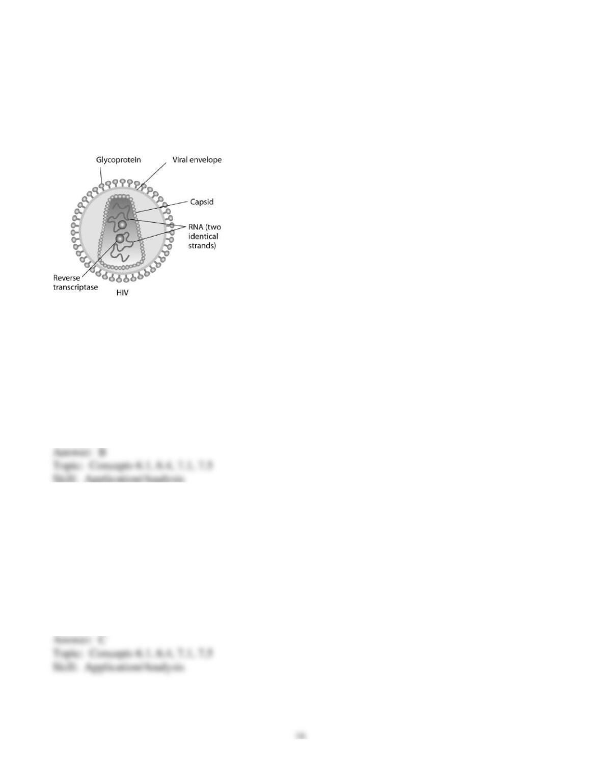
Human immunodeficiency virus (HIV) infects cells that have both CD4 and CCR5 cell surface
molecules. The viral nucleic acid molecules are enclosed in a protein capsid, and the protein capsid is
itself contained inside an envelope consisting of a lipid bilayer membrane and viral glycoproteins. One
hypothesis for viral entry into cells is that binding of HIV membrane glycoproteins to CD4 and CCR5
initiates fusion of the HIV membrane with the plasma membrane, releasing the viral capsid into the
cytoplasm. An alternative hypothesis is that HIV gains entry into the cell via receptor-mediated
endocytosis, and membrane fusion occurs in the endocytotic vesicle. To test these alternative hypotheses
for HIV entry, researchers labeled the lipids on the HIV membrane with a red fluorescent dye.
66) What would be observed by live-cell fluorescence microscopy if the red fluorescent lipid dye-
labeled HIV membrane fuses with the target cell plasma membrane?
A) A spot of red fluorescence will remain on the infected cell’s plasma membrane, marking the site of
membrane fusion and HIV entry.
B) The red fluorescent dye-labeled lipids will diffuse in the infected cell’s plasma membrane and
become difficult to detect.
C) A spot of red fluorescence will move into the infected cell’s cytoplasm.
D) A spot of red fluorescence will remain outside the cell after delivering the viral capsid.
E) Fluorescence microscopy does not have enough resolution to visualize fluorescently labeled HIV
virus particles.
67) What would be observed by live-cell fluorescence microscopy if HIV is endocytosed first, and then
fuses with the endocytotic vesicle membrane?
A) A spot of red fluorescence will remain on the infected cell’s plasma membrane, marking the site of
membrane fusion and HIV entry.
B) The red fluorescent dye-labeled lipids will diffuse in the endocytotic vesicle membrane and become
difficult to detect.
C) A spot of red fluorescence will move into the infected cell’s interior.
D) A spot of red fluorescence will remain outside the cell after delivering the viral capsid.
E) Fluorescence microscopy does not have enough resolution to visualize fluorescently labeled HIV
virus particles.

68) Using live-cell fluorescence microscopy, researchers observed that a red fluorescent spot moved
from the plasma membrane into the interior of target cells when red fluorescent dye-labeled HIV was
added to the cells. What is the best conclusion from these observations?
A) The hypothesis that HIV enters the cell via fusion with the target cell plasma membrane is proved.
B) The hypothesis that HIV enters the cell via fusion with the target cell plasma membrane is not
supported.
C) The hypothesis that HIV enters the cell via endocytosis is proved.
D) The hypothesis that HIV enters the cell via endocytosis is not supported.
E) Neither hypothesis is supported by these results.
69) If HIV first enters the cell in an endocytotic vesicle, instead of directly fusing with the plasma
membrane, then
A) HIV infection should be hindered by microtubule polymerization inhibitors such as nocodazole.
B) HIV infection should be more efficient at lower temperatures.
C) intact cortical actin microfilaments should interfere with HIV infection.
D) cells lacking integrins should be resistant to HIV infection.
E) addition of ligands for other cell-surface receptors to stimulate their endocytosis should increase the
efficiency of HIV infection.
70) In an HIV-infected cell producing HIV virus particles, the viral glycoprotein is expressed on the
plasma membrane. How do the viral glycoproteins get to the plasma membrane?
A) They are synthesized on ribosomes on the plasma membrane.
B) They are synthesized by ribosomes in the rough ER, and arrive at the plasma membrane in the
membrane of secretory vesicles.
C) They are synthesized on free cytoplasmic ribosomes, and then inserted into the plasma membrane.
D) They are synthesized by ribosomes in the rough ER, secreted from the cell, and inserted into the
plasma membrane from the outside.
E) They are synthesized by ribosomes on the HIV viral membrane, which fuses with the plasma
membrane from inside the cell.

Scenario Questions
Cystic fibrosis is a genetic disease in humans in which the CFTR protein, which functions as a chloride
ion channel, is missing or nonfunctional in cell membranes.
71) The CFTR protein belongs to what category of membrane proteins?
A) gap junctions
B) aquaporins
C) electrogenic ion pumps
D) cotransporters
E) hydrophilic channels
72) If the sodium ion concentration outside the cell increases, and the CFTR channel is open, in what
direction will chloride ions and water move across the cell membrane?
A) Chloride ions will move out of the cell, and water will move into the cell.
B) Both chloride ions and water will move out of the cell.
C) Chloride ions will move into the cell, and water will move out of the cell.
D) Both chloride ions and water will move into the cell.
E) The movement of chloride ions and water molecules will not be affected by changes in sodium ion
concentration outside the cell.
73) In the small airways of the lung, a thin layer of liquid is needed between the epithelial cells and the
mucus layer in order for cilia to beat and move the mucus and trapped particles out of the lung. One
hypothesis is that the volume of this airway surface liquid is regulated osmotically by transport of
sodium and chloride ions across the epithelial cell membrane. How would the lack of a functional
chloride channel in cystic fibrosis patients affect sodium ion transport and the volume of the airway
surface liquid?
A) Sodium ion transport will increase; higher osmotic potential will increase airway surface liquid
volume.
B) Sodium ion transport will increase; higher osmotic potential will decrease airway surface liquid
volume.
C) Sodium ion transport will decrease; lower osmotic potential will decrease airway surface liquid
volume.
D) Sodium ion transport will decrease; lower osmotic potential will increase the airway surface liquid
volume.
E) Sodium ion transport will be unaffected; lack of chloride transport still reduces osmotic potential and
decreases the airway surface liquid volume.

74) A patient has had a serious accident and lost a lot of blood. In an attempt to replenish body fluids,
distilled water–equal to the volume of blood lost–is transferred directly into one of his veins. What will
be the most probable result of this transfusion?
A) It will have no unfavorable effect as long as the water is free of viruses and bacteria.
B) The patient’s red blood cells will shrivel up because the blood fluid has become hypotonic compared
to the cells.
C) The patient’s red blood cells will swell because the blood fluid has become hypotonic compared to
the cells.
D) The patient’s red blood cells will shrivel up because the blood fluid has become hypertonic compared
to the cells.
E) The patient’s red blood cells will burst because the blood fluid has become hypertonic compared to
the cells.
75) You are working on a team that is designing a new drug. In order for this drug to work, it must enter
the cytoplasm of specific target cells. Which of the following would be a factor that determines whether
the molecule selectively enters the target cells?
A) blood or tissue type of the patient
B) hydrophobicity of the drug molecule
C) lack of charge on the drug molecule
D) similarity of the drug molecule to other molecules transported by the target cells
E) lipid composition of the target cells’ plasma membrane
End-of-Chapter Questions
The following questions are from the end-of–chapter “Test Your Understanding” section in Chapter 7 of
the textbook.
76) In what way do the membranes of a eukaryotic cell vary?
A) Phospholipids are found only in certain membranes.
B) Certain proteins are unique to each membrane.
C) Only certain membranes of the cell are selectively permeable.
D) Only certain membranes are constructed from amphipathic molecules.
E) Some membranes have hydrophobic surfaces exposed to the cytoplasm, while others have
hydrophilic surfaces facing the cytoplasm.

77) According to the fluid mosaic model of membrane structure, proteins of the membrane are mostly
A) spread in a continuous layer over the inner and outer surfaces of the membrane.
B) confined to the hydrophobic interior of the membrane.
C) embedded in a lipid bilayer.
D) randomly oriented in the membrane, with no fixed inside-outside polarity.
E) free to depart from the fluid membrane and dissolve in the surrounding solution.
78) Which of the following factors would tend to increase membrane fluidity?
A) a greater proportion of unsaturated phospholipids
B) a greater proportion of saturated phospholipids
C) a lower temperature
D) a relatively high protein content in the membrane
E) a greater proportion of relatively large glycolipids compared with lipids having smaller molecular
masses
79) Which of the following processes includes all others?
A) osmosis
B) diffusion of a solute across a membrane
C) facilitated diffusion
D) passive transport
E) transport of an ion down its electrochemical gradient
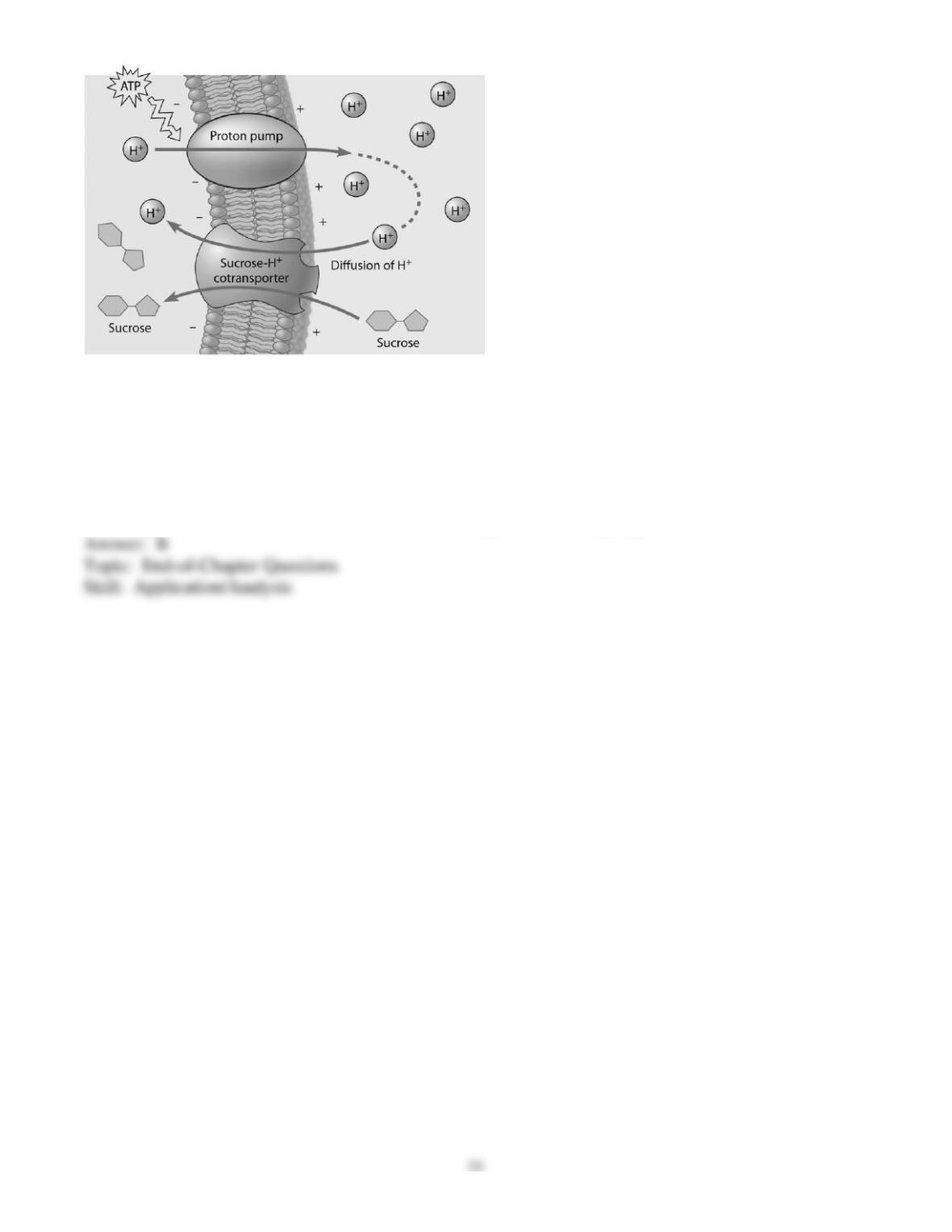
80) Based on the figure above, which of these experimental treatments would increase the rate of
sucrose transport into the cell?
A) decreasing extracellular sucrose concentration
B) decreasing extracellular pH
C) decreasing cytoplasmic pH
D) adding an inhibitor that blocks the regeneration of ATP
E) adding a substance that makes the membrane more permeable to hydrogen ions