Exam
Name___________________________________
MULTIPLE CHOICE. Choose the one alternative that best completes the statement or answers the question.
1)
Which microscope takes advantage of differences in the refractive indexes of cell structures?
1)
A)
compound light microscope
B)
electron microscope
C)
darkfield microscope
D)
phase–contrast microscope
E)
fluorescence microscope
2)
Which of the following pairs is mismatched?
2)
A)
confocal microscope —produces a three–dimensional image
B)
fluorescence microscope —uses a fluorescent light
C)
scanning electron microscope —produces a three–dimensional image
D)
darkfield microscope —uses visible light
E)
scanning tunneling microscope —allows visualization of atoms
B
3)
Which of the following correctly traces the path of light through the compound microscope?
3)
A)
light source; condenser; objective lens; specimen; ocular lens
B)
light source; condenser; specimen; objective lens; ocular lens
C)
condenser; light source; specimen; objective lens; ocular lens
D)
condenser; light source; specimen; ocular lens; objective lens
E)
light source; specimen; condenser; objective lens; ocular lens
B
D
4)
Which of the following places the steps of the Gram stain in the correct order?
1–Alcohol–acetone
2–Crystal violet
3–Safranin
4–Iodine
4)
A)
2–1–4–3
B)
1–2–3–4
C)
2–4–1–3
D)
1–3–2–4
E)
4–3–2–1
5)
You are performing a Gram stain on gram–positive bacteria and you stop after the addition of the
first dye. What is the appearance of the bacteria at this point?
5)
A)
colorless
B)
purple
C)
brown
D)
red
6)
Which of the following is NOT true regarding the acid–fast stain?
6)
A)
Acid–fast cells appear red in a completed acid–fast stain.
B)
Acid–fast cells retain the primary dye after treatment with acid–alcohol.
C)
It is used to identify members of the genus Mycobacterium.
D)
Non–acid–fast microbes appear blue in a completed acid–fast stain.
E)
If cells are acid–fast, they are gram–negative.
7)
The resolution of a microscope can be improved by changing the
7)
A)
wavelength of light.
B)
condenser.
C)
coarse adjustment.
D)
diaphragm.
E)
fine adjustment.
8)
Which type of stain is most useful in helping clinicians to decide which antibiotic to prescribe for a
bacterial infection?
8)
A)
flagella stain
B)
negative stain
C)
endospore stain
D)
Gram stain
E)
simple stain
9)
) In microscopy, the term resolution
9)
A)
is improved when longer wavelengths of light are employed.
B)
refers to the ability to distinguish fine structure and detail in a specimen.
C)
refers to magnification when using the electron microscope.
D)
is the same as the total magnification of a specimen.
E)
is only observed in stained specimens.
10)
Which microscope is used to see detail of a 300–nm virus?
10)
A)
darkfield microscope
B)
electron microscope
C)
fluorescence microscope
D)
DIC microscope
E)
phase–contrast microscope
11)
You are performing a Gram stain on gram–negative bacteria and you stop after the addition of the
mordant. What is the appearance of the bacteria at this point?
11)
A)
red
B)
purple
C)
brown
D)
colorless
12)
What structure does light pass through after leaving the condenser in a compound light
microscope?
12)
A)
specimen
B)
illuminator
C)
objective lens
D)
ocular lens
13)
A microorganism measures 5 µm in length. Its length in mm would be
13)
A)
50 mm.
B)
0.005 mm.
C)
0.5 mm.
D)
500 mm.
E)
0.05 mm.
14)
The purpose of the ocular lens is to
14)
A)
decrease the light.
B)
improve resolution.
C)
decrease the refractive index.
D)
increase the light.
E)
magnify the image from the objective lens.
15)
The negative stain is used to
15)
A)
visualize endospores.
B)
determine Gram reaction.
C)
determine cell size.
D)
visualize capsules.
E)
determine flagella arrangement.
16)
Simple staining is often necessary to improve contrast in which microscope?
16)
A)
fluorescence microscope
B)
darkfield microscope
C)
phase–contrast microscope
D)
compound light microscope
E)
electron microscope
17)
Which microscope is best used for observing the surfaces of intact cells and viruses?
17)
A)
scanning electron microscope
B)
brightfield microscope
C)
phase–contrast microscope
D)
darkfield microscope
E)
fluorescence microscope
18)
Which microscope is used to observe a specimen that emits light when illuminated with an
ultraviolet light?
18)
A)
phase–contrast microscope
B)
darkfield microscope
C)
electron microscope
D)
fluorescence microscope
E)
compound light microscope
19)
Which microscope is used to see intracellular detail in a living cell?
19)
A)
atomic force microscope
B)
two–photon microscope
C)
brightfield microscope
D)
fluorescence microscope
E)
transmission electron microscope
20)
A virus measures 100 nm in length. What is its length in µm?
20)
A)
0.01 µm
B)
1 µm
C)
0.001 µm
D)
10 µm
E)
.1 µm
21)
Which microscope achieves the highest magnification and greatest resolution?
21)
A)
electron microscope
B)
darkfield microscope
C)
phase–contrast microscope
D)
compound light microscope
E)
fluorescence microscope
22)
You are performing a Gram stain on gram–negative bacteria and you stop after the decolorizer
step. What is the appearance of the bacteria at this point?
22)
A)
purple
B)
brown
C)
colorless
D)
red
23)
Which microscope uses two beams of light to produce a three–dimensional color image?
23)
A)
DIC microscope
B)
fluorescence microscope
C)
electron microscope
D)
phase–contrast microscope
E)
darkfield microscope
24)
This microscope produces an image of a light cell against a dark background; internal structures
are NOT visible.
24)
A)
phase–contrast microscope
B)
darkfield microscope
C)
electron microscope
D)
fluorescence microscope
E)
compound light microscope
25)
The purpose of a mordant in the Gram stain is to
25)
A)
make the bacterial cells larger.
B)
make gram–negative cells visible.
C)
remove the simple stain.
D)
prevent the crystal violet from leaving the cells.
E)
make the flagella visible.
26)
Which of the following places the steps in the correct sequence?
1–Staining
2–Making a smear
3–Fixing
26)
A)
3–2–1
B)
2–3–1
C)
1–3–2
D)
1–2–3
E)
The order is unimportant.
27)
Which of the following pairs is mismatched?
27)
A)
methylene blue — simple stain
B)
nigrosin — negative stain
C)
basic dye — negative stain
D)
acidic dye — capsule stain
E)
crystal violet — simple stain
28)
Which step in the Gram stain is the critical step in differentiating gram–positive cells from
gram–negative cells?
28)
A)
alcohol–acetone
B)
iodine
C)
crystal violet
D)
safranin
29)
Assume you stain Bacillus by applying malachite green with heat and then counterstaining with
safranin. Through the microscope, the green structures are
29)
A)
capsules.
B)
endospores.
C)
flagella.
D)
cell walls.
E)
The answer cannot be determined.
30)
Which of the following is NOT equal to 1 mm?
30)
A)
10 6 nm
B)
100 µm
C)
0.1 cm
D)
10–3 m
E)
0.001 m
31)
Which microscope can be used to visualize DNA or botulinum toxin?
31)
A)
scanning tunneling microscope
B)
scanning electron microscope
C)
compound light microscope
D)
confocal microscope
E)
phase–contrast microscope
32)
Which microscope is most useful for visualizing a biofilm?
32)
A)
scanning acoustic microscope
B)
fluorescence microscope
C)
phase–contrast microscope
D)
transmission electron microscope
E)
compound light microscope
33)
You are performing a Gram stain on gram–positive bacteria and you stop after the addition of the
counterstain. What is the appearance of the bacteria at this point?
33)
A)
colorless
B)
brown
C)
purple
D)
red
34)
You are studying a cell structure that is approximately 100 nm in size. Which of the following
provides the lowest magnification you can use to see this structure?
34)
A)
scanning electron microscope
B)
phase–contrast microscope
C)
transmission electron microscope
D)
darkfield microscope
E)
brightfield microscope
35)
Which microscope is used to see internal structures of cells in a natural state?
35)
A)
electron microscope
B)
darkfield microscope
C)
compound light microscope
D)
phase–contrast microscope
E)
fluorescence microscope
36)
You find colorless areas in cells in a gram–stained smear. What should you apply next?
36)
A)
an acid–fast stain
B)
a flagella stain
C)
a capsule stain
D)
a simple stain
E)
an endospore stain
37)
What is the total magnification of a specimen viewed with a 10x ocular lens and a 45x objective
lens?
37)
A)
45x
B)
100x
C)
4.5x
D)
10x
E)
450x
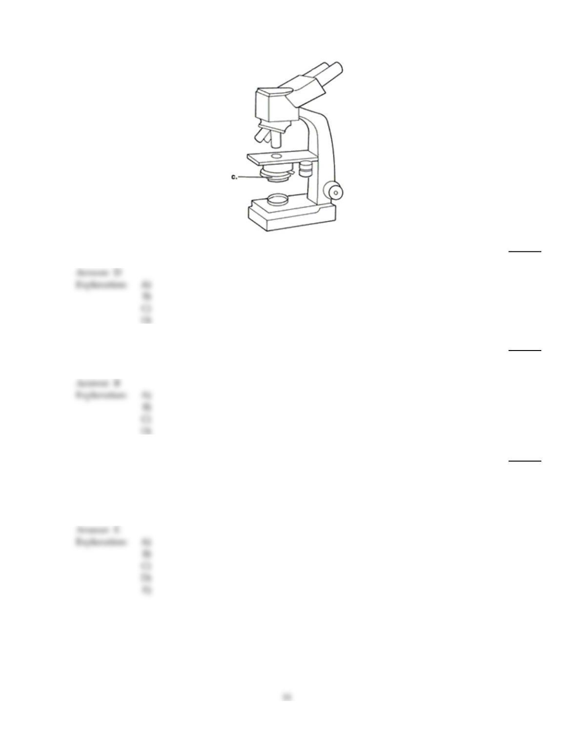
Figure 3.1
38)
In Figure 3.1, line “c” points to the microscope’s
38)
A)
illuminator.
B)
ocular lens.
C)
objective lens.
D)
condenser.
39)
Bacterial smears are fixed before staining to
39)
A)
make the cells visible.
B)
affix the cells to the slide.
C)
make their walls permeable.
D)
accept stain.
40)
In using this microscope, the observer does NOT look directly at an image through a lens.
40)
A)
phase–contrast microscope
B)
fluorescence microscope
C)
compound light microscope
D)
darkfield microscope
E)
electron microscope
41)
Which of the following is NOT useful for observing living cells?
41)
A)
phase–contrast microscope
B)
scanning electron microscope
C)
scanning acoustic microscope
D)
darkfield microscope
E)
brightfield microscope
42)
Which microscope uses visible light?
42)
A)
fluorescence microscope
B)
differential interference contrast microscope
C)
scanning acoustic microscope
D)
scanning electron microscope
E)
confocal microscope
43)
Which microscope is used to observe viruses and the internal structure of thinly sectioned cells?
43)
A)
fluorescence microscope
B)
scanning electron microscope
C)
brightfield microscope
D)
transmission electron microscope
E)
darkfield microscope
44)
Which of the following pairs is mismatched?
44)
A)
safranin — acid dye
B)
iodine — mordant
C)
carbolfuchsin — basic dye
D)
alcohol–acetone — decolorizer
E)
crystal violet — basic dye
45)
A student is looking at a bacterial specimen using the oil immersion lens, but has forgotten to put
immersion oil on the slide. The specimen will appear
45)
A)
larger than it would if immersion oil was used.
B)
to have no color.
C)
the same as it would if the immersion oil was used.
D)
smaller than it would if immersion oil was used.
E)
somewhat fuzzy and have poor resolution.
TRUE/FALSE. Write ‘T’ if the statement is true and ‘F’ if the statement is false.
46)
Both phase–contrast microscopy and differential interference contrast microscopy are used to
view the internal structures of cells without staining.
46)
47)
The limit of resolution of the compound microscope illuminated with visible light is approximately
0.2 µm.
47)
48)
The counterstain used in the Gram stain is a basic dye.
48)
49)
If acid–fast bacteria are stained with the Gram stain, they will stain gram–negative.
49)
50)
The capsules and flagella of bacteria can be observed in gram–stained smears.
50)
51)
In a completed Gram stain, gram–negative bacteria are colorless.
51)
52)
Cells viewed in darkfield microscopy appear similar to those stained with the negative stain.
52)
53)
In a completed Gram stain, gram–positive bacteria are purple.
53)

54)
The greater resolution of the electron microscope compared to the compound microscope is due to
the longer wavelengths of the electrons used to examine specimens.
54)
55)
Scanned probe microscopy is used to examine fine detail of molecular complexes, such as blood
clots, or molecules, such as DNA.
55)
ESSAY. Write your answer in the space provided or on a separate sheet of paper.
56)
You are working in a clinical laboratory and you need to examine an unstained urine sample for the presence of
bacteria. What type of light microscope should you use to observe this specimen? Explain your answer.
57)
In 1877, Robert Koch thought preparing permanently stained slides would be valuable. Why
was his assessment correct?
58)
In 1884, Hans Christian Gram described a method of staining bacterial cells while not staining surrounding
animal tissues. However, he thought that the staining method he developed was faulty because not all bacteria
stained. In a letter to the editor of the journal in which Gram published his findings, write your response to
Gram’s concern.
Answer Key
Testname: C3

Answer Key
Testname: C3