Hall: Guyton and Hall Textbook of Medical Physiology, 12th Edition
Chapter 12: Electrocardiographic Interpretation of Cardiac Muscle and Coronary
Blood Flow Abnormalities: Vectorial Analysis
Test Bank
1. A ventricular depolarization wave when traveling 210 in the frontal plane will cause a
large positive deflection in:
A. aVR
B. aVL
C. Lead II
D. Lead III
E. aVF
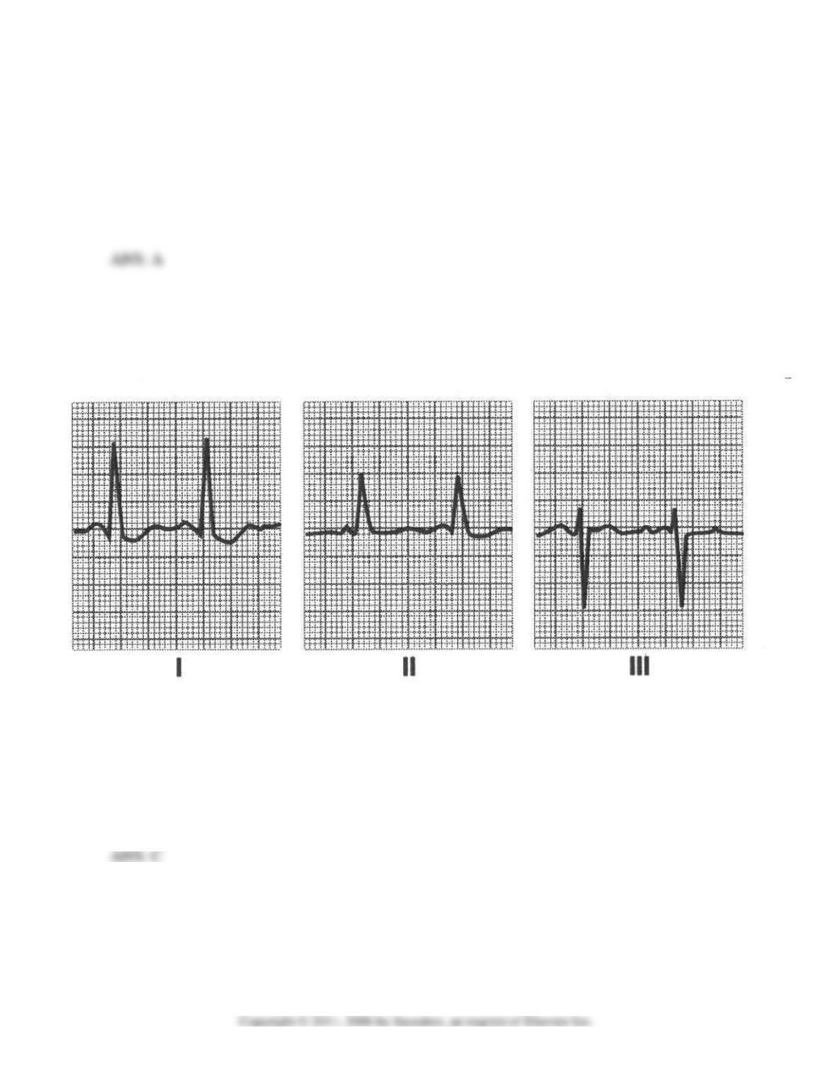
Questions 2-4: A 60-year-old woman had the following EKG recorded at a local emergency
room following an automobile accident. Her weight was 70 kg and her aortic blood pressure
was 140/80 mmHg.
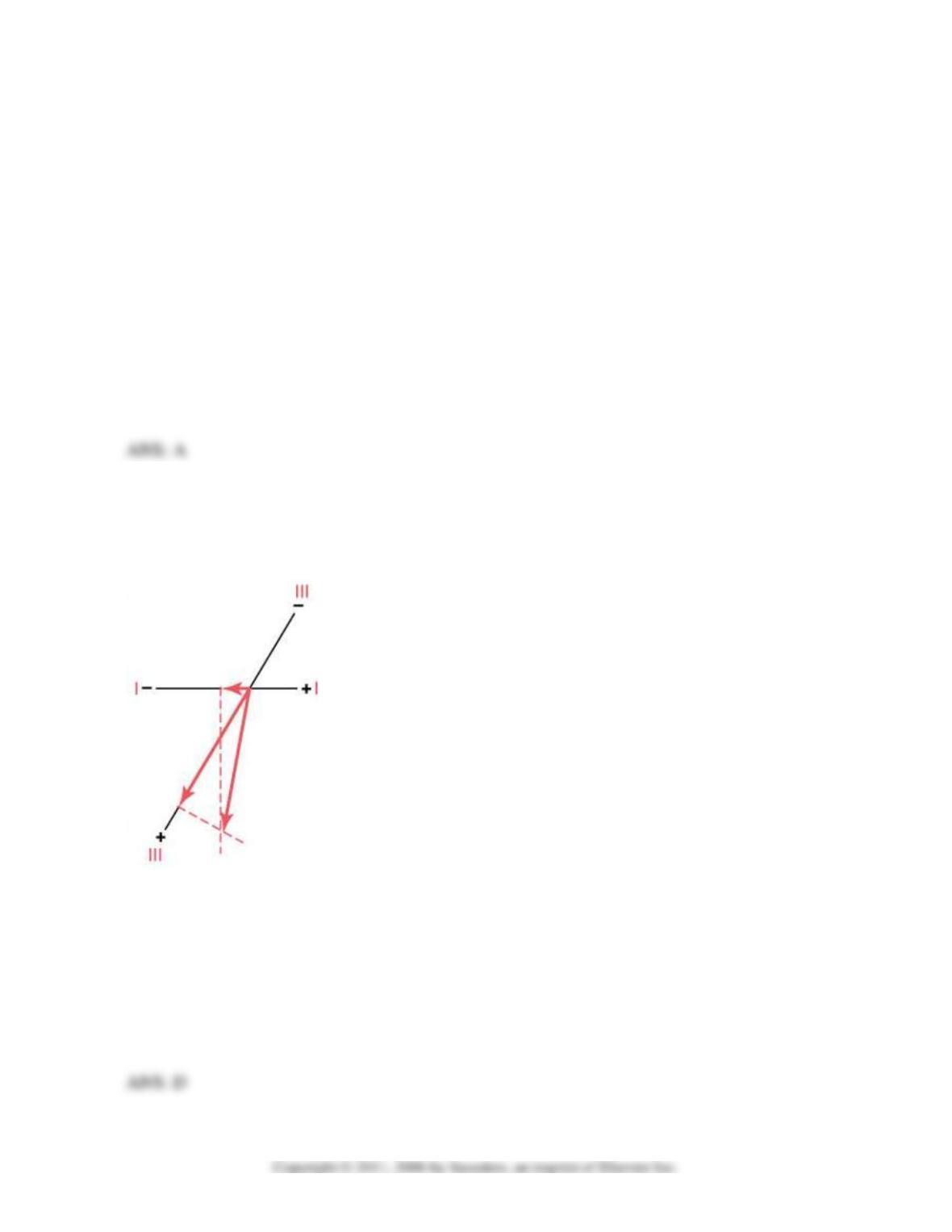
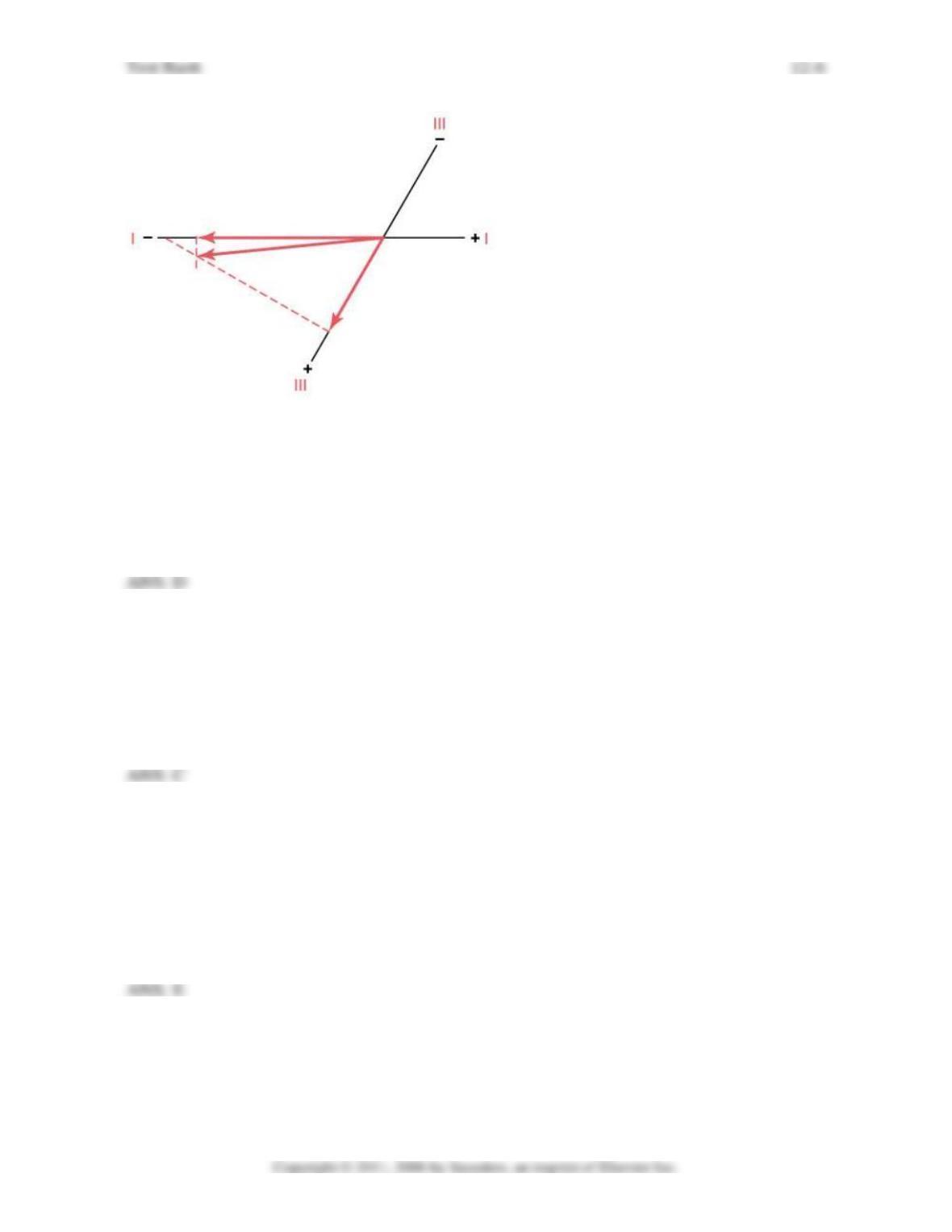
2. What is the mean electrical axis calculated from standard leads I, II, and III shown in the
woman’s EKG?
A. −90
B. −50
C. −12
D. +105
E. +170
Test Bank
12-2
3. What is the heart rate using lead I for the calculation?
A. 70
B. 88
C. 100
D. 112
E. 148
4. What is her likely diagnosis?
A. Mitral valve stenosis
B. Left bundle branch block
C. Pulmonary valve stenosis
D. Right bundle branch block
E. Left ventricular hypertrophy
5. A seventy-five year-old woman came into a local emergency room complaining of chest
pain and shortness of breath. Blood samples were taken for cardiac enzyme
measurements, and an EKG was quickly taken. Based on the EKG tracing shown below,
what is the likely diagnosis?
A. Left bundle branch block
B. Acute anterior wall infarction in the base of the heart
C. Acute anterior wall infarction in the apex of the heart
D. Acute posterior infarction in the base of the heart
E. Acute posterior infarction in the apex of the heart
Test Bank
12-3
6. A ventricular depolarization wave when traveling 90 in the frontal plane will cause a
large positive deflection in:
A. aVR
B. aVL
C. Lead II
D. Lead III
E. aVF
Questions 7-9: A 70-year-old man had the following EKG recorded at his doctor’s office.
His weight was 80 kg and his blood pressure was 160/90 mmHg. He complained that he gets
tired easily.
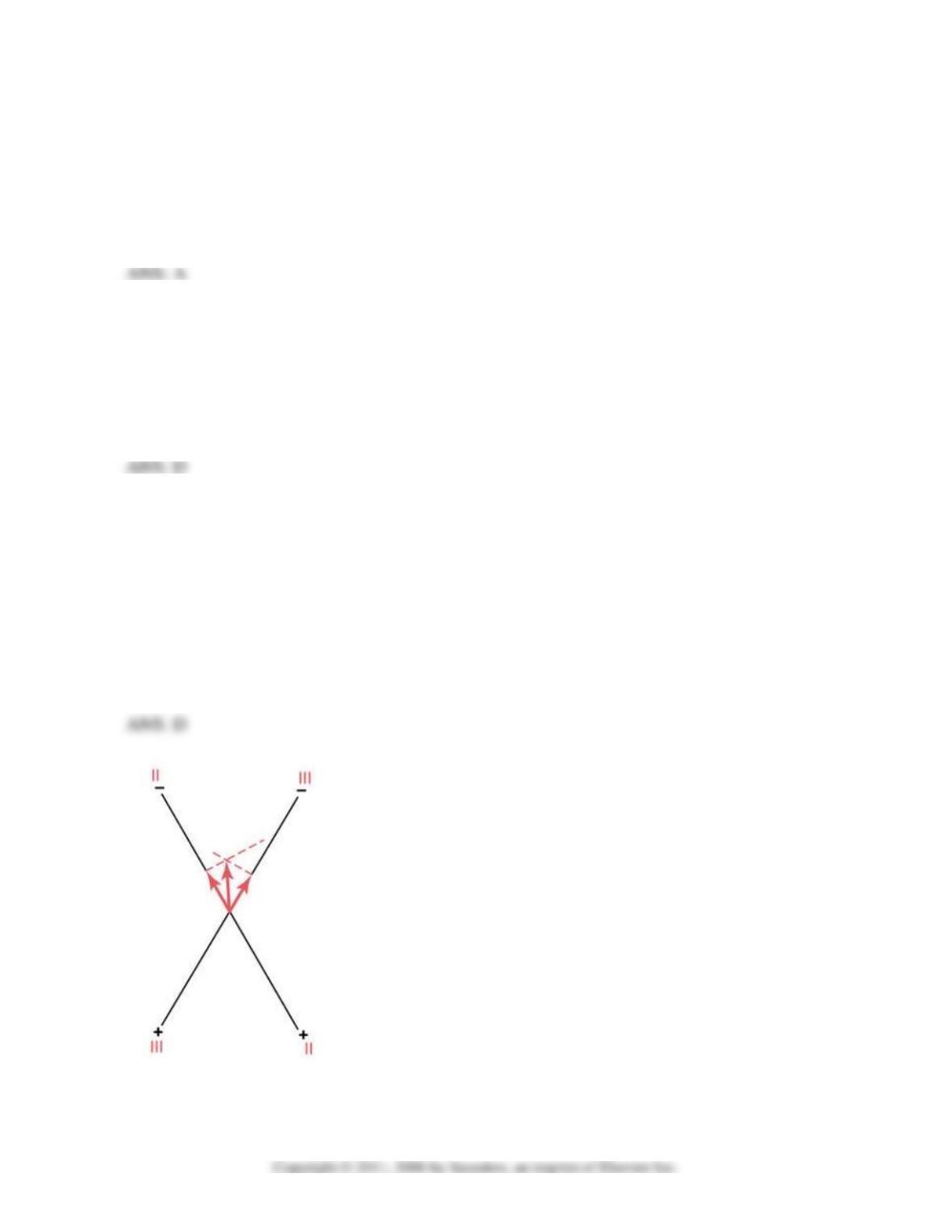
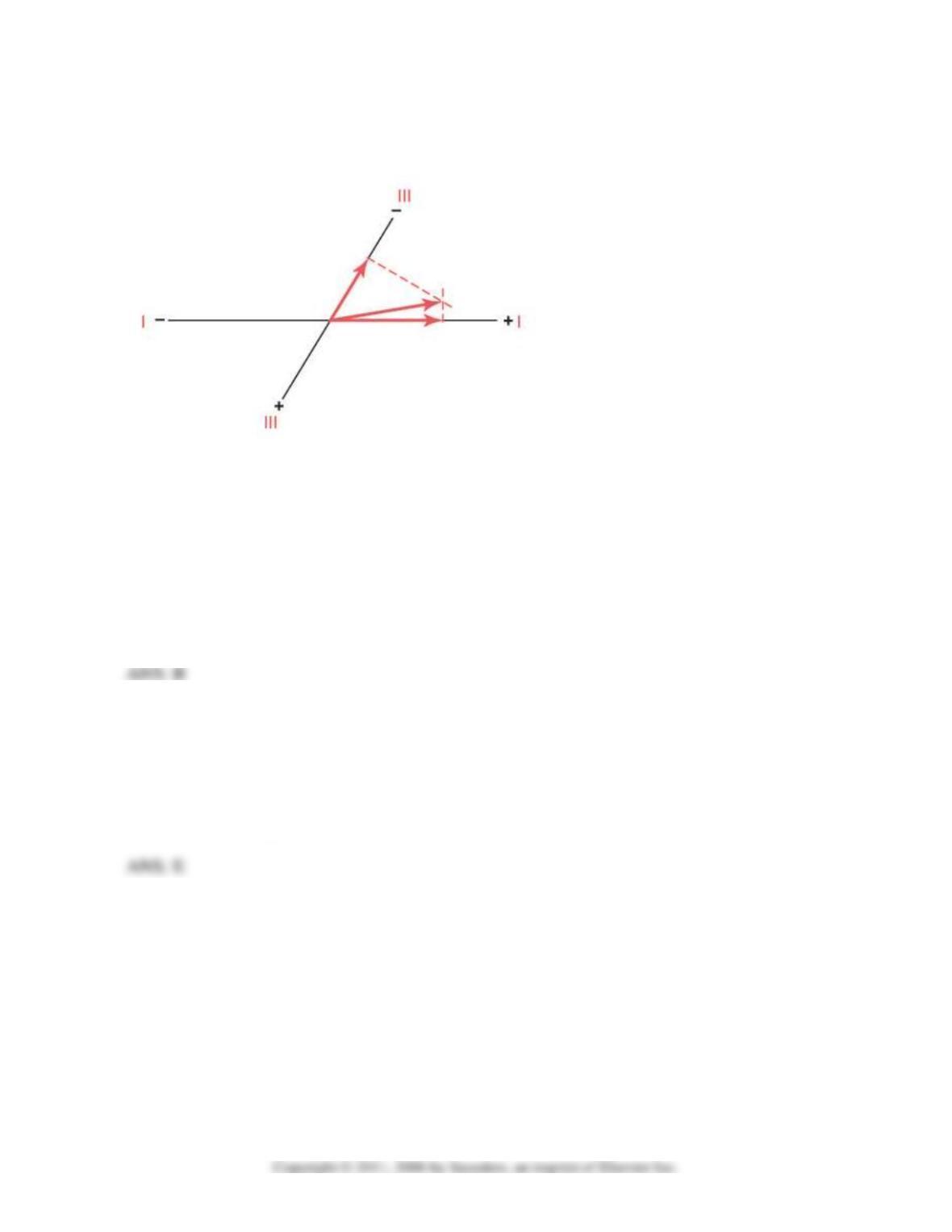
7. What is the mean electrical axis calculated from standard leads I, II, and III shown in the
man’s EKG?
A. −90
B. −60
C. −15
D. +60
E. +170
8. What is the heart rate using lead I for the calculation?
A. 75
B. 88
C. 100
D. 112
Test Bank
12-4
E. 148
9. What is his likely diagnosis?
A. Tricuspid valve stenosis
B. Complete left bundle branch block
C. Pulmonary valve stenosis
D. Pulmonary valve regurgitation
E. Left ventricular hypertrophy
10. A seventy-year-old woman came into the local emergency room complaining of chest
pain and shortness of breath. Blood samples were taken for cardiac enzyme
measurements, and an EKG was quickly taken. Based on the EKG tracing shown below,
what is the likely diagnosis?
A. Left bundle branch block
B. Acute anterior wall infarction in the left ventricle
C. Acute anterior wall infarction in the right ventricle
D. Acute posterior infarction in the left ventricle
E. Acute posterior infarction in the right ventricle
Test Bank
12-5
Questions 11-12: A 65-year-old man went for an annual checkup and had the following EKG
recorded at his physician’s office.
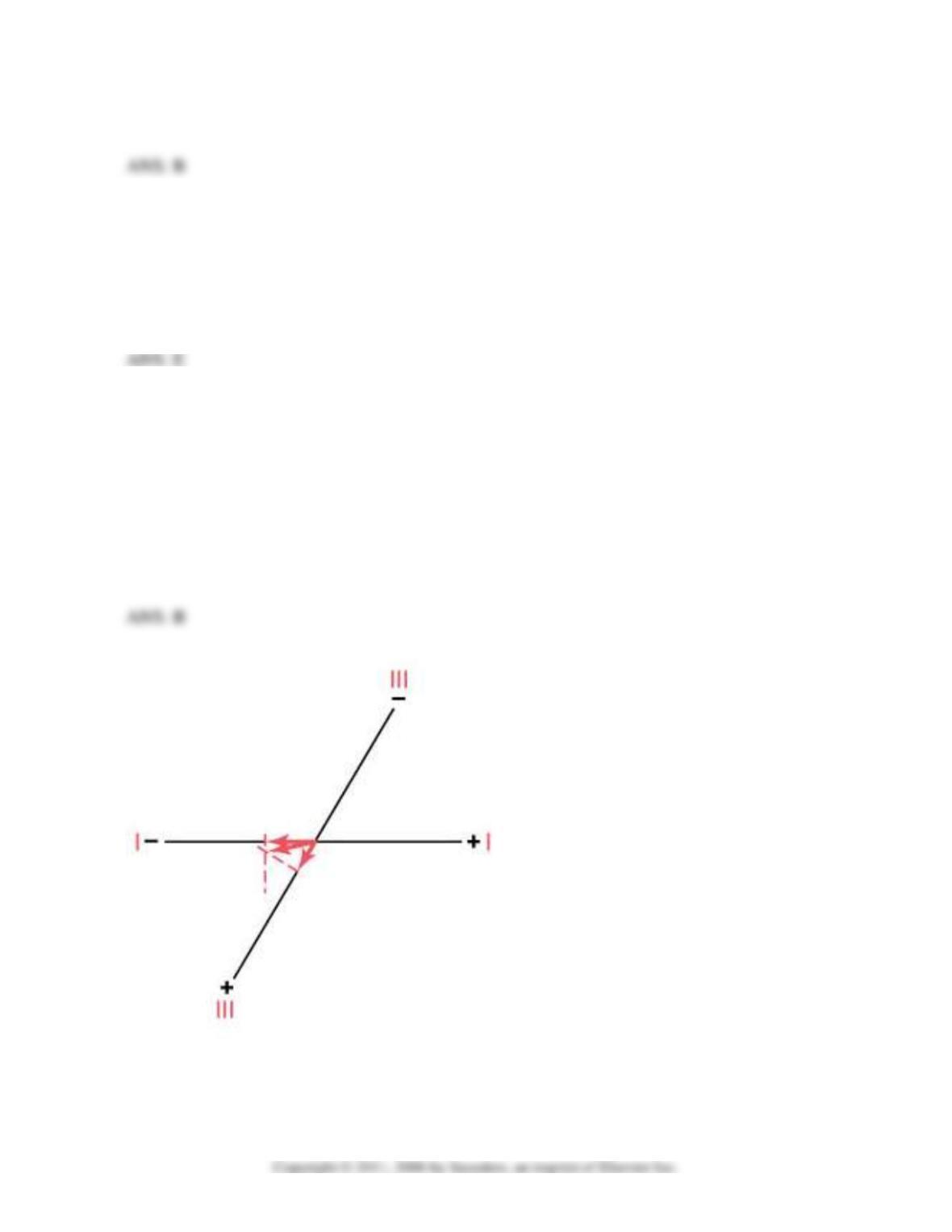
11. What is the mean electrical axis calculated from standard leads I, II, and III shown in his
EKG?
A. −50
B. −15
C. 0
D. +15
E. +50
12. What is the most likely diagnosis?
A. Right ventricular hypertrophy
B. Left bundle branch block
C. Pulmonary valve insufficiency
D. Right bundle branch block
E. Systemic hypertension
Questions 13-14. A 50-year-old woman was admitted to a local emergency room following a
motorcycle accident, and the following EKG was taken.
13. What is her heart rate? Use lead I for the calculation.
A. 56
B. 66
C. 76
D. 103
E. 152
14. What is the most likely diagnosis?
A. Aortic valve insufficiency
B. Left bundle branch block
C. Pulmonary valve stenosis
D. Right bundle branch block
E. Systemic hypertension
15. During the TP interval in an electrocardiogram of a patient with a damaged cardiac
muscle:
A. The entire heart is depolarized
B. All the heart is depolarized except for the damaged cardiac muscle
C. About half the heart is depolarized
D. All of the heart is repolarized
E. All of the heart is repolarized except for the damaged cardiac muscle
16. A 30-year-old woman had an EKG taken, and her QRS complex had a width of 0.20
seconds, her T wave was inverted in lead I, and the R wave had a large negative
deflection in lead III. What is her diagnosis?
A. Right ventricular hypertrophy
Test Bank
12-7
B. Left bundle branch block
C. Pulmonary valve stenosis
D. Right bundle branch block
E. Left ventricular hypertrophy
17. A ventricular depolarization wave when traveling −30 in the frontal plane will cause a
large positive deflection in:
A. aVR
B. aVL
C. Lead II
D. Lead III
E. aVF
Questions 18-19: A 6-year-old boy had the following EKG recorded at his physician’s office.
He had little energy, and during light exercise his respiratory would increase rapidly.
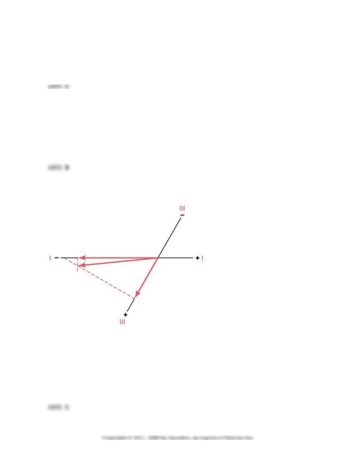
18. What is his mean electrical axis calculated from standard leads I, II, and III shown in his
EKG?
A. −100
B. −20
C. 0
D. +90
E. +170

19. What is his diagnosis?
A. Left ventricular hypertrophy
B. Left bundle branch block
C. Tricuspid stenosis
D. Right bundle branch block
E. Tetralogy of Fallot
20. Mr. Smith had his EKG measured at UMC, but they lost his records. The EKG technician
remembered that the QRS deflection was large and negative in Lead aVF and 0 in Lead I.
His mean electrical axis in the frontal plane is
A. 90
B. 60
C. 0
D. −60
E. −90