CHAPTER 9
Visualizing Cells
Questions
9-1 Your friend has joined a lab that has a light microscope connected to a computer
imaging system. A 30-inch monitor is attached to the computer. Your friend
claims that because he can now enlarge all images obtained using the light
microscope on the large monitor, that he will be able to use the light microscope
to resolve objects that are separated by less than 0.1 µm. Is he right?
9-2 Your friend, Sue, has joined a lab on campus that uses green fluorescent protein
(GFP) as a tool to localize globular proteins to specific compartments in the cell.
Sue creates a fusion protein by fusing GFP to her protein of interest, examines the
fusion protein’s localization in the cell, and finds that it is localized to the plasma
membrane. However, your friend, Bob, points out that the theoretical limit of
resolution for a light microscope is just under 0.2 µm and most globular proteins
are in the 10 nm (0.01 µm) size range and that lipid bilayers are about 5 nm
(0.005 µm) thick. Given that what Bob says is true, explain why Sue can see her
globular protein localized at the plasma membrane.
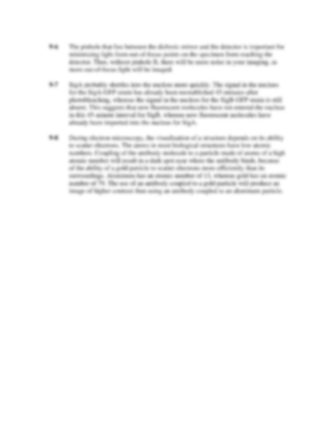
9-3 Figure Q9-3 shows the path of light through a compound microscope to your eye.
If you were to insert a magnifying lens between the light source and the
condenser, would you increase the magnification of the image? Explain.
Figure Q9-3
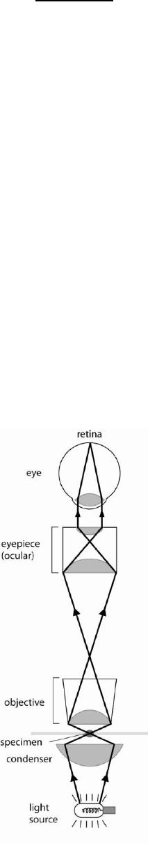
9-4 Differential interference contrast (DIC) microscopy uses the phase differences
that occur as light passes through a cell to generate an image that looks somewhat
three-dimensional. The micrographs in Figure Q9-4 demonstrate the difference
between a bright-field image (on top) and one imaged using DIC (on bottom).
Figure Q9-4
Do the three-dimensional images obtained using DIC represent the actual
geometry of the cell? Explain your answer.
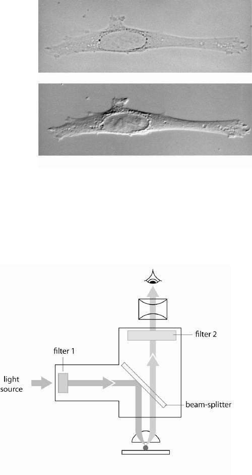
9-5 Figure Q9-5 illustrates the optical system of a fluorescence microscope.
Figure Q9-5
Your friend has discovered a new fluorescent protein that has a peak absorption at
513 nm and a peak emission at 535 nm. She has found eight filters and installed
them on a microscope down the hall, to create four filter sets on it with the
following properties.
Set A. Filter 1: passes wavelengths between 525 and 545 nm;
Filter 2: passes wavelengths between 490 and 520 nm.
Set B. Filter 1: passes all wavelengths above 530 nm;
Filter 2: passes wavelengths between 525 and 545 nm.
Set C. Filter 1: passes wavelengths between 490 and 520 nm;
Filter 2: passes wavelengths between 525 and 545 nm.
Set D. Filter 1: passes all wavelengths above 530 nm;
Filter 2: passes all wavelengths above 500 nm.
Would any of these filter sets be appropriate for your friend’s new fluorescent
protein, and if so, which one? Explain your answer.
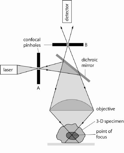
9-6 A confocal microscope is similar to an epifluorescence microscope, except that a
laser is the light source and two pinholes (labeled A and B on Figure Q9-6) are
used.
Figure Q9-6
Unfortunately, somebody has stolen the confocal pinhole that lies between the
dichroic mirror and the detector. You decide to do go ahead and use the
microscope, despite the theft. How will the missing pinhole affect your imaging?
9-7 You are interested in studying the function of two proteins that you believe are
important for cell signaling, called SigA and SigB. Both SigA and SigB are found
in the nucleus and are 95% identical at the amino acid level. You are excited
about the FRAP (fluorescence recovery after photobleaching) technique you have
learned about in class. Thus, you create a cell line that contains SigA fused to
green fluorescent protein (GFP) and a separate cell line strain that contains SigB
fused to GFP, and use these cell lines to perform FRAP experiments. The results
you obtain are diagrammed in Figure Q9-7 (the grey color represents the GFP
signal).
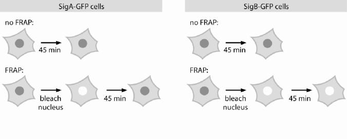
Figure Q9-7
From these results, is SigA or SigB more likely to be imported into the nucleus
more quickly? Explain.
9-8 The subcellular localization of a specific protein can be determined by electron
microscopy if an antibody that recognizes a specific protein is coupled to a gold
particle and this antibody is applied to the sample. Explain why the use of a gold
particle is preferable to an aluminum particle.
Answers