CHAPTER 3
Proteins
Questions
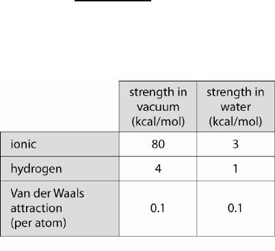
3-1 The table lists the average strengths of noncovalent bonds.
Table Q3-1
Protein Lock binds to protein Key in a reaction with G° = –14 kcal/mol. When
these proteins bind, 350 Å2 of the surface of Lock is closely apposed to the same
area on the surface of Key, so that water is excluded.
A. Estimate how much of the binding energy the van der Waals interactions
contribute. (For this calculation, assume that an average atom has a
surface area of 7 Å2.)
B. If hydrogen bonds contribute the remaining binding energy between
amino acids, estimate how many hydrogen bonds form between Lock and
Key. Does it depend on whether the hydrogen-bonded amino acids occur
within the buried surface or elsewhere in the proteins? Explain.
3-2 Consider a protein X that is known to exist in two slightly different
conformations, called X1 and X2. Proteins A, B, and C are ligands that can bind
to protein X. The sites on X required for binding to A, B, and C are located in
different regions of the protein. On the basis of experimental evidence of protein–
protein complexes, you devise the hypothetical binding reaction scheme shown in
Figure Q3-2. According to the hypothesis, which pairs of binding sites on X
exhibit negative coupling (negative linkage)? Which pairs exhibit positive
coupling (positive linkage)? Does the addition of a high concentration of protein
A increase or decrease the affinity of X for B? Of X for C?
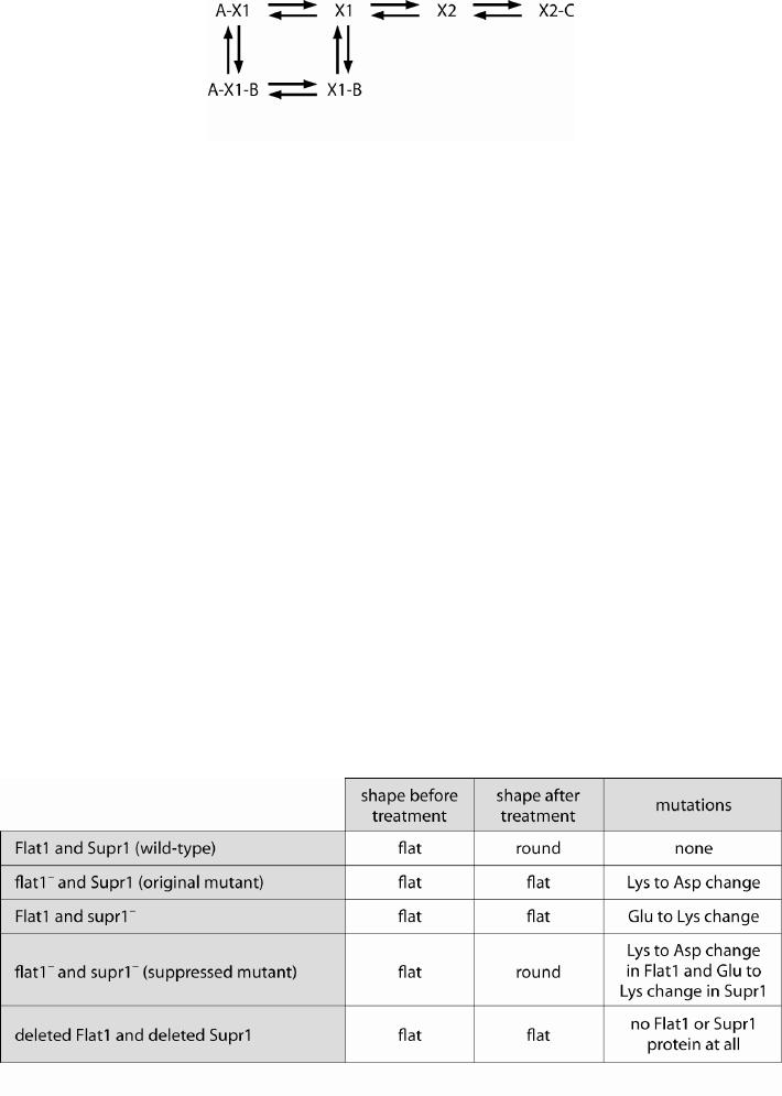
Figure Q3-2
3-3 You are working in a laboratory trying to understand how cells change shape in
response to treatment with a particular chemical. On treatment, normal wild-type
cells convert from a flattened irregular shape to a rounded, nearly spherical shape.
Previously, another student in the laborator identified a mutant that remained flat
after treatment. The gene that encodes the normal protein is called Flat1 and the
mutant gene is designated flat1–. To identify additional genes responsible for this
behavior, you mutagenize flat1– cells and look for those that can respond
normally to stimulation. You identify cells that contain a “suppressor” mutation in
a second gene, called supr1–, that restores the wild-type behavior to cells with the
flat1– mutation. Further genetic manipulation and DNA sequencing reveals the
data shown in the table below.
Initially, you are surprised to find that cells containing a mutation in
supr1– and a normal copy of Flat1 have the same mutant behavior as cells
containing only a flat1– mutation. However, when you examine the nature of the
amino acids that are changed in the mutants, you instantly suggest a hypothesis to
account for the observations. What is your hypothesis? What biochemical
experiment with purified proteins will test your hypothesis? What biochemical
results would support the hypothesis?
Table Q3-3
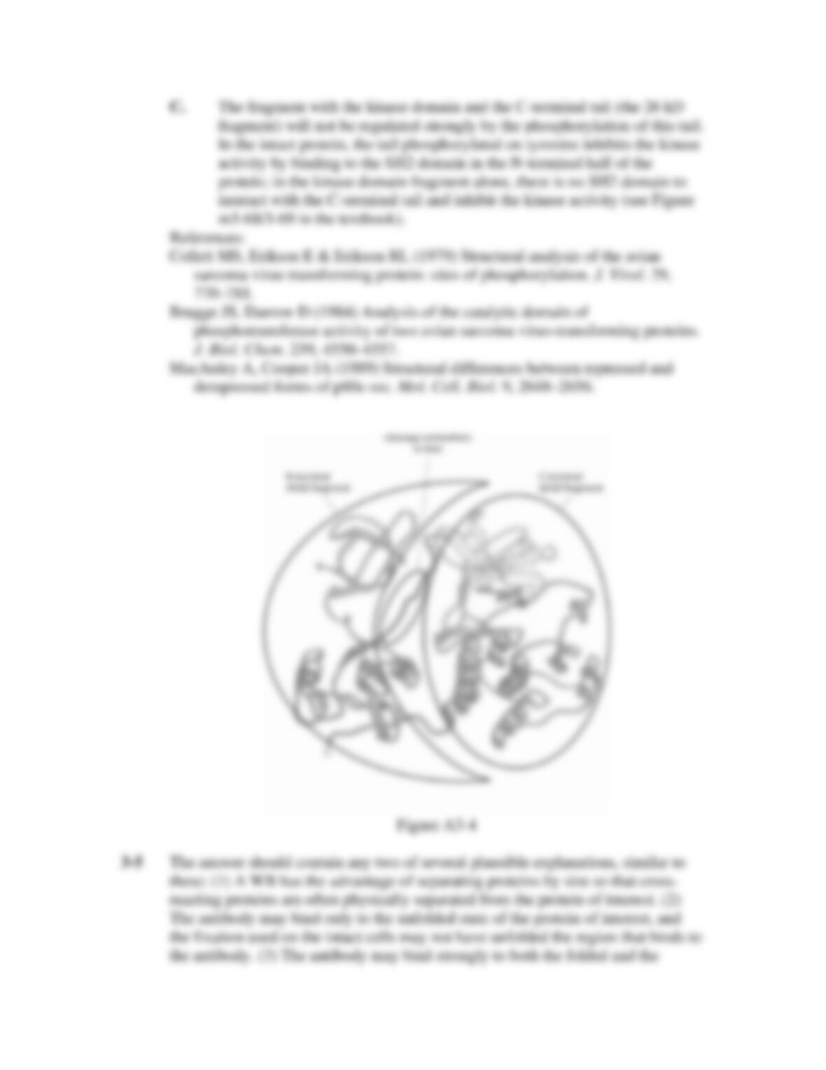
3-4 The Src protein kinase is composed of several domains that interact to control the
behavior of the protein. Figure Q3-4 (A and B) shows the structure of the
repressed conformation of Src. In 1979, long before the x-ray crystal structure of
Src was known, scientists used a technique called partial proteolysis to explore
the structure of Src. Treatment with small amounts of the V8 protease cleaved Src
at a single location to generate two protein fragments. The addition of more V8
protease caused further cleavage of one of the fragments, but in both cases kinase
activity was maintained. Figure Q3-4C shows a protein gel from this experiment.
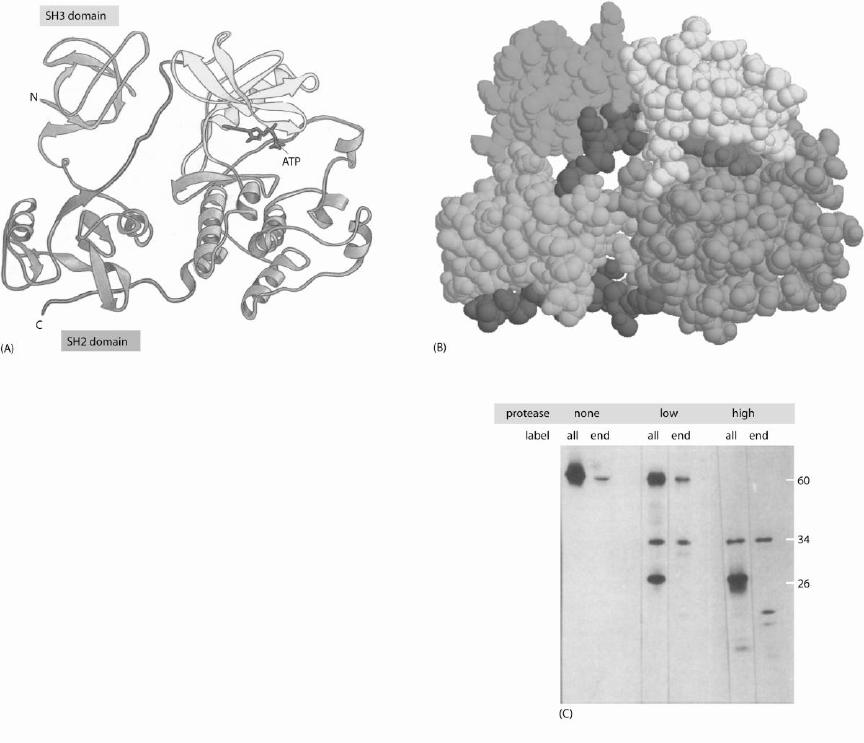
The proteolytic fragments are detected by their radioactivity because the Src
protein contained radioactive atoms; in some reactions Src was radiolabelled
throughout its length (“all”) and in others the radiolabel was only at the N–
terminal end (“end”).
A. Indicate on the ribbon diagram where you think cleavage first occurs to
generate the 34 kD and 26 kD fragments. Circle and label the regions
corresponding to each fragment.
B. Which fragment contains the kinase domain? On the basis of the
regulation of Src explained in the textbook, would you have expected this
fragment to be active as a kinase, as observed? If so, do you expect the
kinase activity be greater than, the same as, or less than that of the intact
protein? Explain.
C. Dephosphorylation, mutation, or deletion of a particular tyrosine residue
in the C-terminal tail of the protein leads to increased activity of the kinase
and promotes cancerous transformation of cells. Do you expect the
proteolytic fragment containing the kinase domain to be regulated strongly
by phosphorylation of the key tyrosine in the C-terminal tail? Explain.
Figure Q3-4
3-5 Antibodies raised against proteins of interest are useful tools for biomedical
research, because they allow the visualization or isolation of individual proteins
from a complex mixture of different proteins. A Western blot (WB) is one method
of visualizing proteins: all proteins are extracted from cells, the proteins are
denatured and separated by size using electrophoresis through a gel, and the
protein of interest is detected by binding to an antibody that is linked to a
fluorescent or luminescent molecule.
Immunofluorescence (IF) provides a way to visualize a particular protein
within a cell using microscopy: the cell is “fixed” (killed and preserved intact),
flooded with an antibody that binds a protein of interest, and washed free of
unbound antibody. Because the antibody is also bound to a fluorescent molecule,
the subcellular location and intensity of the fluorescence indicates the location
and concentration of the protein of interest.
Unfortunately, many antibodies that work well for WB are not useful for
IF. Consider an antibody that yields an intense signal located at the expected
position on a WB. When the WB is performed with cells lacking the protein of
interest, the intense signal is absent but there remain a few faint signals from non-
specific proteins of the wrong size. Yet this antibody is not useful for IF because
in IF the wild-type cells and the cells lacking the protein of interest look the same.
Suggest two possible explanations for the observation that the antibody is more
specific in a WB than in an IF experiment; note that at least five explanations are
possible.
3-6 Immunoprecipitation (IP) is commonly used in biomedical research to examine
whether proteins interact physically with each other. You perform an IP procedure
to determine if a protein called Elm interacts with proteins called Oak and Red.
First, you link an Elm-specific antibody to small beads that settle out of solution
readily. Second, you grind up cells, extract the soluble proteins, add the beads,
and incubate for 2 hours. Third, you centrifuge the mixture to separate the beads
from the unbound proteins. Fourth, you wash the beads three times to remove
weakly bound proteins; to do this, you add a large volume of buffer to the beads,
incubate for 5 minutes, then remove the buffer. Finally, you add detergent to the
beads to denature and dissociate all proteins before separating them on a gel and
revealing them with additional specific antibodies. Because you do not have
antibodies against Oak and Red, you genetically engineer cells in which both of
these proteins are fused to a small protein handle or tag called HA, for which you
purchase specific antibodies. Your gel is shown in Figure Q3-6.
A. Your lab partner Chris looks at your gel and says that you have evidence
that Elm binds directly to Oak and to Red. Do you agree? Is there a
plausible alternative interpretation of your data?
B. Chris says that your data demonstrate that Elm, Oak, and Red form a
three-protein complex. Do you agree? Explain.
C. Assume that you have solid evidence that Elm binds directly to Oak and it
also binds directly to Red. Chris says that Elm has higher affinity for Red
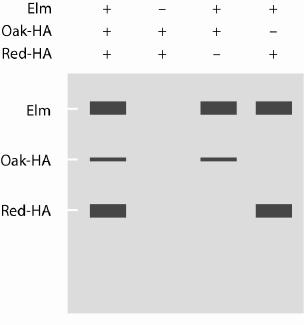
(has a lower Kd) than it has for Oak. Do you agree? Explain, with
reference to an equation for Kd.
Figure Q3-6
3-7 By computational analysis of genome sequences, you discover a new protein
family with members from many different eucaryotic and procaryotic organisms,
but nothing is known about the functions of these proteins. You suspect that these
proteins may share a previously unknown protein fold and thus you wish to
determine the x-ray crystal structure of a member of this family. Because the most
challenging steps in structural determination are the production of very pure,
concentrated protein and the formation of crystals in which protein molecules
pack closely together in a uniform conformation, you need to choose carefully
which member of the family to use for structural experiments. You decide to use a
procaryotic protein, because it is easier to get large amounts of protein. You
narrow the choice to family members from the model organism E. coli, the
pathogenic (disease-causing) organism Yersinia pestis, the thermophilic (heat
loving) organism Thermus aquaticus, and the halophilic (salt loving) organism
Haloquadratum walsbyi. You will overexpress one of these proteins in E. coli
from recombinant DNA. Which one is likely to be easier to purify and to be more
energetically stable and conformationally uniform under the crystallization
conditions? Explain.
3-8 Members of the Mitogen-Activated Protein Kinase (MAPK) family are essential
for many responses to the extracellular environment. A MAPK becomes active
after it becomes phosphorylated by a second kinase. These MAPKs contain a
“docking site” distant from the active site that binds to conserved 10–15 amino
acid “docking motifs” found in a variety of proteins (see Figure Q3-8). Docking
motifs are found in proteins that bind MAPKs (scaffold proteins), proteins that are
substrates of MAPKs, proteins that activate MAPKs by phosphorylating them
(MAPK kinases), and proteins that inactivate MAPKs by dephosphorylating them
(phosphatases). The kinetic properties of a particular MAPK were measured with
a small peptide substrate that contains only the preferred phosphorylation site.
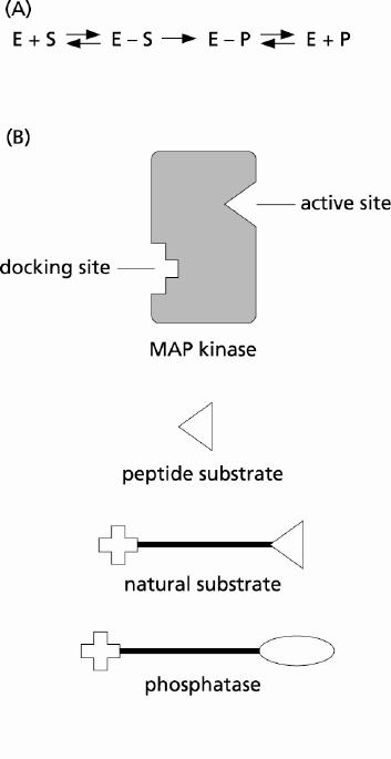
The Km is 300 nM and the kcat is 20/sec. Do you expect the kcat for the natural
protein substrate to be higher or lower than that for the peptide substrate? What
about the Km? Explain, with reference to the reaction shown in panel A below:
Figure Q3-8
Answers