CHAPTER 18
Apoptosis
Questions
18-1 You are working in a laboratory investigating the effects of new chemicals called
X and Y that kill cells. To test whether X and Y cause necrosis or apoptosis, you
briefly treat cultured cells with each chemical and perform several assays.
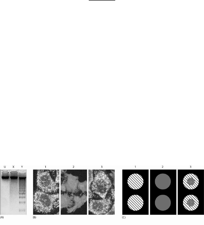
A. First, you examine the chromosomal DNA. You isolate DNA from cells,
label 5 ends with radioactive ATP by using polynucleotide kinase, run the
DNA on a gel, and detect radiolabeled fragments. Your results are shown
in Figure Q18-1A. The lane from the untreated control cells is labeled U.
Does X cause apoptosis? What about Y? Explain the origin of the ladder
in the Y lane.
B. Second, you examine the subcellular localization of cytochrome c using
immunofluorescence microscopy. Unfortunately, during sample
preparation you mixed up the samples. Assign a likely identity (U, X, or
Y) to the samples labeled 1, 2, and 3 in Figure Q18-1B.
C. Third, without making the cells permeable in any way, you stain them
with Annexin V linked to a green fluorophore, which binds
phosphatidylserine, and propidium iodide, which fluoresces red when it
binds DNA. Neither of these compounds can travel across intact plasma
membranes. Again during sample preparation you mixed up the samples.
Assign a likely identity to the samples in Figure Q18-1C. Annexin V
staining is shown as \\ hatching, propidium iodide staining is shown as //
hatching, and staining with both in the same region appears as
crosshatching.
Figure Q18-1
18-2 You want to find out why apoptosis fails to occur in several cell lines derived
from tumors. To identify differences between the tumor cell lines (T1, T2, and
T3) and a control cell line (C) that might prevent tumor cells from undergoing
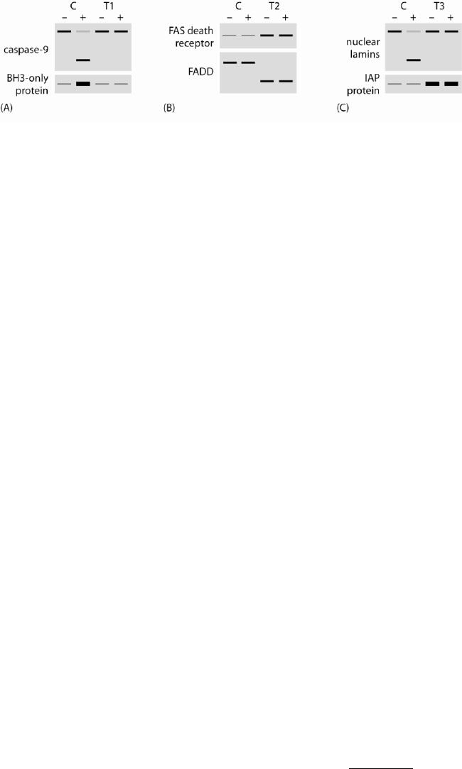
normal apoptosis, you isolate proteins from cells that have (+) or have not (–)
been treated to induce apoptosis. You load the protein samples on gels and
perform immunoblotting with antibodies against proteins involved in apoptosis.
Confusingly, several proteins have altered levels or altered apparent sizes in the
tumor cell lines, as shown in Figure Q18-2. Suggest a hypothesis for each tumor
cell line to explain its apoptosis defect and the immunoblots.
Figure Q18-2
18-3 In patients with autoimmune diseases, the adaptive immune system recognizes
some normal cells or proteins as though they were foreign. Defects in the
regulated apoptosis and clearance of cells are thought to cause or exacerbate some
autoimmune diseases.
A. Explain briefly which cells normally undergo apoptosis and can cause
autoimmune disease if they survive inappropriately.
B. Explain briefly why a defect in clearance or engulfment of apoptotic
bodies might lead to autoimmune disease.
18-4 Cancer cells often exhibit abnormal regulation of the apoptotic program.
Mutations that prevent the normal regulation of apoptosis not only contribute to
carcinogenesis but also affect the response to anticancer therapies.
A. Describe briefly why a defect in apoptosis is an important step in the
transformation of a normal cell into a cancer cell.
B. Describe briefly how a defect in apoptosis will affect the response to
anticancer therapies.
C. Identify a protein whose overexpression in a cancer cell might inhibit
apoptosis.
D. Identify a protein whose elimination or underexpression in a cancer cell
might inhibit apoptosis.
18-5 Effector procaspases acquire high proteolytic activity only after they have been
cleaved by active initiator caspases. In contrast, purified initiator procaspases can
cleave themselves, and this autocatalytic cleavage stimulates their activity only
modestly. Biochemical reconstitution experiments demonstrated that high activity
of the initiator caspase-8 and caspase-9 required their incorporation into the DISC
or apoptosome, respectively. Each of these procaspases has an adaptor domain
(which enables it to assemble with other proteins), a prodomain, a low-affinity
dimerization domain, and a catalytic domain. The catalytic domains of these
caspases are identical at only 34% of the amino acids. Two competing models for
the activation of caspase-9 have been proposed. In the allosteric model, Apaf-1
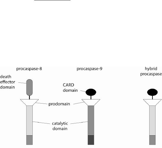
proteins in the apoptosome specifically interact with the catalytic domains of
caspase-9 proteins, thereby triggering a transition to a catalytically active
conformation. In the dimerization model, Apaf-1 proteins simply increase the
local concentration of caspase-9 by binding several adaptor domains
simultaneously, thereby promoting dimerization and coincident activation of
caspase-9. To distinguish between these models, you make a hybrid protein
containing part of caspase-9 and part of caspase-8, as shown in Figure Q18-5.
You test whether this hybrid protein can be activated by Fas ligand or cytochrome
c. What do you expect if the allosteric model is correct? What do you expect if the
dimerization model is correct?
Figure Q18-5
18-6 Several mutational changes to proteins involved in apoptosis are listed below. For
each, indicate whether you expect it to have a dominant-negative phenotype. In
other words, do you expect it to cause a defect in apoptosis even in cells that also
contain the normal protein?
A. Deletion of death domain of Fas death receptor.
B. Mutational change to Fas ligand that prevents its oligomerization.
C. Deletion of the death effector domain of FADD.
D. Deletion of the death effector domain of procaspase-8.
E. Deletion of the CARD domain of Apaf-1.
F. Mutation of the catalytic cysteine of caspase-8 to serine.
G. Mutational change to Bax that prevents its oligomerization with itself and
with Bak.
H. Mutational change to IAP that increases its affinity for caspases.
18-7 A genetic pathway map of the intrinsic pathway of apoptosis is shown in Figure
Q18-7, with empty boxes instead of protein names. Fill in the boxes with the
following proteins: caspase-9, Bcl-2, anti-IAP protein, Apaf-1, cytochrome c,
executioner caspase, BH3-only protein, IAP protein, BH123 protein. A → symbol
indicates activation; –| indicates inhibition.

Figure Q18-7
18-8 You are investigating how the number of nerve cells is regulated in a specific
tissue of your favorite experimental organism. You isolate a mutant that contains
almost none of these nerve cells in the adult animal, and you hypothesize that in
this mutant the nerve cells are either not made or do not connect to their target
cells and therefore fail to get the survival signals they need. You are surprised by
the results of a developmental time course that you perform. You find that in the
mutant more of the nerve cells are made than in controls, and they all connect
correctly to their target cells. But then, at later times, nearly all of the nerve cells
undergo apoptosis. What could be the explanation?
Answers