CHAPTER 17
The Cell Cycle
Questions
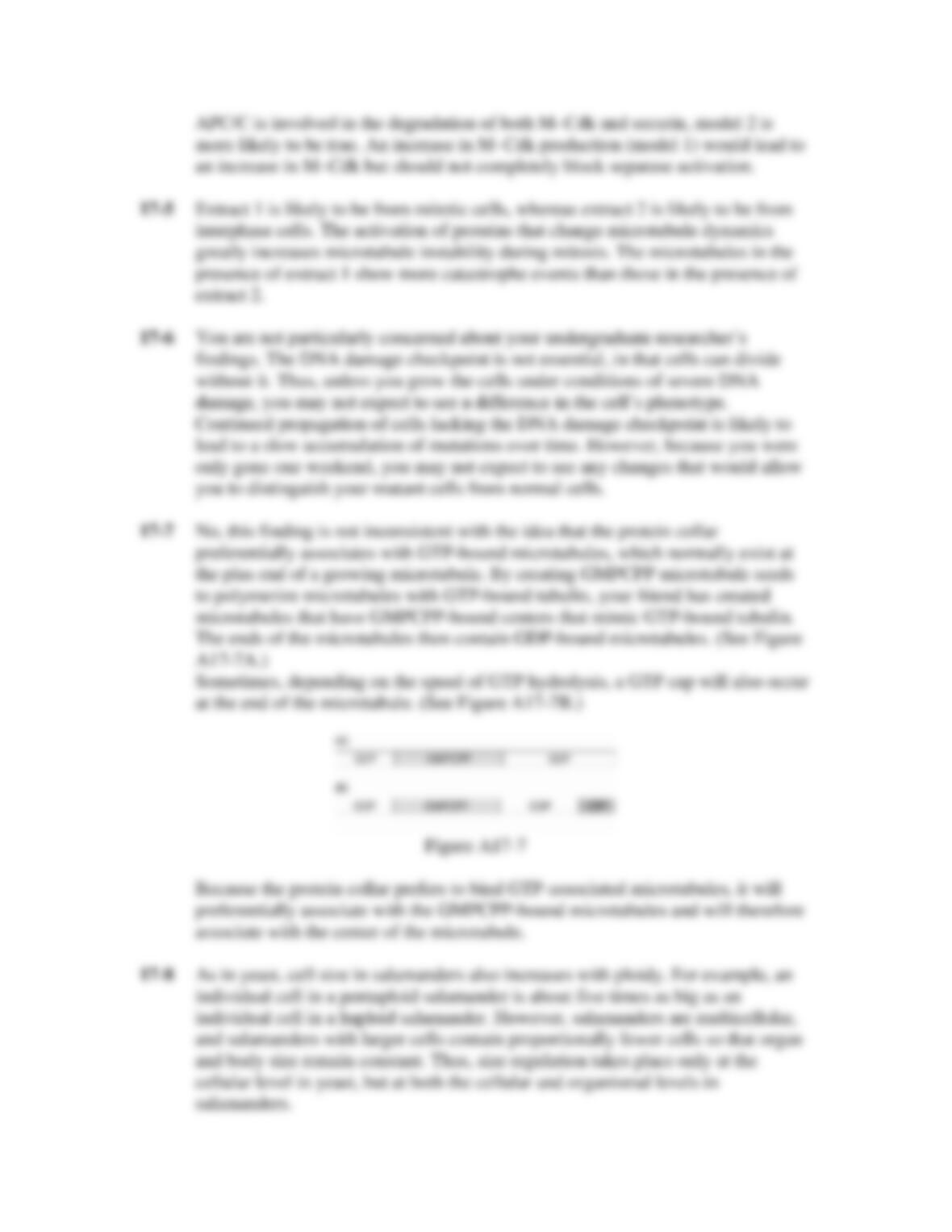
17-1 You want to study the cell cycle in fish embryo cells, in which the cell cycle
length is about 30 min. You examine the amount of M-cyclin in these cells and
see that it varies over the cell cycle. Your friend works in a pharmaceutical
company where they are making drugs that block the cell cycle. She gives you
some of their secret drug, called MI575, and you add it to cells, examine M-
cyclin, and get the results shown in Figure Q17-1.
Figure Q17-1
A. From these data, would you predict that cells treated with MI575 are
arrested in the cell cycle at various stages (G1, S, G2, or M), or do you
predict that cells will be blocked at a particular stage? If you think cells
will be blocked at a particular stage, describe the stage at which you think
the cells would be blocked.
B. From the data, propose a simple molecular explanation for how MI575
acts to block the cell cycle.
17-2 You have isolated a strain of mutant fission yeast that divides normally at 30°C
but arrests in the cell cycle before M phase at 37°C. These mutant yeast cells are
not defective in the production of M–Cdk, because you have isolated the mitotic
cyclin and mitotic Cdk from these mutant yeast and find that both proteins are
normal and can form an M–Cdk complex at both temperatures. Which of the
following types of mutations could be responsible for the behavior of this strain of
yeast? Explain.
A. Inactivation of an enzyme that ubiquitylates M–cyclin.
B. Inactivation of the Wee1 kinase.
C. Inactivation of the CAK kinase.
D. The continuous production of a phosphatase that removes all phosphate
groups from the M–Cdk.
17-3 Your friend works in a lab that studies origin licensing. He is particularly
interested in the pre-replicative complex (pre-RC) and has isolated a temperature-
sensitive yeast mutant that does not seem to assemble the pre-RC at the origins of
replication. However, he has gotten into an argument with a new student in the
lab. The student thinks that this yeast mutant will arrest in late mitosis or early G1,
because that is when the pre-RC is normally assembled. Your friend disagrees.
Who is right, and why?
17-4 You have identified a drug, ID555, that causes M–Cdk protein levels to remain
high in the cell. You have two favorite models for how ID555 works.
Model 1: ID555 activates a transcription factor that stimulates the genes encoding
M–cyclin and M–Cdk, so that excess M–Cdk protein is produced.
Model 2: ID555 inhibits the activity of the Cdc20–APC/C complex that normally
targets M–Cdk for destruction.
Your friend works in a lab with many fancy microscopes and offers to examine
cells that are treated with drug. She sees that cells treated with ID555 assemble a
mitotic spindle and their chromosomes seem to align at the metaphase plate.
However, the chromosome segregation seen during normal anaphase does not
occur. Does this new information cause you to favor model 1 or model 2?
Explain.
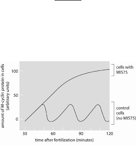
17-5 Your friend comes to you in a panic. He was purifying extracts from interphase
cells as well as mitotic cells. Unfortunately, the labels came off his tubes and he
cannot tell which extract is from which cells. You do an experiment in which you
add a small amount of each extract to fluorescent microtubules you have
polymerized in vitro, and then use video microscopy to follow the behavior of
individual microtubules in the reaction over time. Your results are shown in the
graphs in Figure Q17-5.
Figure Q17-5
Which extract do you think is from mitotic cells and which from interphase cells?
Why?
17-6 You have just read a paper that the addition of caffeine to cells disrupts the DNA
damage checkpoint and you would like to understand why. By searching for
proteins that bind to caffeine, you isolate a protein, Caf1. You believe that Caf1 is
involved in the DNA damage checkpoint because when you delete Caf1 from
yeast cells and add a drug that causes DNA damage, the cells fail to arrest in the
cell cycle.
You give your Caf1-deleted cells to an undergraduate in the laboratory and ask
him to take care of the strain for you while you take the weekend off to celebrate
your finding. Upon your return, you find the poor undergraduate in tears. He
explains that he thinks that he messed up the Caf1 mutant strain while you were
gone because after growing Caf1 and wild-type cells in rich media and examining
them during mitosis, Caf1 mutants looked the same the same as wild-type cells.
Are you concerned about the undergraduate student’s findings? Why?
17-7 Chromosomes are attached to microtubules via the kinetochore. Given the
organization of the mitotic spindle, chromosomes should be attached to the plus
ends of the kinetochore microtubules. Your friend is interested in studying the
protein collar in the kinetochore that is thought to bind to the growing end of a
microtubule. He conducts the following experiment. First, he creates stable
microtubules in the presence of GMPCPP, a non-hydrolyzable analogue of GTP.
Next, he adds GTP-tubulin to the GMPCPP microtubules. Finally, he adds the
kinetochore protein collar to the microtubules to see where they bind. He finds a
large proportion of the protein collars binding to the middle of microtubules and
very few binding to the ends of the microtubules. Is this finding inconsistent with
this protein being part of the kinetochore? Explain.
17-8 Multicellular animals have some mechanism to control cell mass that we do not
understand; for example, salamanders of different ploidies are the same size such
that a haploid, a diploid, and a pentaploid organism are about the same size.
However, a yeast cell differs in size according to ploidy, such that a diploid yeast
cell is about half the size of a tetraploid yeast cell. Given these observations,
explain why yeast and salamanders are not so different.
Answers