CHAPTER 12
Intracellular Compartments and Protein Sorting
Questions
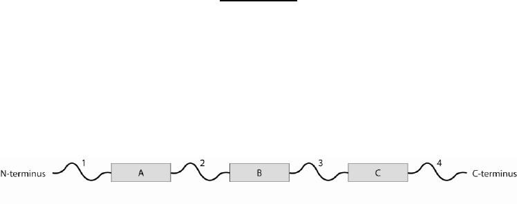
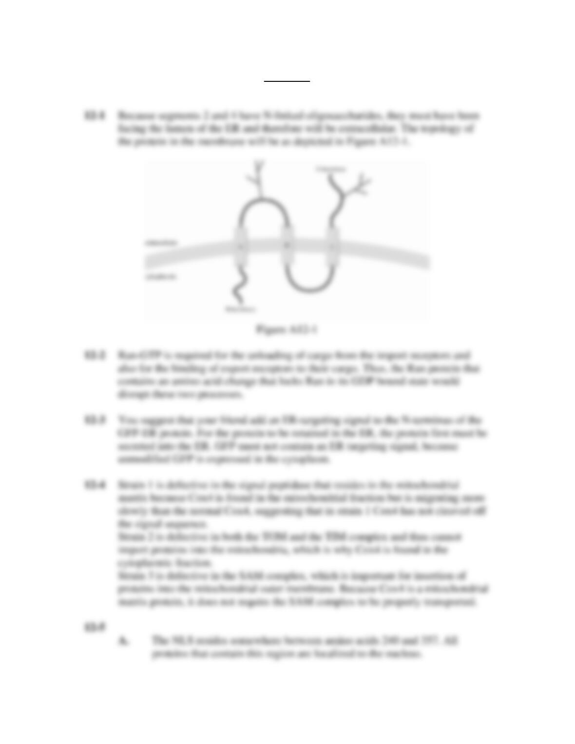
12-1 You are studying a protein that normally resides in the plasma membrane. The
organization of this protein is depicted in Figure Q12-1, where the gray boxes
labeled A, B, and C represent transmembrane regions and the segments labeled 1,
2, 3, and 4 represent the portions of the protein between the transmembrane
regions.
Figure Q12-1
If you also know that segments 2 and 4 contain N-linked oligosaccharides, draw
the topology of the protein as it is inserted in the plasma membrane. Be sure to
label the cytoplasmic and extracellular face of your membrane and the N-terminus
and C-terminus of your protein.
12-2 If you replace the normal Ran protein in the cell with a Ran protein that has an
amino acid change locking Ran in a GDP-bound state, how will this affect nuclear
import and export?
12-3 Your friend is interested in targeting green fluorescent protein (GFP) to the ER.
He decides to create a modified version of GFP that he calls GFP-ER by adding
the ER retention signal (as described in Table 12-3 of the textbook) Lys-Asp-Glu-
Leu to the C-terminus of GFP. He sees that GFP-ER is expressed in cells and that
GFP-ER is four amino acids longer than the unmodified GFP. However, to his
surprise, even though GFP-ER contains an ER retention signal, it is in the
cytoplasm of the cell, just like the unmodified GFP. He comes to you for help in
fixing this problem. What do you suggest he should do?
12-4 The graduate student from the lab next door comes to you in a panic. She had
received three different yeast strains from a lab in Japan, and in the process of
transferring these strains to plates she mixed them up. She knows that she
received three strains:
(A) one defective in the SAM complex,
(B) one defective in both the TOM complex and the TIM23 complex, and
(C) one defective in the signal peptidase that exists in the mitochondrial matrix.
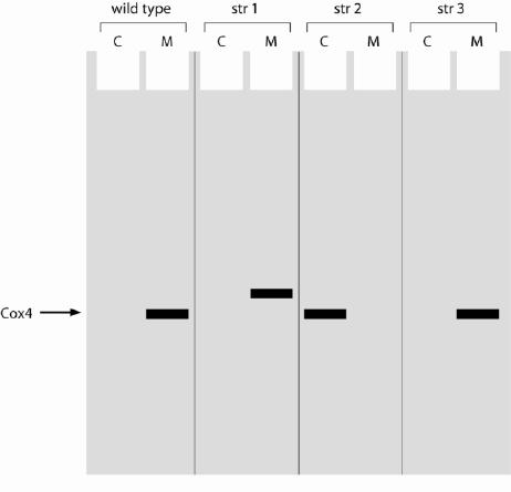
Fortunately, you have antibodies against Cox4, a subunit of cytochrome c
oxidase. Cox4 normally resides in the mitochondrial matrix and is encoded by a
nuclear gene. You conduct some experiments in which you fractionate wild-type
cells and the three unknown strains, and then, for each strain, subject the
cytoplasmic fraction and the mitochondrial fraction to electrophoresis on an SDS
gel. Your results are depicted in Figure Q12-4, in which C indicates the
cytoplasmic fraction and M the mitochondrial fraction.
Figure Q12-4
From these results, identify which strain contains which mutation, and explain
your answers.
12-5 Your friend shows you her data. She has been analyzing a protein to determine
which parts of the protein are responsible for its localization. She has created
various deletion mutations in her protein and examined its localization, under
normal conditions and also in the presence of leptomycin B, an inhibitor of the
nuclear export machinery. Her data are depicted in Figure Q12-5.
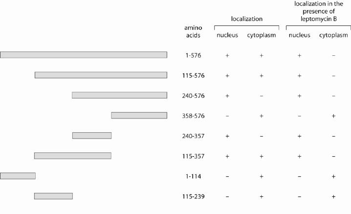
Figure Q12-5
From her data, answer the following questions:
A. Where do you think the nuclear localization signal (NLS) is located?
Explain.
B. Where do you think the nuclear export signal (NES) is located? Explain.
12-6 Your friend sends you two tubes of protein. One tube contains a protein, Crg1,
that contains a nuclear export signal. The other tube contains the nuclear export
receptor, Xpo1, for Crg1. You are excited to have received these proteins, because
you are interested in the biochemistry of nuclear export. You try to get Crg1 to
bind Xpo1 but do not succeed. You complain to your lab partner about how your
friend sent you bad reagents. However, your lab partner has just taken a cell
biology class, and gently reminds you to try one more experiment before you call
your friend. What does your labmate suggest?
12-7 The thylakoid space in chloroplasts contains proteins important for
photosynthesis. Your friend is interested in understanding how proteins get to the
thylakoid space, and is examining the import of the PC protein, a protein that is
encoded by the nucleus and normally resides in the thylakoid space of the
chloroplast. He has defined a 60 amino acid sequence at the N-terminus of the PC
protein that is necessary and sufficient for import into the chloroplast: this 60
amino acid sequence can direct the import of GFP into the chloroplast and the
deletion of these 60 amino acids on PC makes the PC protein localize to the
cytoplasm. Furthermore, all 60 amino acids seem to be required, because tests
using fewer than the 60 amino acids do not show these properties. Excited about
these results, your friend takes the intact PC protein and changes residues 55–60
by replacing the each of the original amino acids with the amino acid alanine, and
calls this mutation PC-ala. He decides to express PC-ala in cells, expecting it to be
cytoplasmic. To his surprise, this protein still goes to the chloroplast, but it does
not enter the thylakoid space. From your understanding of transport to the
thylakoid space, suggest a hypothesis to explain why your friend’s mutants
specifically disrupt this transport.
12-8 State whether the following proteins are being translated on ribosomes attached to
the ER, and explain your answer.
A. A protein that resides in the chloroplast.
B. A protein that resides in an endosome.
C. A protein that will be secreted into the extracellular space.
D. A nuclear protein.
E. A protein linked to the plasma membrane by a GPI anchor.
F. A protein that resides between the inner and outer nuclear membranes.
Answers