CHAPTER 10
Membrane Structure
Questions
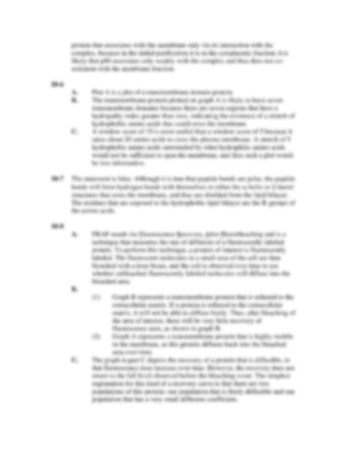
10-1 You are interested in studying the composition of lipid bilayers and how they are
maintained. You discover two uncharacterized phospholipids, which you call
PLX and PLZ. You decide to characterize the behavior of PLX and PLZ by
labeling the head group of each phospholipid. This label is stable when the lipid
resides in the membrane’s outer leaflet but unstable when the lipid resides in the
membrane’s inner leaflet. You incorporate labeled versions of PLX and PLZ into
either the inside or the outside of the cell, and monitor the change in signal
intensity of these lipids in the plasma membrane. Your data are presented in the
graphs in Figure Q10-1.
Figure Q10-1
A. Where in the plasma membrane are PLX and PLZ normally located?
B. Are there flippases in the cell for either of these phospholipids? Why?
10-2 You are interested in studying lipid rafts. You have devised a method to create an
artificial lipid bilayer that contains small patches of lipids that are straighter and
longer than those found in the rest of the bilayer. How would you expect the
fluidity of these small patches to compare with the rest of the lipid bilayer?
Explain.
10-3 It is thought that excess lipids in the cell are packaged into lipid droplets in the
endoplasmic reticulum. Explain why lipid droplets are surrounded by a
phospholipid monolayer and not a phospholipid bilayer, like other vesicles that
bud from the endoplasmic reticulum.
10-4 Your friend has isolated plasma membranes and reassembled the membranes into
small vesicles. Using fluorescently labeled lectin, he sees that some of his vesicles
are fluorescently labeled and some are not. Recall that lectin binds to
carbohydrates. Furthermore, his labeled lectin cannot permeate membranes.
A. Which population of vesicles has a surface similar to that of the cell?
Why?
B. How do you explain the other population of vesicles?
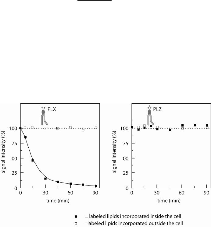
10-5 Your friend is working on a protein that he calls p125, because of its molecular
mass. He knows that p125 is a transmembrane protein with three membrane-
spanning domains. It has been previously reported that p125 interacts with three
proteins called p175, p80, and p50 (again, on the basis of their apparent sizes on
an SDS polyacrylamide gel). These four proteins are thought to exist as a protein
complex in the cell. To determine how these proteins interact with the membrane,
you perform a set of experiments in which you first lyse the cells and save some
of your lysate, which you run in the input lane (labeled “I” in Figure Q10-5). The
lysate is then subjected to a low-speed centrifugation so that you separate out the
membrane fraction (which ends up in the pellet, “P”) from the cytoplasm (which
is in the supernatant, “S”). You then wash the pellet from the first extraction with
a high-salt wash that does not disrupt the lipid bilayer, and save a little bit to run
on the gel. After the high-salt wash, you centrifuge the pellet again. Your results
are illustrated on the gel in Figure Q10-5. From these data, explain the nature of
the association of these proteins with cellular membranes.
Figure Q10-5
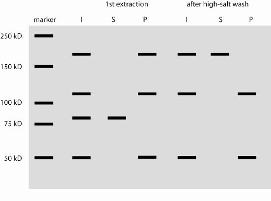
10-6 Examine the two hydropathy plots in Figure Q10-6.
Figure Q10-6
A. Which is a plot of a protein that contains -helices that cross the
membrane?
B. For the protein containing a transmembrane domain, how many
transmembrane domains would you predict there are? Why?
C. The plots in Figure Q10-6 were created with a “window size” of 19. In
creating a hydropathy plot, a value is assigned to an amino acid based on
how hydrophobic it is deemed to be. The window size determines the
number of amino acids over which a hydrophobicity value is averaged.
Once a hydrophobicity value is calculated for a particular window, a
hydrophobicity value is then calculated starting at the next amino acid for
the next window. Explain why a “window size” of 19 is more useful for
calculating a hydrophobicity plot for examining transmembrane proteins
as opposed to a smaller “window size” such as 5.
10-7 Is the following sentence true or false? Explain.
Peptide bonds are polar and thus must be covalently modified before proteins can
be inserted into the membrane.
10-8 Your friend is examining the mobility of transmembrane proteins using FRAP.
A. Explain what FRAP stands for and how it is performed.
B. Figure Q10-8A and B depicts two typical graphs your friend has obtained
from her FRAP studies. Which of these graphs would best represent the
following types of proteins? Explain your reasoning.
(1) A transmembrane protein that is tethered to the extracellular matrix.
(2) A transmembrane protein that is highly mobile in the membrane.
C. Your friend has also obtained a third graph from her experiments, shown
in Figure Q10-8C. Explain how you might get a reading such as this from
a FRAP experiment examining transmembrane proteins.
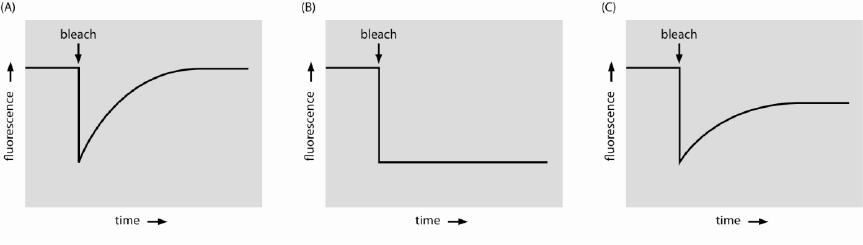
Figure Q10-8
Answers