Lecture Learning Objectives for Unit 1 – BIO 210 (fall 2016)
(Adapted from Julie Haugsness-White)
Martini, Timmons, and Tallitsch, 8th edition
****Please note that you are expected to review LLOs #1-4 (from your prerequisite biology
course) on your own. There are biology and anatomy books in the library (Campbell’s
General Biology or Garrett’s Getting Ready for A & P).
REVIEW OF BIOCHEMISTRY AND CYTOLOGY (from your prerequisite course)
TEXT READING– Chapter 2
1. List the four main classes of large biological molecules, their respective
monomers/components, and their major functions in the human body.
Carbohydrates(polymer): an example is fat and the functions are cell membranes and
energy storage. Lipids(monomer): two examples are starch and sugar and their functions
are energy store and structure. Proteins(polymer): an example is trypsin and the function
would be cell machinery. Nucleic acids(polymer): two examples are DNA and RNA and the
function is to store genetic material.
2. Learn the name and specific function of each cellular organelle listed in Table 2.1.
Cytoskeleton(Microtubule/Microfilament): strengthen and support; movement of cellular
structures and materials. Microvilli: increase surface area to facilitate absorption of
extracellular materials. Centrosome: essential for movement of chromosomes during cell
division; organization of microtubules in cytoskeleton. Cilia: movement of materials over
cell surface. Ribosomes: protein synthesis. Mitochondria: produce 95% of ATP required
by the cell. Nucleus: control of metabolism; storage and processing of genetic information;
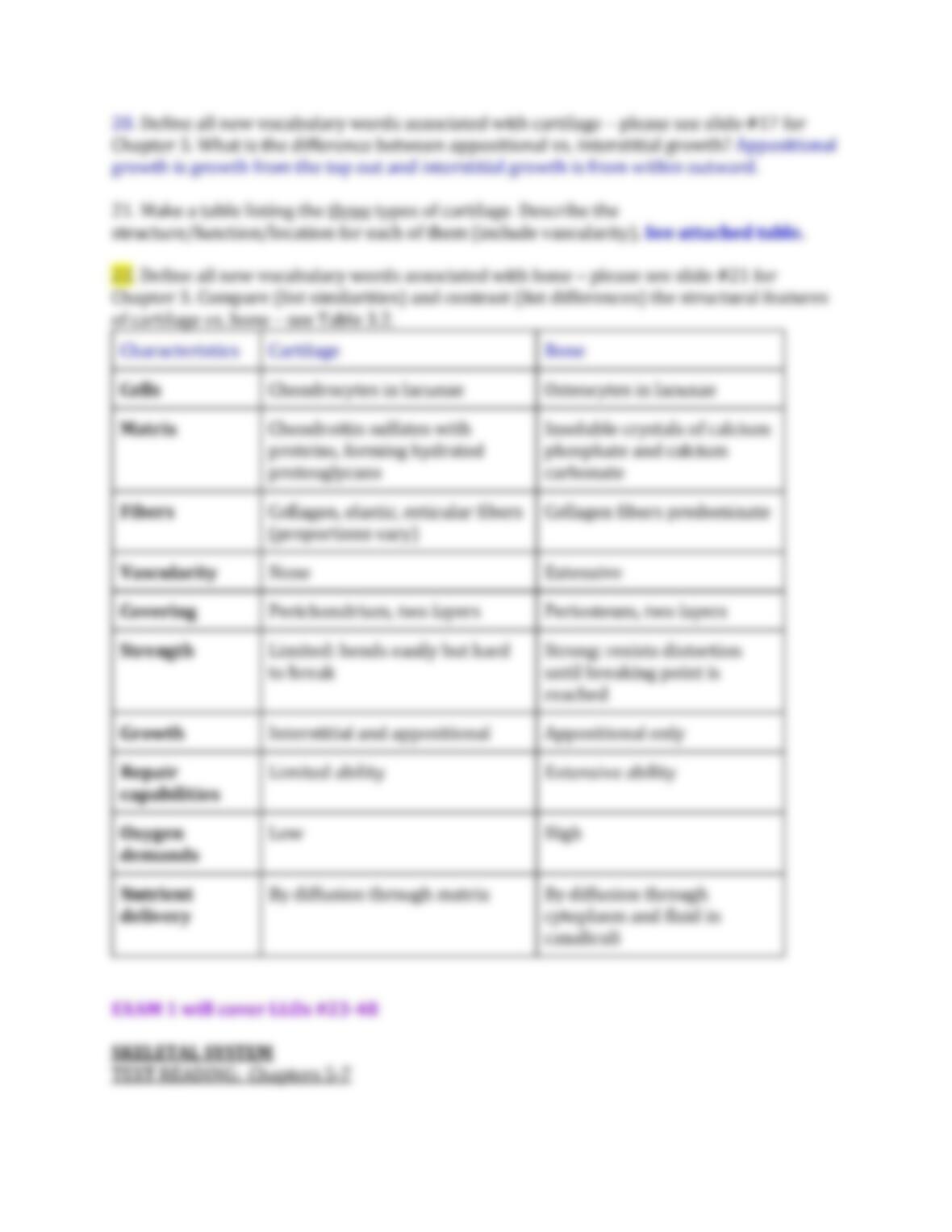
control of protein synthesis; site of rRNA synthesis and assembly of ribosomal subunits.
Endoplasmic reticulum (ER): Synthesis of secretory products; intercellular storage and
transport; modification and packaging of newly synthesized proteins; lipid, steroid, and
carbohydrate synthesis; calcium ion storage. Golgi apparatus: storage, alteration, and
packaging of secretory products and lysosomal enzymes. Lysosome: intracellular removal
of damaged organelles or of pathogens. Peroxisome: catabolism(breakdown complex to
simple) of fats and other organic compounds; neutralization of toxic compounds generated
in the process.
3. Describe the structure/function of the plasma membrane with respect to the transport
mechanisms listed in Figure 2.4 (hint – why is it selectively permeable – why are transport
processes required?). Hydrophobic tail and hydrophilic head: ions needs protein to go
through. Selectively permeable because what a certain amount of things coming in. Also,
polar and charged ions need transport to get across.
Diffusion allows molecular movement of solutes and the direction of which is determined
by relative concentrations(from high to low). The rate is affected by the magnitude of the
concentration gradient; size, molecular size, charge, lipid protein solubility, and
temperature which affect the rate. Osmosis(water only) permits movement of water
(solvent) molecules toward higher solute concentrations. The rate is affected by the size of
the solute concentration gradient and opposing pressure. Facilitated diffusion allows the
carrier molecules transport materials down a concentration gradient and requires
membrane. Consists of concentration gradient, opposing pressure and the availability of
carrier protein. Active transport: permit the carrier molecules work despite opposing
concentration gradients and consist of the availability carrier, substrate and ATP.
Endocytosis: allows the formation of membranous vesicles (endosomes) containing fluid
or solid material at the plasmalemma. Consisting of stimulus and mechanism and requires
ATP. Exocytosis permits the fusion of vesicles containing fluids and/or solids with the
plasmalemma. Which consists of stimulus and mechanism and requires ATP and calcium
ions.
4. Which organelle would you expect to be abundant in a muscle cell? In a pancreatic beta
cell (which secretes the protein, insulin)? In a cell found in the ovaries or testes (which
secretes a sex hormone)? Mitochondria in a muscle cell because you need ATP for the
contraction of the overall muscle, and mitochondria synthesize ATP. Rough endoplasmic
reticulum in abundance in a pancreatic beta cell (secretes insulin). Smooth endoplasmic
reticulum abundant in a cell found in the ovaries or testes (secretes sex hormones).
NEW MATERIAL STARTS HERE
5. Compare and contrast the structure/function/location of the three major types of
intercellular junctions (tight junction, desmosome, gap junction).
No quiz ? Intercellular junctions: Communicating/gap junction: function-
”channel”(rigid signaling: electrical+chemical), structure-membrane proteins, location-
cardiac muscle tissue. Adhering junctions: tight and anchoring. Tight junction: function:
“belt”(diffusion barrier to prevent leakage), structure-membrane proteins, location-
epithelial tissue(stomach). Anchoring junction: function-”rivet”(resist tearing +
stretching), structure-desmosome and membrane proteins, location-epithelial
tissue(stomach). Gap junction: connects cytoplasm together, in heart, rapid signals
chemical and electrical.
Foundations – An Introduction to Anatomy
TEXT READING – Chapter 1
6. Define the following terms: microscopic anatomy (cytology Study of cells vs. histology
Study of tissue); gross anatomy (surface anatomy vs. regional anatomy vs. systemic
anatomy); homeostasis. Microscopic anatomy: need a microscope to see it. Cytology:
study of cells. Histology: study of tissue. Gross anatomy: can see with the unaided eye.
Surface anatomy: study of external anatomical forms and markings . Regional anatomy:
an approach to anatomic study based on regions, parts, or divisions of the body, emphasis
of the relationships of various systemic structures with that area. Systemic anatomy:
study of specific organ system. Homeostasis: balance of inner body, ambience.
7. List and define the levels of organization in the human body and give examples of each
(atom, molecule, organelle, cell, tissue, organ, organ system).
Organism level: multicellular organism composed of 5 levels of organization, an example
would be a human. Organ system: a group of organs that work together to enact one or
more functions; example-cardiovascular system, which includes the heart, the blood, and
blood vessels. Organ: part of an organism that is self-contained and has a specific vital
function such as the heart, which is a three-dimensional organ. Tissue: specialized material
made of cells, an example would be cardiac muscle tissue. Cell: smallest unit of life that can
replicate independently, an example would be heart muscle cells. Organelle: “small
organs,” which are the metabolic machinery of the cell, and that are highly organized to
carry out specific functions for the cell as a whole, an example would be the nucleus.
Molecule: a group of atoms joined by chemical bonds, an example would be H2O, when
two hydrogen atoms combine with one oxygen atom. Atom: smaller particles, an example
would be the element carbon, C.
8. Describe anatomical position while standing vs. lying down (prone or supine). The
supine position is a position of the body lying with the face up, the dorsal side is down and
the ventral side is up. While, prone position is a position with the face down; dorsal side up
and the ventral side is down.
9. Use directional terminology to describe the relative position of body parts (see Lab
Activity #1).