Species-Specific PCR aids Bacterial Identification of Coagulase-Negative Staphylococci
Jessica Cyr
Emporia State University
Cyr 1
Key Words: Coagulase-negative staphylococci, multiplex primers, superoxide dismutase (sodA), species-
specific PCR
Introduction: Microbial forensics faces any classification of crime with microorganisms or biological
influences. Before we can combat biological crimes, bacteria must be reliably identified. Staphylococcus
aureus is a coagulase-positive staphylococci that is viewed as the core for bacterial pathogens leading to
sickness and disease. As more research evolves, common coagulase-negative staphylococci species have
been shown to be the cause of other bacterial diseases. Identifying the origin of these pathogens is
crucial for preventative measures and treatments. Some coagulase-negative staphylococci include S.
epidermidis, S. capitis, S.caprae, and S. warneri. More coagulase-negative staphylococci need to be
identified and more information is needed about their behavior. There is also a wide variety within the
species totaling 74 sequencing types (Otto, 2009). In forensic microbiology, we need to identify these
rarely studied species for scientists to be able to discern between them. In humans, DNA is almost
identical but there can be variability among its microbial flora. There are many approaches to coagulase-
negative staphylococci identification. Some other techniques include: matrix-assisted laser desorption
ionization time-of-flight mass spectrometry (MALDI-TOFMS), API Staph test, and whole-genome DNA-
DNA hybridization analysis (Kim, 2018). Yet, these approaches are either too costly, too labor intensive
or have a lower identification rate. 16S rRNA is a common method for bacterial identification. However,
the 16S rRNA PCR has limitations between differentiating species that are too similar. It also needs
further analyses for clear identification within the Staphylococcus genus (Blaoitta, 2005). The goal of my
research is to reliably identify bacteria, which is incapable using 16S. Due to high similarity in genotypes,
S. pasteuri and S. warneri are two species in a conserved area that are difficult to distinguish. Superoxide
dismutase (sodA) is a gene used in species-specific PCR for genotypic identification. Kim (2018)
experiments further after species-specific PCR including DNA sequencing and homology comparisons. To
be able to differentiate these species, I used multiplex primers and species-specific PCR to confirm the
presence of each strain (Kim, 2018).
Materials and Methods: DNA was isolated using whole-cell polymerase chain reaction (PCR) with
freshly grown cells on agar. Samples were obtained, prior to this study, from random DNA skin cells
swabbed from the inner elbow, behind the ear, or on the forehead and stored at –80⁰ C. Once taken out
of storage, the samples were plated onto liquid broth plates and grown overnight at 37⁰ C. A DNA
colony collected from the agar plate and was mixed into a microfuge tube with 100 μL of TE buffer. Once
mixed 5 μL of sample was put in each GoTaq tube, as well as 5 μL of primer and primer mixes. GoTaq
consists of heat-stable DNA Polymerase, dNTP’s, dye, buffer and MgCl2. Primer sequences were specific
to each species based on the sodA gene, listed below. The stock concentration for the primers was 2.5
mmol. When the primers or primer mixes were added to the reaction, it totaled a concentration of 0.25
mmol. The primers and mixes are listed below show which primers bind to the correlating species. For
initial samples with 16S rRNA PCR, 1% agarose gels were used to observe the DNA samples showing the
presence of DNA with 100-bp DNA markers. DNA sequencing was completed by an outside facility,
Molecular Cloning Laboratories (MCLAB). After reviewing chromatogram peaks and base pairs, the
edited sequences of the samples were put into basic local alignment search tool (BLAST) analysis for
identification. Higher-percent agarose gels are used to observe differences in species that are within 50-
100 bp of each other based on their amplicon size. Amplicon size of tested species was found in Table 2
of Improved multiplex PCR primers for rapid identification of coagulase-negative staphylococci (Kim,
2018). 2% agarose gels were used when observing samples that were tested in species-specific PCR with
Cyr 2
multiplex primers. 50–bp DNA markers were used in 2% agarose gels, as well as corresponding positive
controls (pure DNA) for S. epidermidis, S. pasteuri and S. warneri. No positive control was used during
testing for S. capitis and S. caprae due to unavailability. The agarose gels were observed under the
ultraviolet light transilluminator. Samples identified as S. pasteuri and S. warneri by 16S rRNA PCR used
the STAPH-1 PCR conditions below. Samples identified as S. epidermidis, S. capitis, and S. caprae by 16S
rRNA PCR used STAPH-2 PCR conditions below.
Agarose gel recipe
1%: 0.3g of agarose, 30mL of TAE buffer, 2 μL of ethidium bromide
(EtBr) mixed into an Erlenmeyer flask, microwaved until dissolved and
cool.
2%: 0.7g of agarose, 30mL of TAE buffer, 2 μL of ethidium bromide
(EtBr) mixed into an Erlenmeyer flask, microwaved until dissolved and
cool.
Amplicon size (bp)
16S: 1500
S. epidermidis: 194
S. capitis: 103
S. caprae: 252
S. warerni: 110
S. pasteuri: 237
LB agar plate recipe
Add 11 g/mL of agar and 10 g/mL of LB Broth, Lennox to 500 mL of H2O. Autoclave, cool and pour into
plates.
Primer sequences from Integrated DNA Technologies
8F for 16S: 5’AGA GTT TGA CTC AG 3’
1492R (L) for 16S: 5’ GGT TAC CTT GTT ACG ACT T 3’
164 CT103R S. capitis: 5’ CTA CTT CAC CTT TTT CTT CAG A 3’
165 CT103F S. capitis: 5’ TCA GAT ATT CAA ACT GCA GTA G 3’
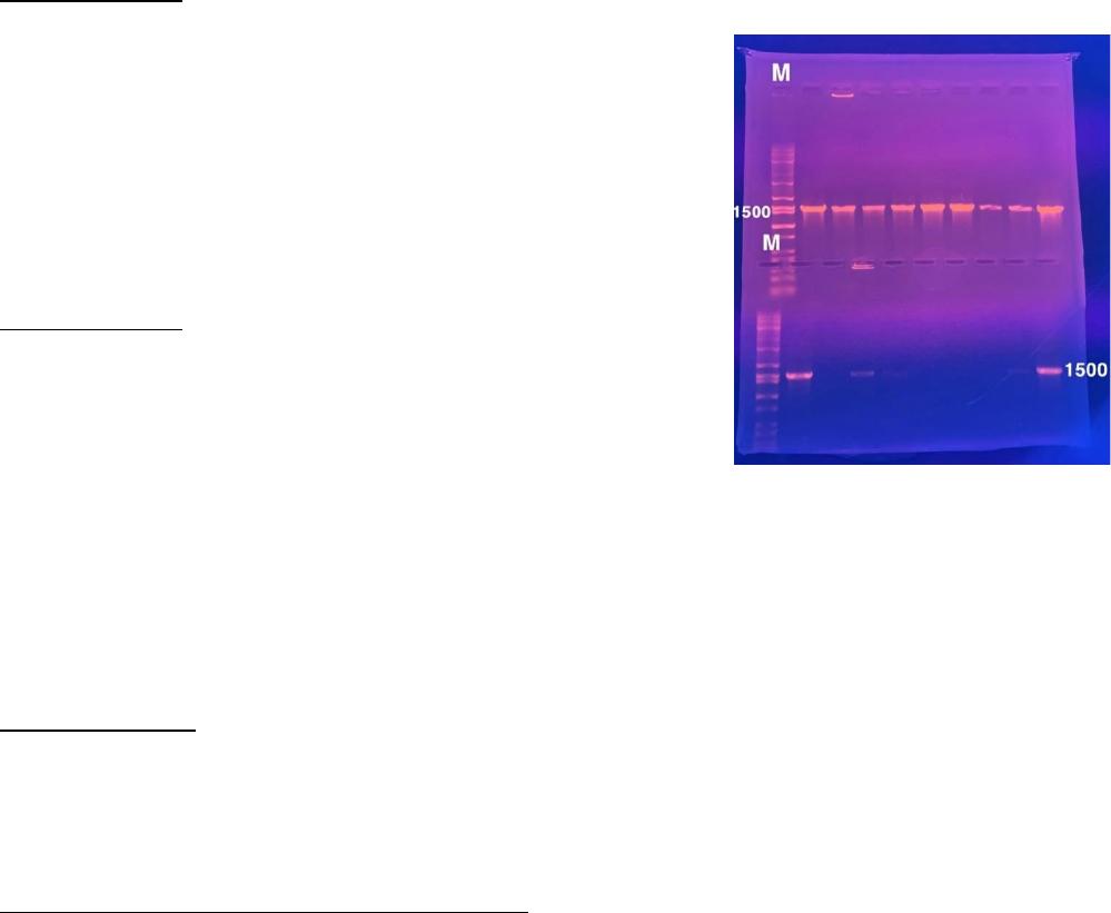
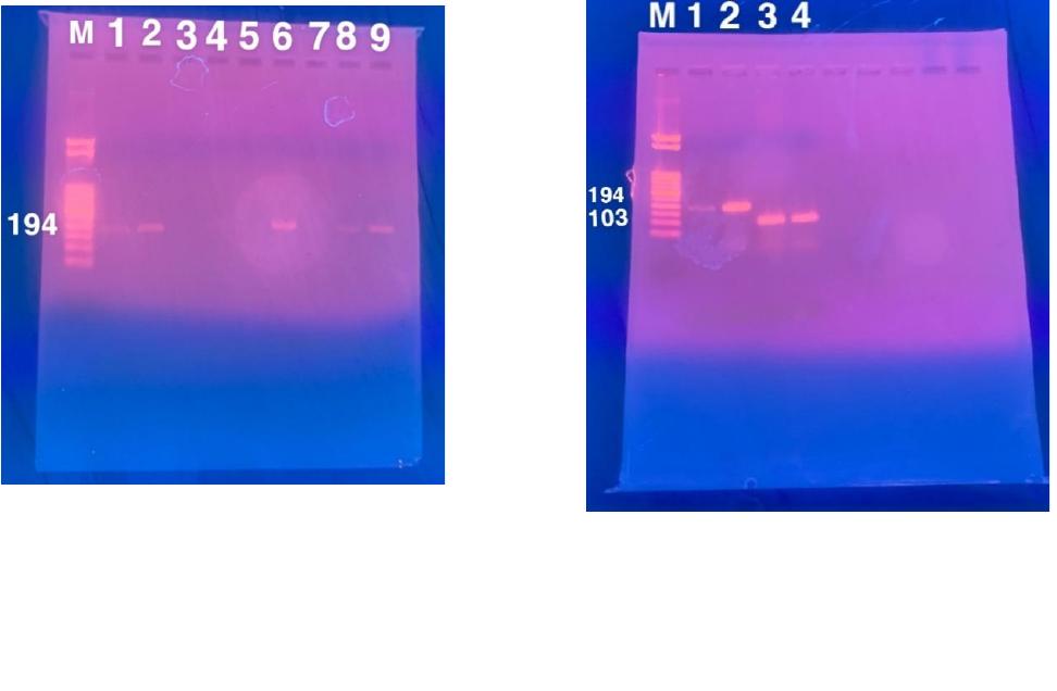
Figure 1: 1% agarose gel of 16S rRNA showing
initial presence of DNA samples alongside
DNA markers (M).

Cyr 3
166 EpiR S. epidermidis: 5’ TGG CTA ATG GTT TGT CAC CA 3’
167 EpiF S. epidermidis: 5’ GGC AAA TTT GTG GGT CAA GA 3’
172 SW110R S. warneri: 5’ TCT TAC TGC AGT TTG AAT ATC AGA 3’
173 SW110F S. warneri: 5’ GTA ACA AAA TTA AAT GCA GCT G 3’
174 PA237R S. pasteuri: 5’ GCC CGT TAT TTA CTA CTA ACC A 3’
175 PA237F S. pasteuri: 5’ GCT AAT TTA GAC AGT GTA CCT TCT G 3’
PCR conditions for 16S rRNA primer
Cycle 1: 94⁰ C for 1 minute
Cycle 2: 94⁰ C for 1 minute
Cycle 3: 50⁰ C for 1 minute
Cycle 4: 72⁰ C for 1 minute and 30 seconds
Cycle 5: Go back to step 2 and repeat cycles 2-4 29 times until moving to cycle 6
Cycle 6: 72⁰ C for 5 minutes
Cycle 7: 12⁰ C hold indefinitely
PCR conditions for STAPH-1 with 172,173,174,175 primer mix
Cycle 1: 95⁰ C for 5 minutes
Cycle 2: 95⁰ C for 30 seconds
Cycle 3: 55⁰ C for 30 seconds
Cycle 4: 72⁰ C for 30 seconds
Cycle 5: Go back to step 2 and repeat cycles 2-4 29 times until moving to cycle 6
Cycle 6: 72⁰ C for 30 seconds
Cycle 7: 12⁰ C hold indefinitely
PCR conditions for STAPH-2 with 164, 165, 166,167 and 166,167 primer mix
Cycle 1: 95⁰ C for 5 minutes
Cycle 2: 95⁰ C for 30 seconds
Cycle 3: 55⁰ C for 30 seconds
Cycle 4: 72⁰ C for 30 seconds
Cycle 5: Go back to step 2 and repeat cycles 2-4 29 times until moving to cycle 6
Cycle 6: 72⁰ C for 7 minutes
Cycle 7: 12⁰ C hold indefinitely
Cyr 4
Results and Discussion:
Figure 1 above is an example of positive and negative results for presence of bacteria using 16S. The
fluoresced bands show DNA in the agarose gel. Below are the top four results 16S BLAST analysis of
JC362. As you can see the percent identity is 100% for both S. warneri and S. pasteuri.
Staphylococcus warneri strain DK131 16S ribosomal RNA gene, partial sequence
439 439 100% 2e-119 100.00% MT642942.1
Staphylococcus pasteuri strain ML029 16S ribosomal RNA gene, partial sequence
439 439 100% 2e-119 100.00% MT636753.1
Staphylococcus pasteuri strain BCVME2 16S ribosomal RNA gene, partial sequence
439 439 100% 2e-119 100.00% MT598013.1
Staphylococcus warneri strain 513 16S ribosomal RNA gene, partial sequence
439 439 100% 2e-119 100.00% MT573107.1
There could be presence of both species in the sample’s skin
cells, but we want a reliable confirmation if there is only one
species since they are commonly misidentified as each other.
The advantages of whole-cell PCR include: it is more cost-
efficient than DNA extraction from a kit, it is quick and dirty to
plate on the agar plates, and easily isolated when replated
again. Blaiotta et al. (2004) sought after species-specific PCR
while also confirming identification, but they used singleplex
PCR rather than multiplex PCR. S. epidermidis, S. capitis, and S.
caprae are also misidentified species of coagulase-negative
staphylococci. Ghebremedhin, Layer, Konig, & Konig establish
that the sodA gene found 94% similarity between S. capitis and
S. caprae (2008). Table 1 shows the data regarding the
identified species. The 16S rRNA PCR column shows which
bacterial identification sequenced with BLAST that 16S could
do. Species-specific PCR in the next column shows the
superiority it has over 16S for reliable identification. With the
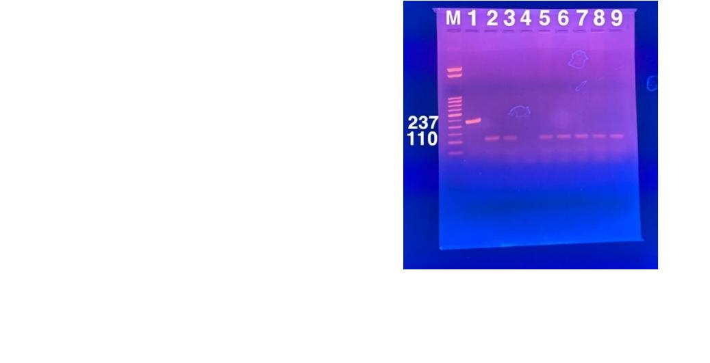
use of the 172,173,174,175 multiplex primers, S. warneri was
identified in samples JC362, JC126, JC131, JC135, JC137, and
JC140, seen in Figure 2. In Figure 2, lane 1 is the positive control for S. pasteuri, lane 2 is the positive
control for S. warneri and the samples that fluoresced show the identification of S. warneri. Previously
identified S. epidermidis samples from 16S: JC361, JC367, JC373, JC374, JC376, JC377, JC401, JC404,
JC407, JC411, JC412, JC413, JC414, JC417, JC421, JC121, JC122, and JC124 were confirmed as S.
epidermidis with the use of the 166,167-multiplex primers in PCR, shown in Figure 3. The presence of
bands in Figure 3 confirm the bacterial identification as S. epidermidis. Sample JC110 was identified as S.
epidermidis with 164, 165, 166, and 167 multiplex primers. Samples JC111 and JC113 were identified as
S. capitis with 164, 165, 166, and 167 multiplex primers. These positive results can be seen in Figure 4.
Although there were no positive controls for S. capitis or S. caprae, the amplicon size was used for S.
capitis and there were no S. caprae samples that were present at 252 bp. Unconfirmed species using
species-specific PCR were not visually present on agarose gels but were tested twice. There could be a
Figure 2: 2% Agarose gel electrophoresis. M: DNA markers; 1:
S.pasteuri positive control; 2: S. warneri positive control; 3:
JC362 S. warneri; 4: JC365 Unconfirmed; 5: JC126 S. warneri; 6:
JC131 S. warneri; 7: JC135 S. warneri; 8: JC137 S. warneri; 9:
JC140 S. warneri
Cyr 5
variety of reasons why a sample that was tested in PCR does not work. Whole-cell PCR does not have
the purest of DNA being used, experimental errors can occur, and some DNA samples are more difficult
to grow and test. 16S can still provide a baseline of identification that can be further confirmed. The
positive results show that 2% agarose gel electrophoresis with multiplex species-specific PCR can
confirm and differentiate previously identified coagulase-negative staphylococci from 16S rRNA PCR and
my research goal was achieved. Reliable identification of our microbial flora makes it possible to find the
variability from person to person if there is variability between species.
Figure 3: 2% Agarose gel electrophoresis. M: DNA
markers; 1: S. epidermidis positive control; 2: JC
401 S. epidermidis; 3: JC403 unconfirmed; 4: JC404
S. epidermidis; 5: JC405 unconfirmed; 6: JC407 S.
epidermidis; 7: JC411 S. epidermidis; 8: JC412 S.
epidermidis; 9: JC413 S. epidermidis
Figure 4: 2% Agarose gel electrophoresis. M: DNA
markers; 1: S. epidermidis positive control; 2: JC 110
S. epidermidis; 3: JC111 S. capitis; 3: JC113 S. capitis

Cyr 6
Samples
16S RNA PCR
Species-Specific PCR
JC361
S. epidermidis
S. epidermidis
JC362
S. warneri/pasteuri
S. warneri
JC363
S. epidermidis
Unconfirmed
JC365
S. warneri/pasteuri
Unconfirmed
JC366
S. epidermidis
Unconfirmed
JC367
S. epidermidis
S. epidermidis
JC373
S. epidermidis
S. epidermidis
JC374
S. epidermidis
S. epidermidis
JC376
S. epidermidis
S. epidermidis
JC377
S. epidermidis
S. epidermidis
JC401
S. epidermidis
S. epidermidis
JC403
S. epidermidis
Unconfirmed
JC404
S. epidermidis
S. epidermidis
JC405
S. epidermidis
Unconfirmed
JC407
S. epidermidis
S. epidermidis
JC411
S. epidermidis
S. epidermidis
JC412
S. epidermidis
S. epidermidis
JC413
S. epidermidis
S. epidermidis
JC414
S. epidermidis
S. epidermidis
JC417
S. epidermidis
S. epidermidis
JC421
S. epidermidis
S. epidermidis
JC110
S. epidermidis/capitis
S. epidermidis
JC111
S. capitis/caprae/sp.
S. capitis
JC113
S. capitis/sp.
S. capitis
JC121
S. epidermidis
S. epidermidis
JC122
S. epidermidis
S. epidermidis
JC124
S. epidermidis
S. epidermidis
JC126
S. warneri/pasteuri
S. warneri
JC131
S. warneri/pasteuri
S. warneri
JC135
S. warneri/pasteuri
S. warneri
JC137
S. warneri/pasteuri
S. warneri
JC140
S. warneri/pasteuri
S. warneri
Table 1: Comparison of identification with 16S RNA PCR and species-specific PCR

Cyr 7
References:
Blaiotta, G., Casaburi, A., & Villani, F. (2005). Identification and differentiation of Staphylococcus
carnosus and Staphylococcus simulans by species-specific PCR assays of sodA genes. Systematic and
Applied Microbiology, 28(6), 519–526. doi: 10.1016/j.syapm.2005.03.007
Budowle, B. (2003). PUBLIC HEALTH: Building Microbial Forensics as a Response to Bioterrorism. Science,
301(5641), 1852–1853. doi:10.1126/science.1090083
Ghebremedhin, B., Layer, F., Konig, W., & Konig, B. (2008). Genetic Classification and Distinguishing of
Staphylococcus Species Based on Different Partial gap, 16S rRNA, hsp60, rpoB, sodA, and tuf Gene
Sequences. Journal of Clinical Microbiology, 46(3), 1019–1025. doi:10.1128/jcm.02058-07
Kim, J., Hong, J., Lim, J.-A., Heu, S., & Roh, E. (2017). Improved multiplex PCR primers for rapid
identification of coagulase-negative staphylococci. Archives of Microbiology, 200(1), 73–
83. doi:10.1007/s00203-017-1415-9
Kiratisin, P., Li, L., Murray, P. R., & Fischer, S. H. (2003). Identification of Bacteria Recovered from Clinical
Specimens by 16SrRNA Gene Sequencing. European Journal of Clinical Microbiology & Infectious
Diseases, -1(1), 1–1. doi:10.1007/s10096-003-1003-6
Mignard, S., & Flandrois, J. P. (2006). 16S rRNA sequencing in routine bacterial identification: A 30-month
experiment. Journal of Microbiological Methods, 67(3), 574–581. doi: 10.1016/j.mimet.2006.05.009
Otto, M. (2009). Staphylococcus epidermidis — the “accidental” pathogen. Nature Reviews Microbiology,
7(8), 555–567. doi:10.1038/nrmicro2182
Cyr 8
Appendix A:
The following results were obtained by 16S rRNA PCR and BLAST analysis:
JC361
Raw Sequence:
NNNNNNNNNNNNNNNGNGNNNNTACNTGCAGTCGAGCGAACAGACGAGGAGCTTGCTCCTCTGACGTTAGCG
GCGGACGGGTGAGTAACACGTGGATAACCTACCTATAAGACTGGGATAACTTCGGGAAACCGGAGCTAATACCG
GATAATATATTGAACCGCATGGTTCAATAGTGAAAGACGGTTTTGCTGTCACTTATAGATGGATCCGCGCCGCATT
AGCTAGTTGGTAAGGTAACGGCTTACCAAGGCAACGATGCGTAGCCGACCTGAGAGGGTGATCGGCCACACTGG
AACTGAGACACGGTCCAGACTCCTACGGGAGGCAGCAGTAGGGAATCTTCCGCAATGGGCGAAAGCCTGACGGA
GCAACGCCGCGTGAGTGATGAAGGTCTTCGGATCGTAAAACTCTGTTATTAGGGAAGAACAAATGTGTAAGTAAC
TATGCACGTCTTGACGGTACCTAATCAGAAAGCCACGGCTAACTACGTGCCAGCAGCCGCGGTAATACGTAGGTG
GCAAGCGTTATCCGGAATTATTGGGCGTAAAGCGCGCGTAGGCGGTTTTTTAAGTCTGATGTGAAAGCCCACGGC
TCAACCGTGGAGGGTCATTGGAAACTGGAAAACTTGAGTGCAGAAGAGGAAAGTGGAATTCCATGTGTAGCGGT
GAAATGCGCAGAGATATGGAGGAACACCAGTGGCGAAGGCGACTTTCTGGTCTGTAACTGACGCTGATGTGCGA
AAGCGTGGGGATCAAACAGGATTAGATACCCTGGTAGTCCACGCCGTAAACGATGAGTGCTAAGTGTTNGGGGT
TTCCGCCCCTTAGTGCTGCAGCTAACGCATTAAGCACTCCGCCTGGGAGTACGACCGCAAGGTTGAAACTCAAAG
GGAATTGACGGGGACCCGCACAAGCGGTGGNGCATGTGGTTTAATTTCGAAGCAACGCGAANAANNTTANCNAA
ATCTTGACATCCTCTGACCCTCTAGAGATAGAGTTTTCCCCTTCGGGGGANNGANTGACNNNNNNGCATGNNNN
NTCGTCAGCTCNNGTCGNGGANATNNNNNNNNNTCNNNNNNCGANNNCNNNNNNNNNCTNNNNNNCNTCN
TTAANNNNNNNNCTNTAANNTNNNCNNNNNNNNNN
Edited Sequence:
GGTTCAATAGTGAAAGACGGTTTTGCTGTCACTTATAGATGGATCCGCGCCGCATTAGCTAGTTGGTAAGGTAACG
GCTTACCAAGGCAACGATGCGTAGCCGACCTGAGAGGGTGATCGGCCACACTGGAACTGAGACACGGTCCAGAC
TCCTACGGGAGGCAGCAGTAGGGAATCTTCCGCAATGGGCGAAAGCCTGACGGAGCAACGCCGCGTGAGTGATG
AAGGTCTTCGGATCGTAAAACTCTGTTATTAGGGAAGAACAAATGTGTAAGTAACTATGCACGTCTTGACGGTACC
TAATCAGAAAGCCACGGCTAACTACGTGCCAGCAGCCGCGGTAATACGTAGGTGGCAAGCGTTATCCGGAATTAT
TGGGCGT
Staphylococcus epidermidis strain HP01 16S ribosomal RNA gene, partial sequence
691 691 100% 0.0 100.00% MT586029.1
Staphylococcus epidermidis strain 3039 16S ribosomal RNA gene, partial sequence
691 691 100% 0.0 100.00% MT613456.1
Staphylococcus epidermidis strain ISP111A 16S ribosomal RNA gene, partial sequence
691 691 100% 0.0 100.00% MT605366.1
Staphylococcus epidermidis strain ISP111B 16S ribosomal RNA gene, partial sequence
691 691 100% 0.0 100.00% MT605363.1
Cyr 9
Alignment: Query: None Query ID: lcl|Query_16603 Length: 382
>Staphylococcus epidermidis strain HP01 16S ribosomal RNA gene, partial
sequence
Sequence ID: MT586029.1 Length: 815
Range 1: 113 to 494
Score:690 bits(764), Expect:0.0,
Identities:382/382(100%), Gaps:0/382(0%), Strand: Plus/Plus
Query 1 GGTTCAATAGTGAAAGACGGTTTTGCTGTCACTTATAGATGGATCCGCGCCGCATTAGCT 60
||||||||||||||||||||||||||||||||||||||||||||||||||||||||||||
Sbjct 113 GGTTCAATAGTGAAAGACGGTTTTGCTGTCACTTATAGATGGATCCGCGCCGCATTAGCT 172
Query 61 AGTTGGTAAGGTAACGGCTTACCAAGGCAACGATGCGTAGCCGACCTGAGAGGGTGATCG 120
||||||||||||||||||||||||||||||||||||||||||||||||||||||||||||
Sbjct 173 AGTTGGTAAGGTAACGGCTTACCAAGGCAACGATGCGTAGCCGACCTGAGAGGGTGATCG 232
Query 121 GCCACACTGGAACTGAGACACGGTCCAGACTCCTACGGGAGGCAGCAGTAGGGAATCTTC 180
||||||||||||||||||||||||||||||||||||||||||||||||||||||||||||
Sbjct 233 GCCACACTGGAACTGAGACACGGTCCAGACTCCTACGGGAGGCAGCAGTAGGGAATCTTC 292
Query 181 CGCAATGGGCGAAAGCCTGACGGAGCAACGCCGCGTGAGTGATGAAGGTCTTCGGATCGT 240
||||||||||||||||||||||||||||||||||||||||||||||||||||||||||||
Sbjct 293 CGCAATGGGCGAAAGCCTGACGGAGCAACGCCGCGTGAGTGATGAAGGTCTTCGGATCGT 352
Query 241 AAAACTCTGTTATTAGGGAAGAACAAATGTGTAAGTAACTATGCACGTCTTGACGGTACC 300
||||||||||||||||||||||||||||||||||||||||||||||||||||||||||||
Sbjct 353 AAAACTCTGTTATTAGGGAAGAACAAATGTGTAAGTAACTATGCACGTCTTGACGGTACC 412
Query 301 TAATCAGAAAGCCACGGCTAACTACGTGCCAGCAGCCGCGGTAATACGTAGGTGGCAAGC 360
||||||||||||||||||||||||||||||||||||||||||||||||||||||||||||
Cyr 10
Sbjct 413 TAATCAGAAAGCCACGGCTAACTACGTGCCAGCAGCCGCGGTAATACGTAGGTGGCAAGC 472
Query 361 GTTATCCGGAATTATTGGGCGT 382
||||||||||||||||||||||
Sbjct 473 GTTATCCGGAATTATTGGGCGT 494
JC362
Raw Sequence:
NNNNNNNNGGNNGCNNNTANTGCAGTCGAGCGACAGATAAGGAGCTTGCTCCTTTGACGTTAGCGGCGGACG
GGTGAGTAACACGTGGATAACCTACCTATAAGACTGGGATAACTTCGGGAAACCGGAGCTAATACCGGATAACAT
ATTGAACCGCATGGTTCAATAGTGAAAGGCGGCTTTGCTGTCACTTATAGATGGATCCGCGCCGTATTAGCTAGTT
GGTAAGGTAACGGCTTACCAAGGCAACGATACGTAGCCGACCTGAGAGGGTGATCGGCCACACTGGAACTGAGA
CACGGTCCAGACTCCTACGGGAGGCAGCAGTAGGGAATCTTCCGCAATGGGCGAAAGCCTGACGGAGCAACGCC
GCGTGAGTGATGAAGGTCTTCGGATCGTAAAACTCTGTTATCAGGGAAGAACAAATGTGTAAGTAACTGTGCACA
TCTTGACGGTACCTGATCAGAAAGCCACGGCTAACTACGTGCCAGCAGCCGCGGTAATACGTAGGTGGCAAGCGT
TATCCGGAATTATTGGGCGTAAAGCGCGCGTAGGCGGTTTTTTAAGTCTGATGTGAAAGCCCACGGCTCAACCGT
GGAGGGTCATTGGAAACTGGAAAACTTGAGTGCAGAAGAGGAAAGTGGAATTCCATGTGTAGCGGTGAAATGCG
CAGAGATATGGAGGAACACCAGTGGNGAAGGCGACTTTCTGGNCTGTAACTGACGCTGATGTGCGAAAGCGTGG
GGATCAACAGGGATTAGATACCCTGGNAGTCCACGNCGTAAACGATGANTGCTAGNGTNGGGGGTTTCNGCCCC
TTANNGCTGCAGCTAACGCATTNAGCNNCTCCGNCTGGGNAGTACGACNNNNGGNNGANNTCANNNNNTGAC
GGGGACCNGCNNNGCGGTGNAGCNTNNNNNNNNNNNNAANNNANNNNAANNANNNNNNNATCTTGNNNN
NNNNGACCNNNNNNNNNAANNNNAGNTNNNNNNNNNNGNNNNNNNNNNNGNNNNTGNNNNCNGNNN
Edited Sequence:
GGGAGGCAGCAGTAGGGAATCTTCCGCAATGGGCGAAAGCCTGACGGAGCAACGCCGCGTGAGTGATGAAGGT
CTTCGGATCGTAAAACTCTGTTATCAGGGAAGAACAAATGTGTAAGTAACTGTGCACATCTTGACGGTACCTGATC
AGAAAGCCACGGCTAACTACGTGCCAGCAGCCGCGGTAATACGTAGGTGGCAAGCGTTATCCGGAATTATTGGGC
GTAAAGCGCGCGTAGGCGG
Staphylococcus warneri strain DK131 16S ribosomal RNA gene, partial sequence
439 439 100% 2e-119 100.00% MT642942.1
Staphylococcus pasteuri strain ML029 16S ribosomal RNA gene, partial sequence
439 439 100% 2e-119 100.00% MT636753.1
Staphylococcus pasteuri strain BCVME2 16S ribosomal RNA gene, partial sequence
439 439 100% 2e-119 100.00% MT598013.1
Staphylococcus warneri strain 513 16S ribosomal RNA gene, partial sequence
439 439 100% 2e-119 100.00% MT573107.1
Cyr 11
Alignment1:
Query: None Query ID: lcl|Query_57565 Length: 243
>Staphylococcus pasteuri strain ML029 16S ribosomal RNA gene, partial
sequence
Sequence ID: MT636753.1 Length: 943
Range 1: 269 to 511
Score:439 bits(486), Expect:2e-119,
Identities:243/243(100%), Gaps:0/243(0%), Strand: Plus/Plus
Query 1 GGGAGGCAGCAGTAGGGAATCTTCCGCAATGGGCGAAAGCCTGACGGAGCAACGCCGCGT 60
||||||||||||||||||||||||||||||||||||||||||||||||||||||||||||
Sbjct 269 GGGAGGCAGCAGTAGGGAATCTTCCGCAATGGGCGAAAGCCTGACGGAGCAACGCCGCGT 328