Jenna Costa Deedy
NATURE’S ULTIMATE SUPER
POWER
How Animal Perceive the World
Jenna Costa Deedy
Rivier University
April 11th, 2017
Sensation and Perception
Is it true that it’s possible
that we could actually tell how
animals see? Or what if it was
actually possible to move behind
the lenses of animals in order to
see the world through their own
eyes? Well, what if I told you
that it’s really possible that we
can be able to perceive the
world through the eyes of
animals like dogs, dolphins,
parrots, and snakes. This is
because while many animals
have adapted to develop vision
that would enable them to see the world through their own perspective, only a very few can be
able to do so in the same way as people do (Sinclair, 1, 1985). In the animal kingdom alone,
vision is considered to be the most important link to their world. For example, some animals,
such as sharks and bears, may be more dependent on their sense of smell, touch or taste, while
others such as lions, elephants, primates, and owls, based on the scale of the nature of
evolution, rely more and more on their eyes as they serve the only direct opening of the central
nervous system to the world (Sinclair, 1, 1985). It’s really no surprise that from the extraordinary
diversity of animal life, nature has no doubt, experimented with all of its living creations in
order to give each and every one of them a chance to survive in all sorts of climate and terrains
across the globe.
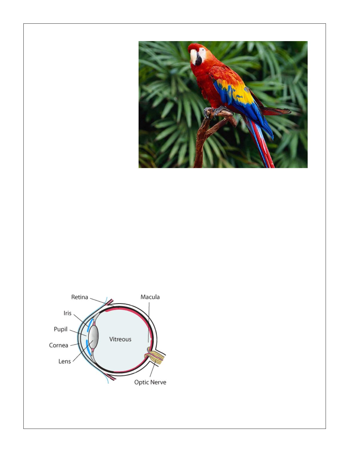
To understand how animals perceive the world through vision, we first must understand
the inner workings of the eye itself. Keep in
mind that nature has evolved different ways
of forming images that would enable us
humans to perceive our own world. However,
let’s start with the human eye to see how it
enables people to perceive their own world.
First, the light rays have to be bent so that
they could focus directly on the retina.
Second, the curved cornea bends light into
the eyes while the lens changes shape to
bring everything into focus (Museum of
Vision, 2, 1997). Once light reaches the retina, it is then picked up by the millions of cones and

rods that would then
convert these light
waves into color,
shape, and motion,
which is then sent to
the brain through the
optic nerve. From
there, the retina
begins to code visual
information that would allow one to recognize objects, people, and places (Museum of Vision,
2, 1997). For animals, however, they have three different ways to perceive their own world. The
first is a camera-like eye that can bring images into focus on the retina. The second is the
compound eye, which is found in insects, crustaceans, and mollusks. It’s a fixed focus eye that
has individual refractive units called ommatidias, which are all responsible for a portion of the
visual field (Sinclair, 1, 1985). Dragonflies for example, have as many as 28,000 individual
ommatidias, given that they have an almost spherical field of vision. In the case of compound
eyes, lens and corneas are one and light that are focused by the many small lenses on individual
photoreceptors. The third type of eye scans like TV cameras, building pictures for successive
impression. It’s the least common form of vision (Sinclair, 1, 1985).
In nature, color is probably one of the most
obvious and pervasive qualities for all animals who
live in their environment (Goldstein, Brockmole,
195, 2017). For us people, we use color to help use
note traffic lights, choose outfits that are color
coordinate, or be able to appreciate the colors of
paintings. In animals, particularly mammals, only a
very few non-human animal species have
developed the ability to have color vision,
particularly, Tasmanian Devils, crocodiles, rabbits, rats, monkeys, apes, pigeons, cats, and dogs
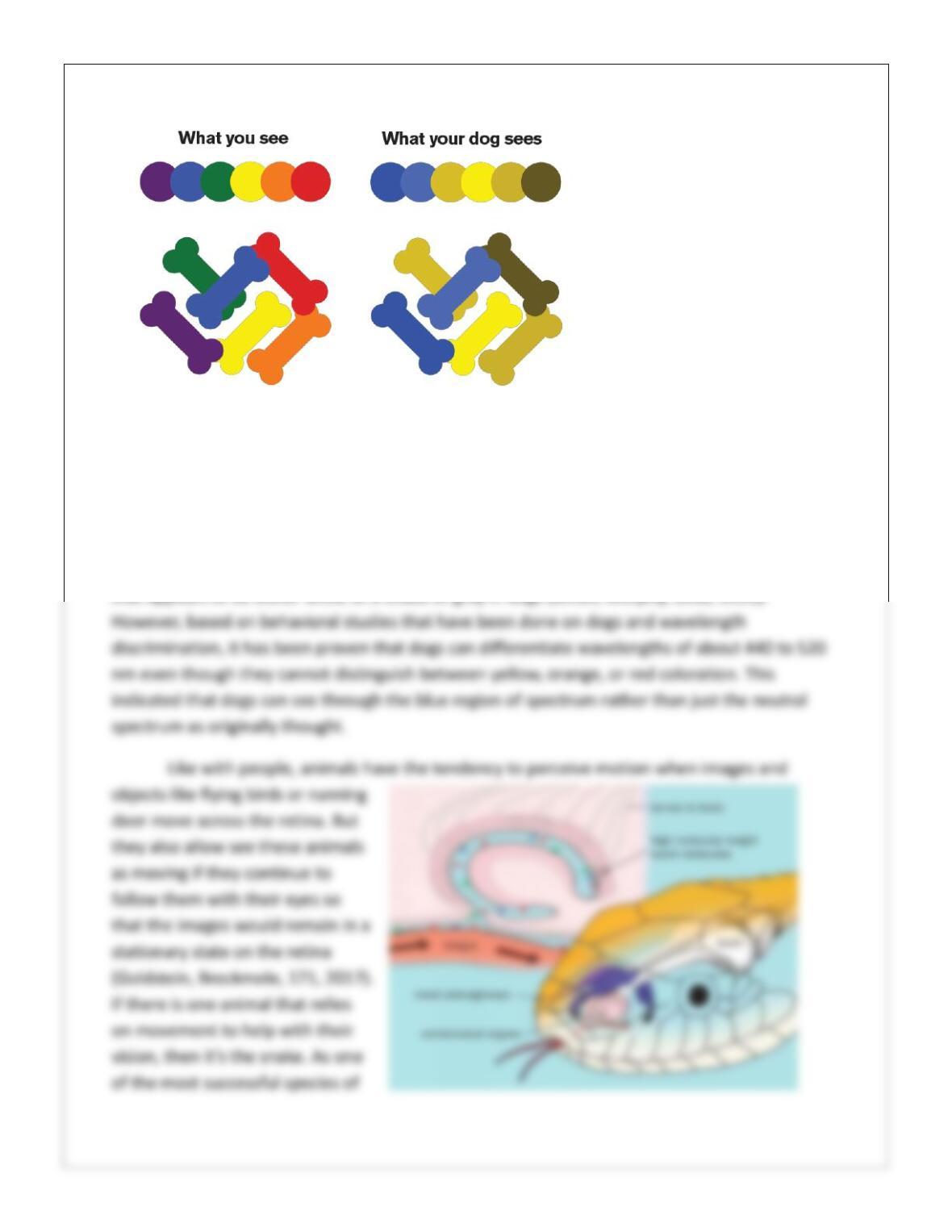
(Sinclair, 116, 1985). In the case of dogs, they do tend to have a completely different view of the
world than humans do. Yet, their peripheral vision is far sharper than our own (Robinson, 1,
2012). The color sensitive cones are found in the canine retina which means that there is at
least, an anatomic potential for dogs to have some sort of color vision (Miller, Murphy, 1631,
1995). Studies that relied on the use of antibodies to cone outer segments indicate that dogs
have two different types of cones even though they only comprise a minority of photoreceptors
in the central area of the canine retina. This does suggest that these cones are slightly more
concentrated in the central area than they are in the peripheral portion of the retina (Miller,
Murphy, 1632, 1995). In other
two studies that have been
done on color vision in dogs,
the distribution pattern is
virtually uniform. The first
cone is maximally sensitive to
light with wavelengths of
around 429 to 435 nm, which
appears to be violet to people
who have a healthy color
vision. The second cone has
the maximally sensitivity to
light with wavelengths of about 555 nm, which to people, appears to be yellow-green (Miller,
Murphy, 1632, 1995). While it’s not known if dogs have the ability to perceive their world in
these two colors in the same way that people do, it is suggested that the visible spectrum in
dogs is divided into two hues, which are one in the violet and blue-violet range and one in the
green-yellow, yellow and red range. In addition, dogs also have narrow regions of visible
spectrum that appear to be colorless and light that ranges in wavelengths of about 475-485 nm,