I. Genetics
A. Structure and Function of DNA
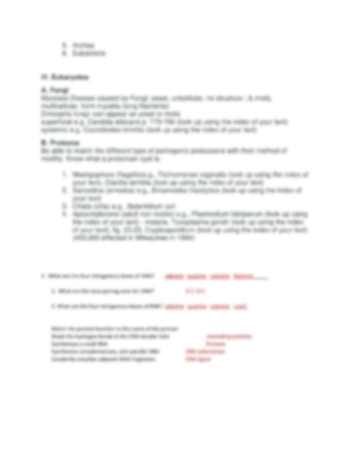
DNA, double stranded (DS), anti-parallel, base pairing rules: A-T, G-C
ATP is the energy molecule prominent in chapter 5, it is also the precursor monomer
nucleotide in RNA, the related molecule dATP is the precursor monomer nucleotide for
DNA
DNA instructions are transcribed into RNA
B. Structure and Function of RNA
RNA, single stranded (SS), base pairing rules A-U, G-C,
The three classes of RNA cooperate together to make the proteins
C. Replication (DNA)
Figure 8.5 summarizes this best:
1. unwinding proteins break the hydrogen bonds holding the two DNA strands
together
2. RNA polymerase (enzyme that makes the polymer, RNA) binds a portion of
the SS (single stranded) DNA and synthesizes a short complementary, anti–
parallel RNA (synthesis is in the 5′ to 3′ direction).
3. DNA polymerase (enzyme that makes the polymer, DNA) binds the short
RNA primer and is able to extend off it, new DNA, anti-parallel and
complementary to the SS DNA it is working from, synthesis is in the 5′ to 3′
direction
4. DNA polymerase can excise RNA primers and replace the RNA with DNA
thus completing a DNA complementary strand. Since this occurs
simultaneously on both strands at completion there are two complete DS
DNA molecules.
mRNA
messenger, information molecule, which directs the ribosome to put in place the particular
sequence of amino acids for a polypeptide chain
tRNA
transfer, transport molecule, each of twenty different tRNAs carry a specific amino acid to the
mRNA/ribosome complex
rRNA
ribosomal, enzymatic molecule, along with ribosomal proteins forms the ribosome

5. DNA ligase seals the adjacent DNA fragments after DNA polymerase has
finished excising the RNA primer and refilling with DNA nucleotides
While DNA polymerase generally has high fidelity (few mistakes), still mistakes are
made and these altered sequences are called mutations which ‘fuel’ evolution.
D. Transcription
RNA polymerase binds the start of a gene, and synthesizes a SS RNA complementary
to one of the DNA strands. Synthesis is complementary, anti-parallel and in the 5′ to 3′
direction
E. Translation
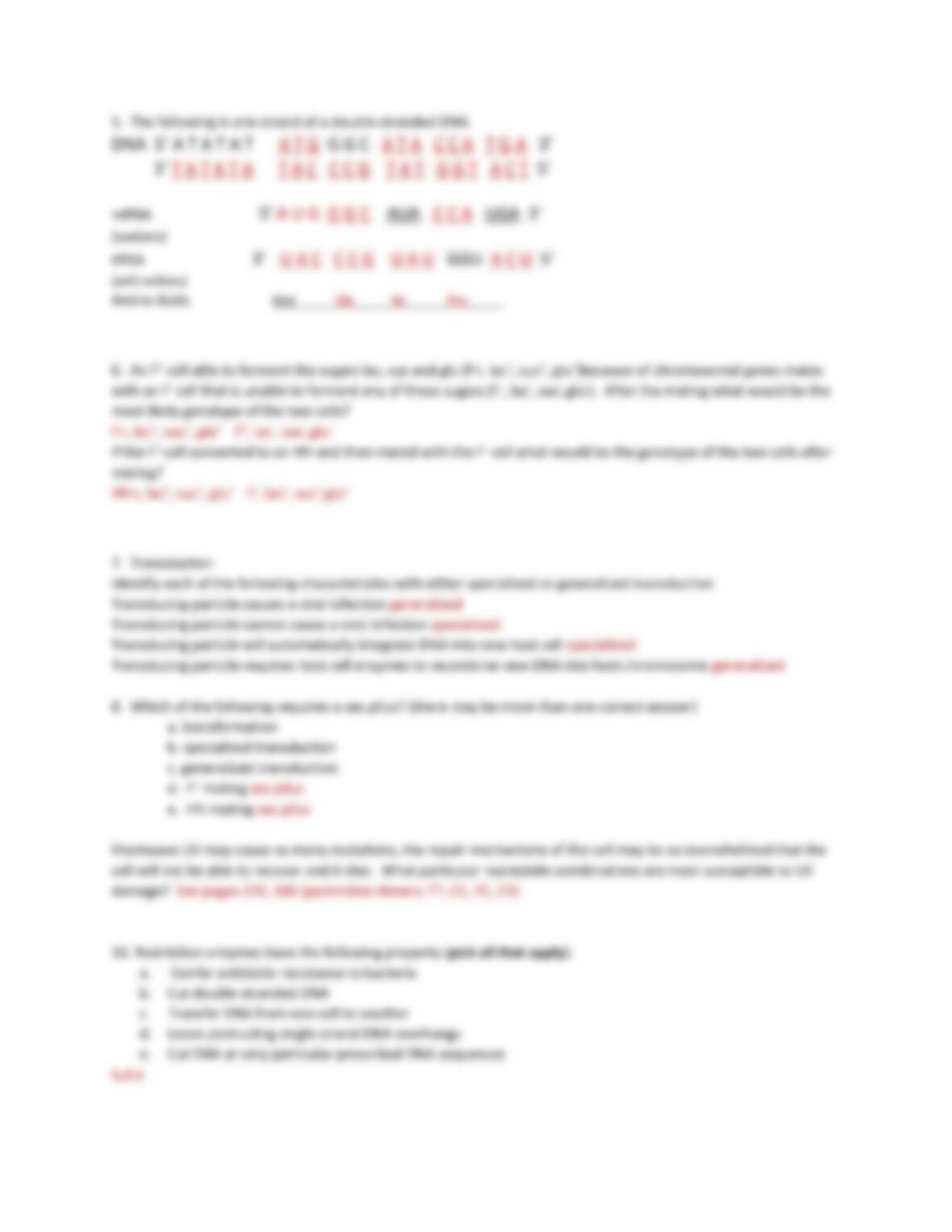
1. The ribosome binds the mRNA and synthesizes a polypeptide chain
according to the mRNA directions
2. the ribosome binds the 5′ end of the mRNA and moves ‘downstream’ until it
finds the first AUG, this triplet of nucleotides is called a codon. AUG is always
the first codon, subsequent codons of mRNA are gene specific.
3. a tRNA that has a complementary anti-codon for the codon and carries a
specific and corresponding amino acid (in the case of AUG codon, the anti-
codon on the tRNA is UAC and the amino acid carried is methionine (fig. 8.9).
4. a second tRNA with a complementary anti-codon for the 2nd triplet of
nucleotides on the mRNA (i.e., the second codon) binds the second codon.
5. the ribosome excises the first amino acid off the first tRNA and covalently
attaches it (peptide bond) to the second amino acid on the second tRNA.
6. the ribosome shifts three nucleotides down the mRNA, the first tRNA falls
away and a third tRNA ‘sits’ down on the third codon. Of course this tRNA has
a complementary anti-codon, and carries an amino acid corresponding to the
codon. The dipeptide is cleaved from the second tRNA and is attached to the
amino acid on the third tRNA.
7. the ribosome continues down the mRNA similarly, attaching the growing
polypeptide chain to the next amino acid.
You can look to the codon table on in chapter 8 to see which amino acid will be brought
to each of the 64 codons (any of three stop codons signal the ribosome to stop).
F. Gene Regulation (lac operon)
Some genes are always on (constitutive). However, natural selection works
powerfully to ensure optimization and no waste. Genes that are not needed are
turned off; when needed they are turned on. Make sure you understand that this
set of genes is under dual control; repression and activation at the same time in
order to finely tune the genes only being expressed when there is a need and
there is lactose present.

G. Mutation
1. Types of mutations
1. spontaneous, errors in DNA replication, uncorrected.
2. chemical, DNA damage repaired with error(s)
3. radiation, DNA damage repaired with error(s)
2. Outcomes from mutation:
1. silent (due to redundancy in the genetic code, i.e., more that one codon may
call for the same amino acid, for e.g., DNA AAA would yield codon UUU
calling for tRNA carrying phenylalanine but, DNA mutation to AAG would yield
mRNA codon UUC and we would still get phenylalanine.
2. neutral we may have in fact a new amino acid due to a DNA change but
sometimes substitution of one amino acid for another does not effect the
function of the protein
3. harmful protein has less function or causes some deleterious result
4. beneficial rare but powerful, these mutations are more likely preserved in later
generations than harmful ones. Why? If a shark mutates to less sharp teeth
or slower swimming perhaps this individual will have less reproductive
success and these mutation may not be represented very well in future
generations but a mutation that leads to the opposite will have a different
effect on the representation of that gene in future generations. This was the
great contribution that Charles Darwin made in biology, natural selection. This
is something that is ongoing in all populations.
II. Microbial Genetics
A. plasmid: these small, circular, autonomously replicating DNAs have vast importance
in that they can be easily transmitted from some cells to others thus accelerating the
different ‘types’ of individuals in a population
B. Mechanisms for taking on new genetic information (note how this is different
from mutation which is an alteration of existing DNA)
1. Transformation DNA is absorbed out of the environment, presumably from some
other dead cell that has released its’ DNA to the environment. Reading carefully fig.
8.27 allows one to see the first discovery of transformation. Steptococcus pneumoniae
with a capsule is deadly (bacteria are able to escape phagocytosis, proliferate, cause
disease, kill host). The use of the mouse in the experiment is an interesting element.
Transformation is relatively rare and inefficient so identifying the one smooth colony
(capsule former) from the rest would be too difficult to spot on agar plates if its’
occurrence were only once in say a million. But the mouse acts like a selective
medium! Live Steptococcus pneumoniae unable to produce capsule mixed with remains
(including DNA) from capsule producing Steptococcus pneumoniae are put into a
mouse instead of onto an agar plate. The mouse selects for the one cell in a million that
has been transformed (i.e., escaped the mouse’s immune system). Brilliant. We will see
the principles of transformation exploited in the next chapter on biotechnology.
2. Conjugation DNA is transferred from one cell to another by direct contact. F system
in E. coli is a particularly well studied system. F stands for fertility. F+ cells have the F
DNA as a separate double stranded (ds) circular DNA that is outside the chromosome
(like a plasmid). The difference between F DNA which is an episome and a plasmid
which is not is that F DNA and episomes in general can integrate into the cells’
chromosome. When this happens the cell harboring the integrated F DNA, is
called Hfr. Conjugation is different with F+ cells and Hfr cells.
Similarities: F+ cells and Hfr cells both:
a. have a pilus and,
b. know how to use it (they both can transfer DNA to a F- cell –a cell that has no F
DNA).
Differences:
a. F+ cell only transfers F DNA (see figures in text) and does so with near certainty, thus
changing the F- cell to a F+ cell.
b. Hfr which stands for high frequency of recombination transfers only a very small
piece of F DNA and then some of the cells’ chromosomal DNA. The recipient cell will
almost never receive enough DNA to become Hfr or F+ but with high certainty will