Chapter 3 – Structure and Function of Living Cells
This lab is considered a “dry” lab because you will not be performing any experiments…instead, you will be
completing this worksheet and visiting several websites as you are instructed here to experience all there is to
offer there.
Part I – Cellular Organelles and their Function. You need to use your textbook or the interactive models at
https://www.cellsalive.com/cells/cell_model.htm to fill in the function of the following cellular components below.
Indicate with a letter whether it is found in Animal (A), Plant (P) and Bacteria (B). You need to highlight those
components in the first column, that are found in all cells (prokaryotic and eukaryotic)
Component
A, P, B
Function
Plasma membrane
A,P,B
Protect cell from surroundings
Cytoplasm
A,P,B
Contains all organelles and cell parts
Nucleus
A,P
Contains the cell’s genetic materials and forms chromosomes
Nuclear envelope
A,P
Provides compartmentalization
Nucleolus
A,P
Rewrite rRNA and combine it with proteins
Nuclear pores
A,P
Transport of molecules across the nuclear envelope
DNA
A,P,B
Long-term storage of information
Rough Endoplasmic
Reticulum
A,P
Assembly of many proteins
Soft Endoplasmic Reticulum
A,P
Has ribosomes for protein synthesis
Golgi body
A,P
Receives synthesized proteins from the endoplasmic reticulum
Mitochondria
A,P
Produce ATP
Lysosome
A,P
Digest excess or worn out organelles
Ribosome
A,P,B
Repairing damages or chemical process
Nucleoid
B
Contains Proteins, RNA, and Enzymes
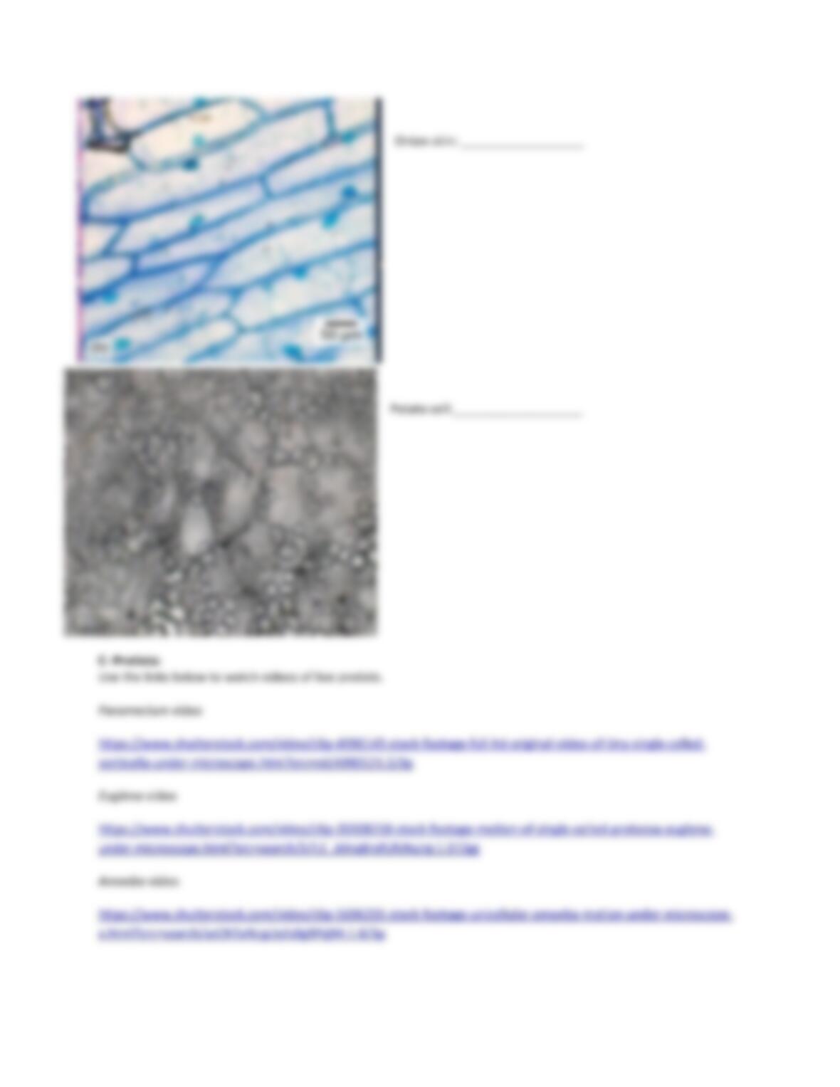
Chloroplast
P
Convert light energy into sugars
Cytoskeleton
A,P
Provides structural framework for cell shape
Central vacuole
A,P
Maintain proper pressure
Vesicles
A,P
Breakdown food particles
Cell wall
P
Structural support and protection
Part II – Types of Cells:
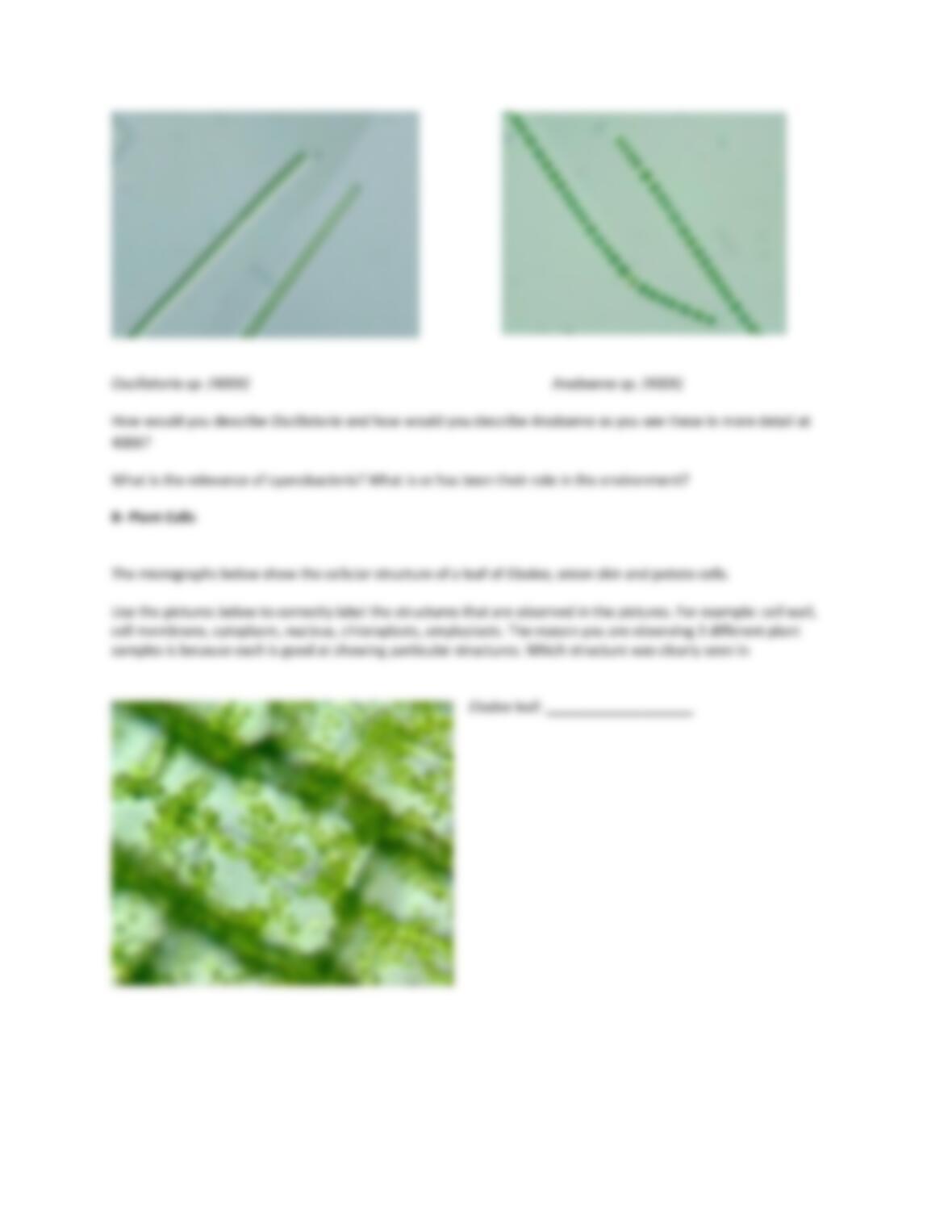
A- Prokaryotes:
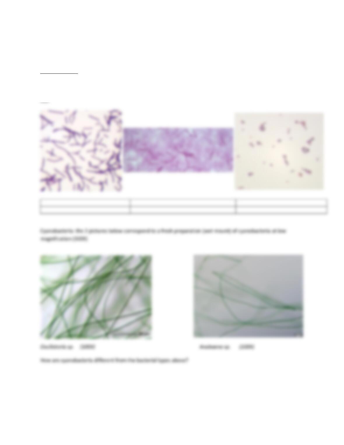
Bacterial types: the 3 pictures below correspond to a prepared slide of different types of bacteria as observed at
400X total magnification
Use the 1st line to label the corresponding bacterial shape in the micrograph, use the 2nd line to describe what you