Lab Exercise: 20 points
Bonus Opportunity: Flow Diagram for Next Lab (5 points)
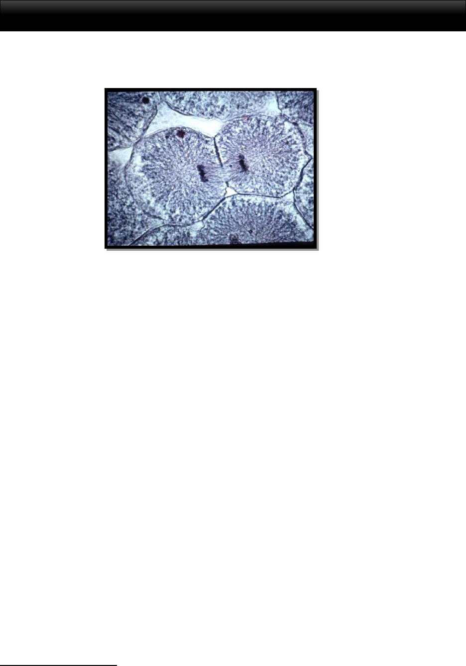
Figure 1: Image of a white fish cell in the telophase cycle of cell division 1
Cell Division in Eukaryotes
Life can take on various forms and biologists attempt to define and categorize different
lifeforms by grouping them in ways that make logical sense. One way in which we can start to
divide these groups into categories is to note how the basic cell structures may differ from
organism to organism. Lifeforms that have similar features can be grouped together. The first
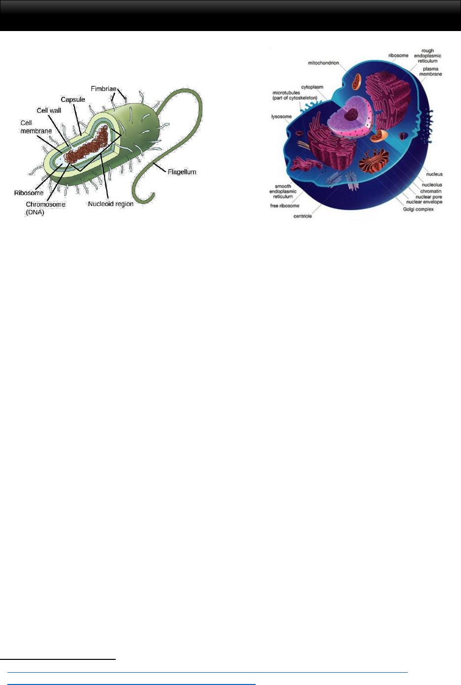
step in observing a cell is to determine whether or not the lifeform is a prokaryote or a eukaryote.
A prokaryote is an organism that doesn’t have membrane enclosed organelles inside of its cell.
These types of lifeforms include bacteria and an archaic form of bacteria-like organisms called
archaea. The cells that we will observe in lab today will be eukaryotic, which means that they
do include organelles that have membranes. These types of lifeforms include all animals, plants
and fungi, and single-celled microscopic organisms called protists. The cell shapes, the
placement of the DNA, and the general features of eukaryotic cells are vastly different from
those of prokaryote cells (See Figure 2). This is why we separate these types of lifeforms out into
two different large groups.
1 Paul D. Camp Community College. Suolk. Whitesh Telephase. Retrieved 24 March 2017.
h!p://www.pdcfaculty.org/science1/biology-labmanual/lab7mitmei/white)shtelo.jpg
1
HUMANS & THE ENVIRONMENT: MITOSIS
vs
Figure 2: Image of a prokaryote cell2 (left) versus a eukaryote cell (right)3
One notable feature of a eukaryotic cell is the nucleus, which is often visible as a dark
circular spot under a microscope (See figure 3). The nucleus is where we would find our DNA, a
double-stranded helix consisting of genes that is home to our genetic information. The visibility
of the nucleus and the shape of the DNA is important in identifying the different stages of normal
cell division, or mitosis. Is the DNA loose or tightly coiled? Can we see the familiar “X” shape
of a chromosome? Has the membrane around the nucleus disappeared? These are all clues for
how to identify stages of mitotic cell division.
The cells in Figure 3 show what we would expect to see in a typical animal cell or a
typical plant cell in between cell division cycles. We sometimes refer to these cells as “pausing”
or being “at rest” even though they are performing normal cellular functions, such as
metabolizing. We simply use these terms to indicate that no cell division is taking place. These
pictures are extreme close-ups of the cells and allow us to see organelles is great detail. A
specialized microscope called a scanning electron microscope was used to capture this level of
detail. You’ll note as you perform this lab with a compound light microscope that you won’t be
able to see most of these smaller features. Just try to keep an eye on the nucleus and the DNA
inside to identify the phases.
2 h!ps://ka-perseus-images.s3.amazonaws.com/95cd645b33b4a8883218ce52a0b;5ade93f8d52.png
3 h!p://techhydra.com/wp-content/uploads/eukaryo<c-cell.jpg
2
HUMANS & THE ENVIRONMENT: MITOSIS

vs
Figure 3: Image of a typical animal cell4 (left) versus a typical plant cell (right). Note that both are eukaryotic
lifeforms. Further note that an animal cell is surrounded by a plasma membrane while the plant cell has a thicker
wall-like structure we call a cell wall.
Stages of Mitosis5
Cells will continuously cycle through different stages until they die. Some of those
stages are considered steps in cell division while other stages are not. For now, we’ll just focus
specifically on mitosis. Mitosis is a type of cell division that passes along the same number of
chromosomes to each daughter cell. To recap, the chromosome is a tight coil of DNA that
contains genetic information. It forms an easily recognizable “X” shape inside the cells.
Humans have 46 chromosomes and inherit 23 from each parent. When cell division is not taking
place, we generally say that the cell is experiencing a cycle called Interphase.
Our first indication that cell division is about to take place is the G2 Stage of Interphase,
sometimes called the growth phase of the cell cycle. This stage can be difficult to spot under a
compound light microscope, but when we can find it we should expect to see many strands of
loose DNA. This is because the individual strands of DNA have been synthesized. Careful
scanning can allow one to spot small fractures in the membrane, or envelope, surrounding the
nucleus (See figure 4).
Nuclear Envelope
4 h!ps://ka-perseus-images.s3.amazonaws.com/95cd645b33b4a8883218ce52a0b;5ade93f8d52.png
5 Reece, J.B., et el (2016). Glossary. Campbell Biology. Pp G1 – G35. New York, NY. Pearson.
3
HUMANS & THE ENVIRONMENT: MITOSIS
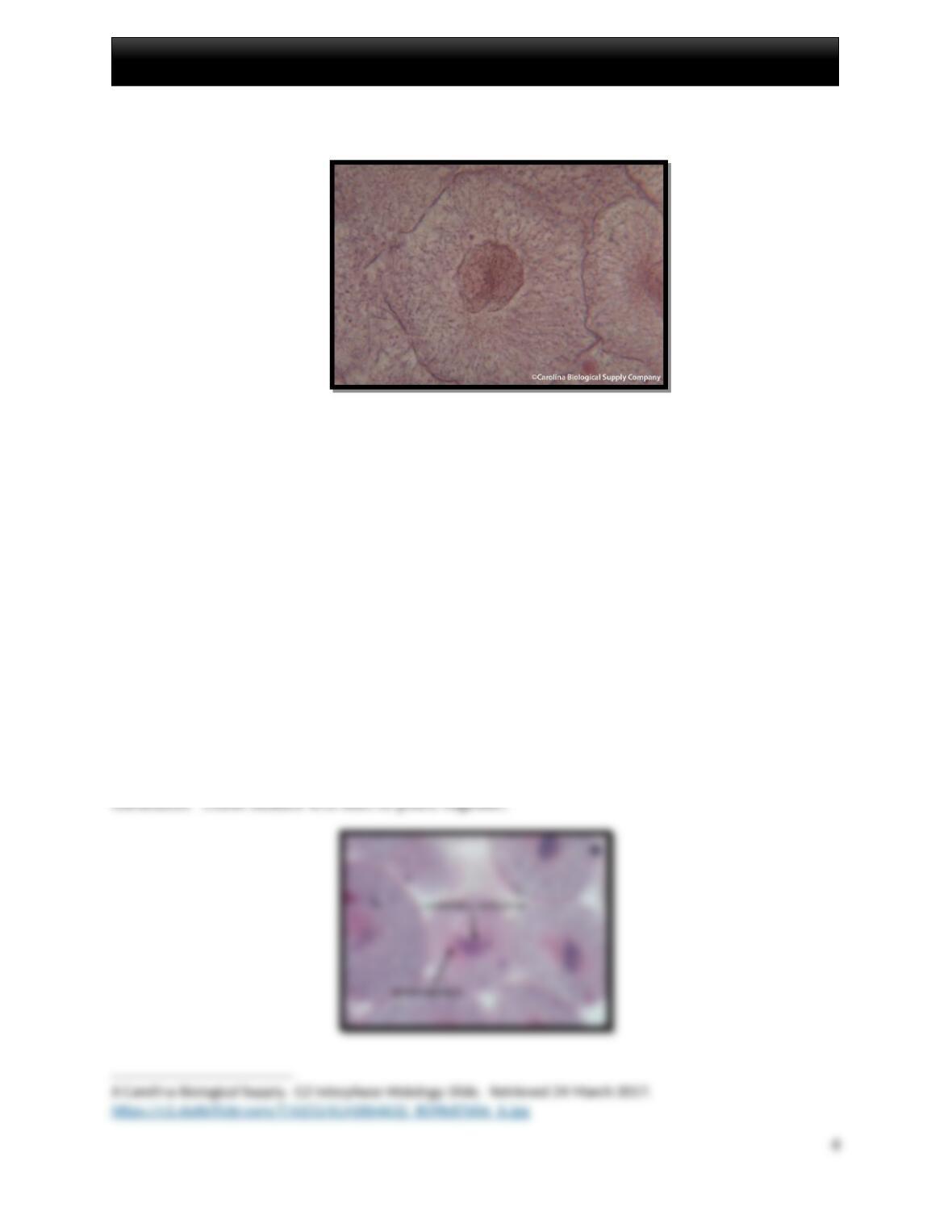
Figure 4: Image of G2 Stage of Interphase in a White Fish cell. 6
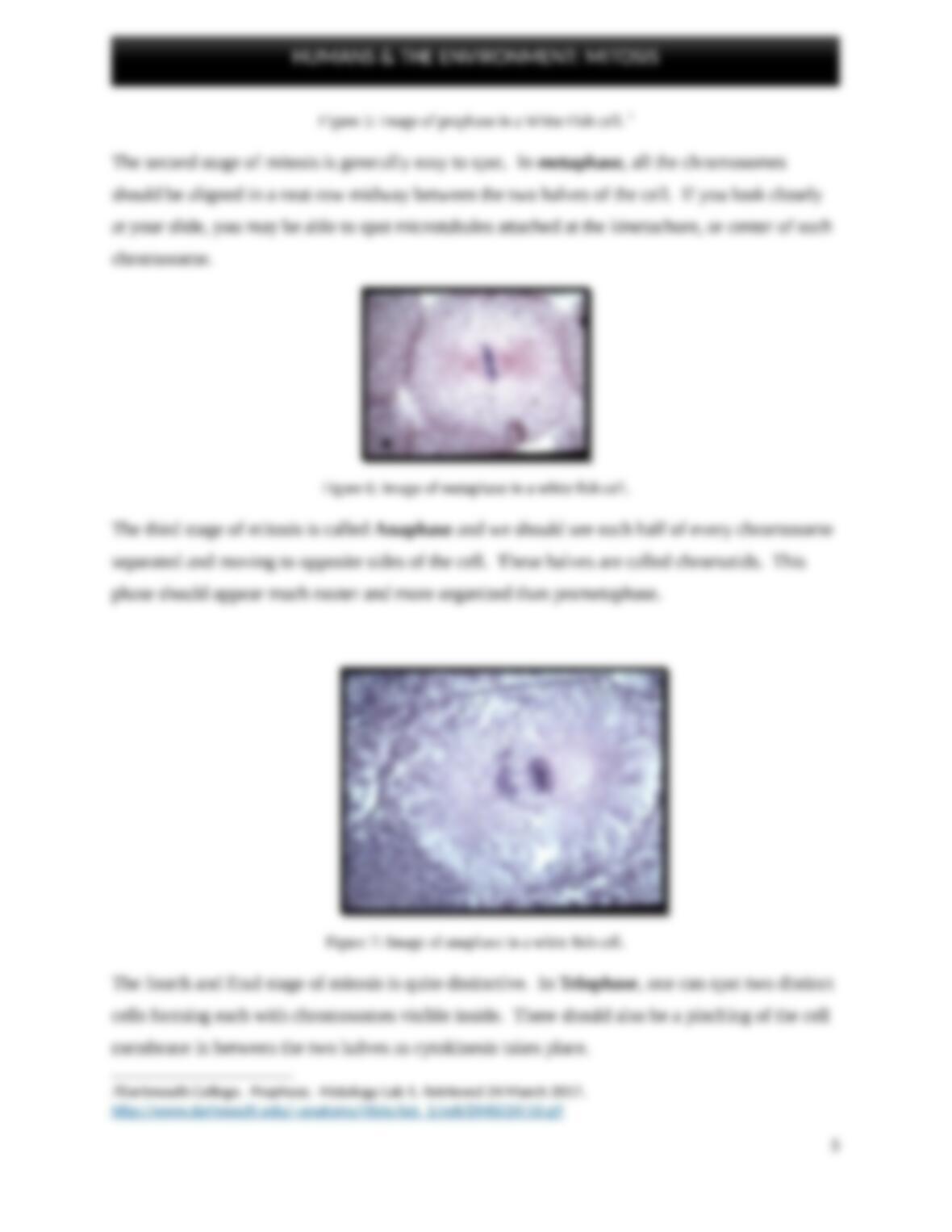
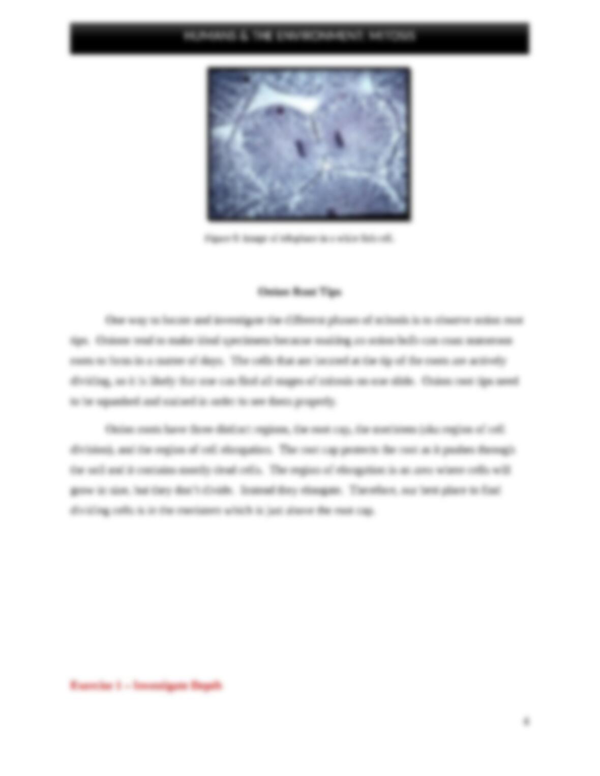
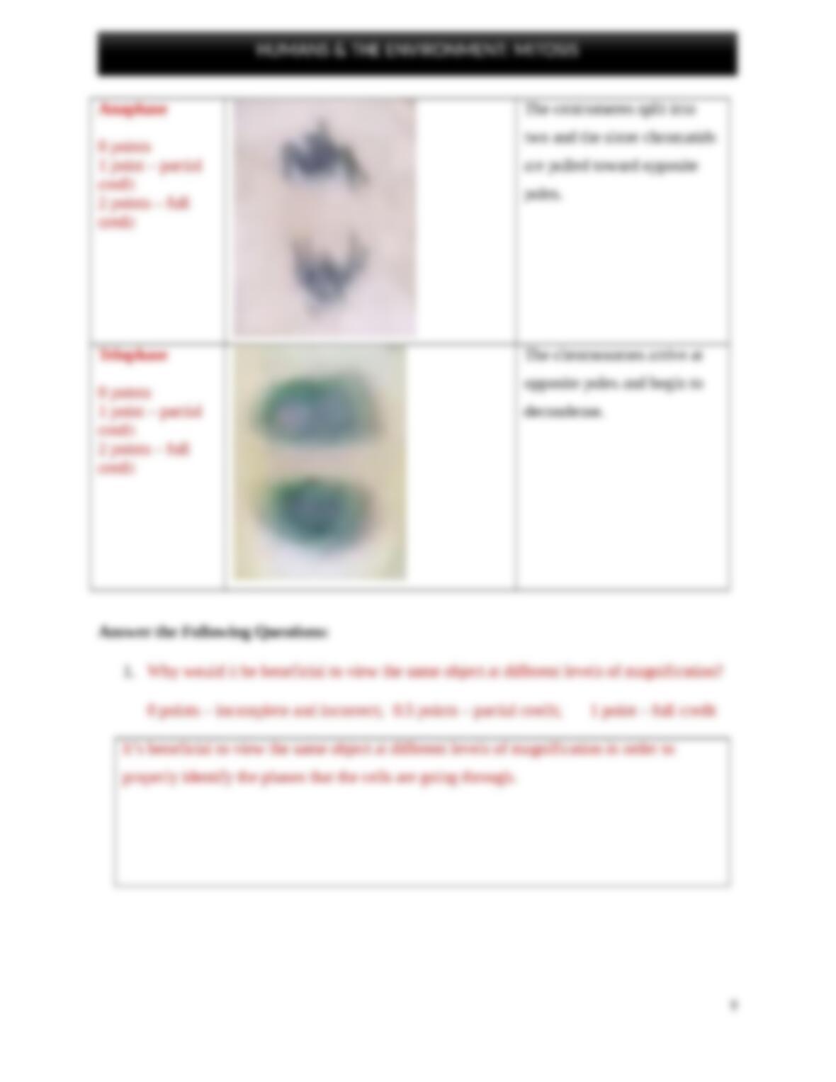
The G2 stage of Interphase is followed by the first official stage of mitosis; prophase. In
Prophase the nucleolus inside the nucleus will disappear and the DNA will condense into
HUMANS & THE ENVIRONMENT: MITOSIS