1
Chapter 4: Prenatal Development
Chapter Objectives:
1. Describe changes that occur within each of the three periods of prenatal
development
2. Recognize the risks to prenatal development posed by exposure to teratogens.
3. Evaluate different types of prenatal assessment.
4. Describe complications associated with pregnancy
5. Evaluate infertility options
Prenatal Development
Now we turn our attention to prenatal development which is divided into three periods:
The germinal period, the embryonic period, and the fetal period. The following is an
overview of some of the changes that take place during each period.
The Germinal Period
The germinal period (about 14 days in length) lasts from conception to implantation of
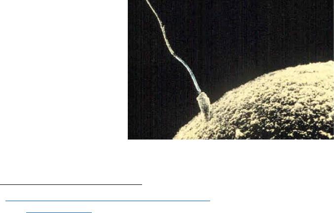
the fertilized egg in the lining of the uterus. At ejaculation millions of sperm are released
into the vagina, but only a few reach the egg and typically only one fertilizes the egg.
Once a single sperm has entered the wall of the egg, the wall becomes hard and
prevents other sperm from entering. After the sperm has entered the egg, the tail of the
sperm breaks off and the head of the sperm, containing the genetic information from the
father, unites with the nucleus of the egg. It is typically fertilized in the top section of the
fallopian tube and continues its journey to the uterus. As a result, a new cell is formed.
This cell, containing the combined genetic information from both parents, is referred to
as a zygote.
1
1
Lifespan Development: A Psychological Perspective by Martha Lally and Suzanne Valentine-French is licensed
under CC BY-NC-SA 3.0
2
Sperm and ovum at conception
2
During this time, the organism begins cell division through mitosis. After five days of
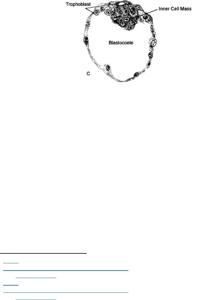
mitosis there are 100 cells, which is now called a blastocyst. The blastocyst consists of
both an inner and outer group of cells. The inner group of cells, or inner cell mass will
become the embryo, while the outer group of cells, or trophoblast, becomes the
support system which nourishes the developing organism. This stage ends when the
blastocyst fully implants into the uterine wall (U.S. National Library of Medicine, 2015).
3
Image of the blastocyst which shows the inner cell mass, which will become the fetus, the trophoblast
which will form the placenta, and the blastocoel, a fluid-filled cavity.
4
Mitosis is a fragile process and fewer than one half of all zygotes survive beyond the
first two weeks (Hall, 2004). Some of the reasons for this include the egg and sperm do
not join properly, thus their genetic material does not combine, there is too little or
damaged genetic material, the zygote does not replicate, or the blastocyst does not
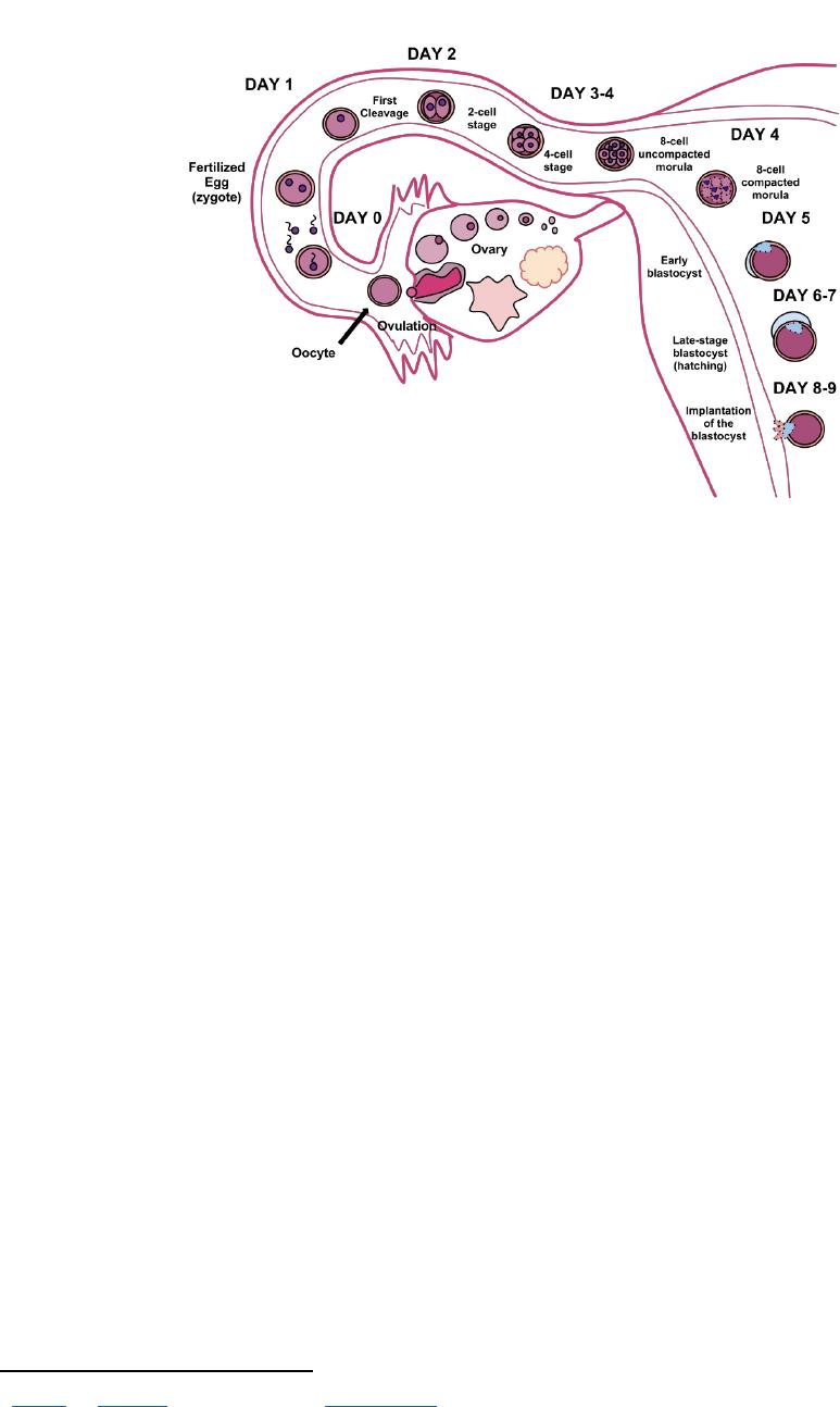
implant into the uterine wall. The failure rate is higher for in vitro conceptions. The figure
below illustrates the journey of the ova from its release to its fertilization, cell duplication,
and implantation into the uterine lining.
5
2
Image is in the public domain
3
Lifespan Development: A Psychological Perspective by Martha Lally and Suzanne Valentine-French is licensed
under CC BY-NC-SA 3.0
4
Image is in the public domain
5
Lifespan Development: A Psychological Perspective by Martha Lally and Suzanne Valentine-French is licensed
under CC BY-NC-SA 3.0
3
Human Fertilization. The sperm and ovum unite through fertilization creating a zygote that (over the
course of 8-9 days) will implant in the uterine wall, where it will reside over the course of 9 months.
6
The Embryonic Period
Starting the third week, the blastocyst has implanted in the uterine wall. Upon
implantation this multi-cellular organism is called an embryo. Now blood vessels grow
forming the placenta. The placenta is a structure connected to the uterus that provides
nourishment and oxygen from the mother to the developing embryo via the umbilical
cord.
During this period, cells continue to differentiate. Growth during prenatal development
occurs in two major directions: from head to tail called cephalocaudal development and
from the midline outward referred to as proximodistal development. This means that
those structures nearest the head develop before those nearest the feet and those
structures nearest the torso develop before those away from the center of the body
(such as hands and fingers). You will see that this pattern continues after birth.
The head develops in the fourth week and the precursor to the heart begins to pulse. In
the early stages of the embryonic period, gills and a tail are apparent. However, by the
end of this stage they disappear and the organism takes on a more human appearance.
6
Image by Ttrue12 is licensed under CC BY-SA 3.0
4
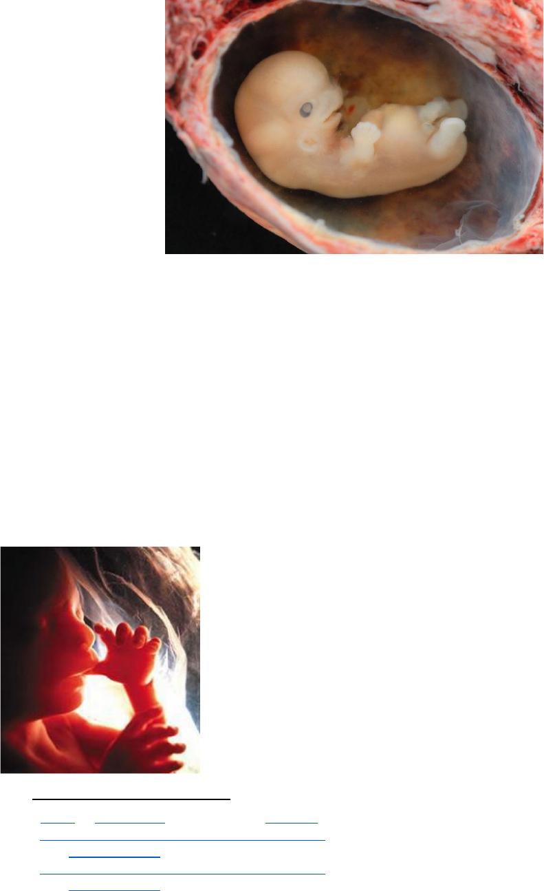
Human embryo – Approximately 8 weeks estimated gestational age
7
About 20 percent of organisms fail during the embryonic period, usually due to gross
chromosomal abnormalities, often before the mother even knows that she is pregnant. It
is during this stage that the major structures of the body are taking form, making the
embryonic period the time when the organism is most vulnerable to the greatest amount
of damage if exposed to harmful substances. Prospective mothers are not often aware
of the risks they introduce to the developing embryo during this time. The embryo is
approximately 1 inch in length and weighs about 4 grams at the end of eight weeks. The
embryo can move and respond to touch at this time.
The Fetal Period
From the ninth week until birth (which is forty weeks for a full-
term pregnancy), the organism is referred to as a fetus. During
this stage, the major structures are continuing to develop. By
the third month, the fetus has all its body parts including
external genitalia. The fetus is about 3 inches long and weighs
about 28 grams. In the following weeks, the fetus will develop
hair, nails, teeth and the excretory and digestive systems will
continue to develop.
8
Fetus sucking thumb
9
7
Image by lunar caustic is licensed under CC BY 2.0
8
Lifespan Development: A Psychological Perspective by Martha Lally and Suzanne Valentine-French is licensed
under CC BY-NC-SA 3.0
9
Lifespan Development: A Psychological Perspective by Martha Lally and Suzanne Valentine-French is licensed
under CC BY-NC-SA 3.0

5
During the 4th – 6th months, the eyes become more sensitive to light and hearing
develops. The respiratory system continues to develop, and reflexes such as sucking,
swallowing and hiccupping, develop during the 5th month. Cycles of sleep and
wakefulness are present at this time as well. The first chance of survival outside the
womb, known as the age of viability is reached at about 24 weeks (Morgan,
Goldenberg, & Schulkin, 2008). Many practitioners hesitate to resuscitate before 24
weeks. The majority of the neurons in the brain have developed by 24 weeks, although
they are still rudimentary, and the glial or nurse cells that support neurons continue to
grow. At 24 weeks the fetus can feel pain (Royal College of Obstetricians and
Gynecologists, 1997).
Between the 7th – 9th months, the fetus is primarily preparing for birth. It is exercising its
muscles and its lungs begin to expand and contract. The fetus gains about 5 pounds
and 7 inches during this last trimester of pregnancy, and during the 8th month a layer of
fat develops under the skin. This layer of fat serves as insulation and helps the baby
regulate body temperature after birth.
At around 36 weeks the fetus is almost ready for birth. It weighs about 6 pounds and is
about 18.5 inches long. By week 37 all of the fetus’s organ systems are developed
enough that it could survive outside the mother’s uterus without many of the risks
associated with premature birth. The fetus continues to gain weight and grow in length
until approximately 40 weeks. By then the fetus has very little room to move around and
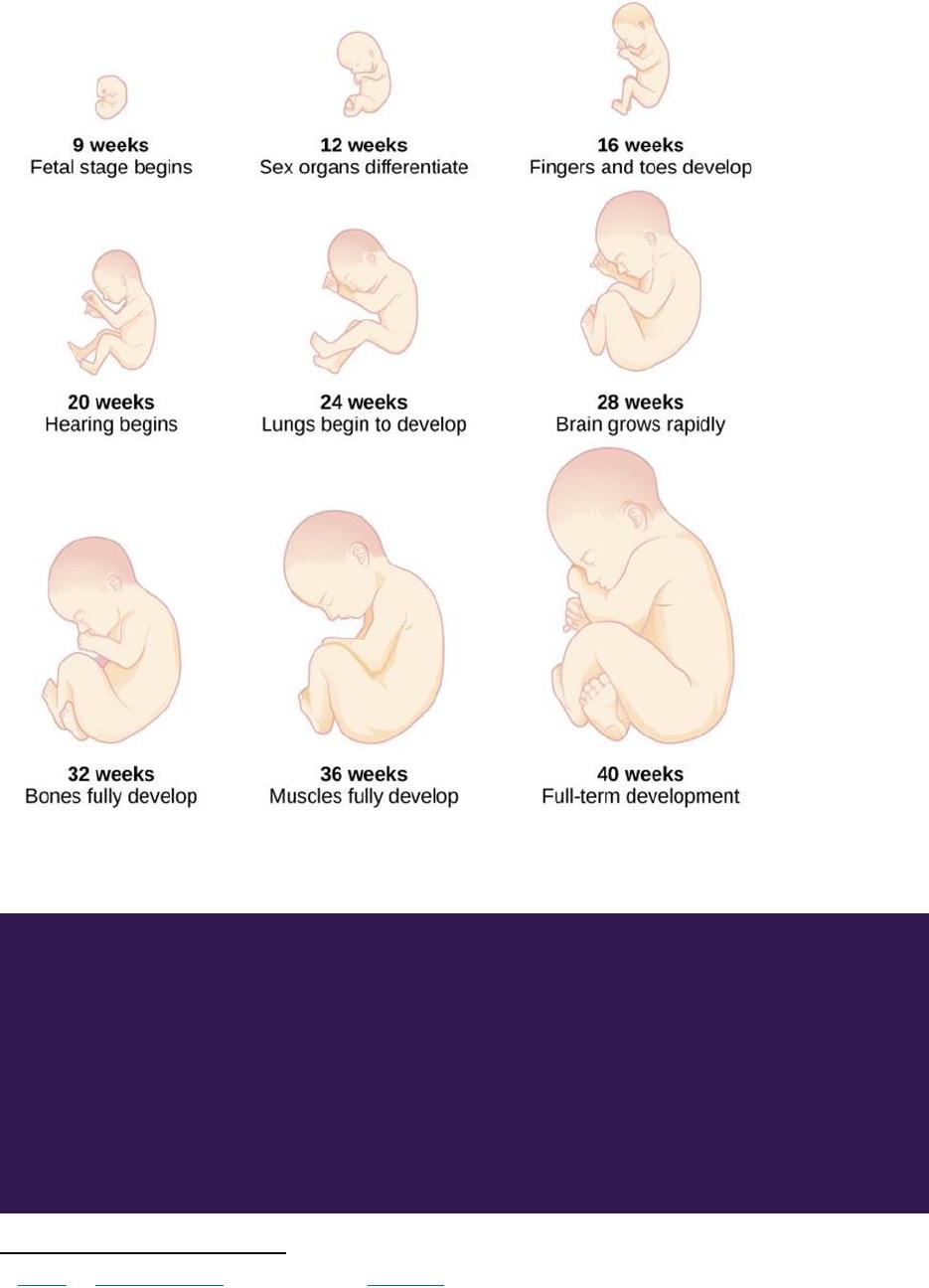
birth becomes imminent. The progression through the stages is shown in the following
figure.
10
10
Lifespan Development: A Psychological Perspective (pages 44–45) by Martha Lally and Suzanne Valentine-French
is licensed under CC BY-NC-SA 3.0
6
Prenatal development of a fetus
11
Monozygotic and Dizygotic Twins
Monozygotic or identical twins occur when a fertilized egg splits apart in the first two weeks of
development. The result is the creation of two separate, but genetically identical offspring. That is, they
possess the same genotype and often the same phenotype. About one-third of twins are monozygotic
twins.
Sometimes, however, two eggs or ova are released and fertilized by two separate sperm. The result is
dizygotic or fraternal twins. These two individuals share the same amount of genetic material as would
any two children from the same mother and father. In other words, they possess a different genotype
and phenotype.
11
Image by CNX Psychology is licensed under CC BY 4.0

Older mothers are more likely to have dizygotic twins than are younger mothers, and couples who use
fertility drugs are also more likely to give birth to dizygotic twins.12
Monozygotic Twins13
Dizygotic Twins14
Figure 2.12
Figure 2.13
Prenatal Brain Development
Prenatal brain development begins in the third gestational week with the differentiation
of stem cells, which are capable of producing all the different cells that make up the
brain (Stiles & Jernigan, 2010). The location of these stem cells in the embryo is
referred to as the neural plate. By the end of the third week, two ridges appear along the
neural plate first forming the neural groove and then the neural tube. The open region in
the center of the neural tube forms the brain’s ventricles and spinal canal. By the end of
the embryonic period, or week eight, the neural tube has further differentiated into the
forebrain, midbrain, and hindbrain.