Bovine Tuberculosis: The Often Forgotten Pathogen
C. R. Long
Missouri Valley College, Marshall, MO 65340
Introduction
In the 1900’s approximately 150,000 people died from tuberculosis (O’Brien et al., 2013). Tuberculosis
not only affects people but cattle as well. Every year over one million head of cattle have been sampled
or tested for bovine tuberculosis according to the USDA APHIS (2014). Bovine tuberculosis is a pathogen
that has many harsh effects to its hosts. This paper will discuss what bovine tuberculosis is, how the
disease is transmitted, prevention of TB, and economic effects that this pathogen causes.
Discussion
Bovine Tuberculosis
Bovine tuberculosis (TB) is an infectious disease caused by Mycobacterium bovis and is
characterized by the formation of tubercles (Konyha, 1981). Cattle are the primary host of M. bovis,
however any warm blooded animal can contract the disease (USDA APHIS, 2014). Bovine TB typically
involves the lungs but can be spread to other organs (USDA APHIS, 2014). The USDA APHIS (2014) states
that bovine tuberculosis is hard to identify solely using physical exam. In the beginning there are no
visible symptoms however, as the infection progresses symptoms could include: lethargy, weight loss,
weakness, a low-grade fever, and chronic pneumonia with possible enlarged lymph nodes (USDA APHIS,
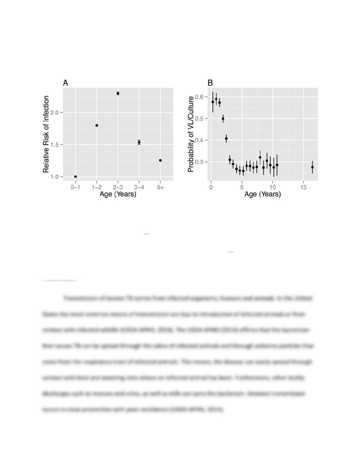
2014). In Fig. 1 graph A the risk of infection is shown to be the highest between two and three years of
age (Conlan et al., 2015). Almost all infected herds are identified by tracing back of infected animals to
the herd of origin (Scanlan, 2003). In order to confirm a diagnosis of bovine TB, lesions found within the
cattle are cultured and sent for testing (Scanlan, 2003). According to Dr. Scanlan the lesions found are
called tubercles and they can be found in any organ that has reticuloendothelial tissue present. He
continues, the primary sites of infection are correlated with the form of M. bovis contracted whether
that be inhalation or ingestion. Multiple organs are effected when a lesion erodes into a blood vessel,
causing whole body infection (Scanlan, 2003).
Fig 1. Age-stratified patterns of reactors in Great Britain. A The relative risk of infection by age RR(a) measured relative to the risk for 0–1 year
old cattle, Error bars denote 95% credible intervals. RR(a) is calculated as the ratio of the force of infection for each age group divided by the
estimate for the 0–1 year old group. The force of infection is a combined estimate for all national herds (beef and dairy) calculated from reactor
cattle reported between 2004–2009 as described in [21]. B The probability of “confirmation” of reactor animals by the presence of visible
lesions or culture as a function of age at slaughter, estimated as the proportion of reactor animals within each (200 day) age-class that
demonstrated visible lesions or a positive culture result. Estimates are calculated using all reactor animals from within our study population of
herds. The qualitative pattern is robust between different test-types and parish testing intervals [21].