Lecture 13:
Circulatory system:
• Heart- pump that makes pressure to move blood to tissue
• Blood vessels- blood travels
o Perfusion- blood moving through BV
• Blood- nutrients, water, gases enter body
o Materials move from cell to cell
o Wastes eliminated by cell
Blood Vessels:
• Arteries- blood AWAY from heart
• Capillaries- vessels of exchange between blood & air/cells
• Veins- blood TOWARD heart
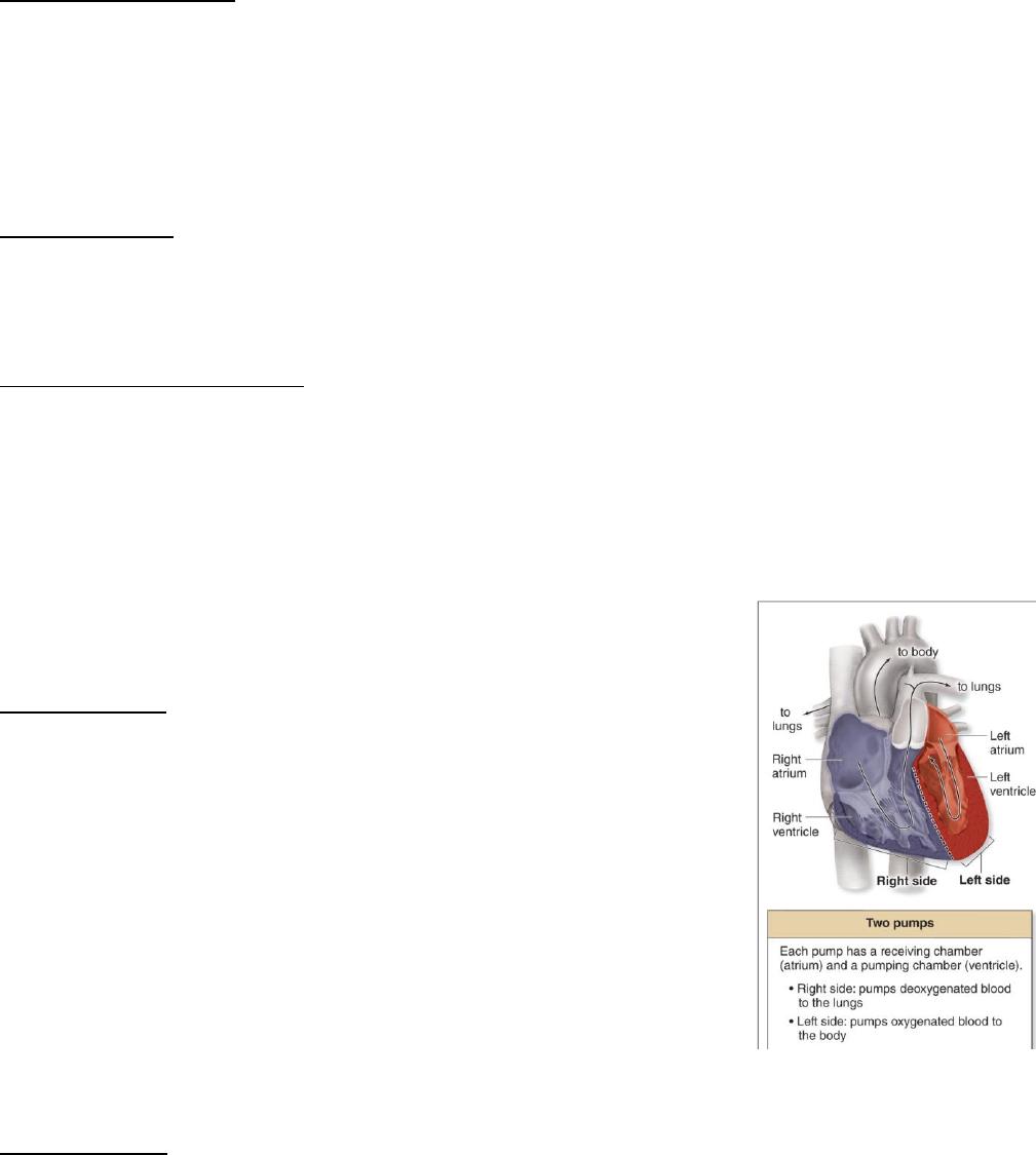
Heart/2 pump structure:
• 4 chambered organ, 2 pairs of valves
• Right pump– pulmonary (pump to lung)
o Deoxygenated blood from body & propels to lungs
• Left pump– systemic (pump to systematic)
o Oxygenated blood from lung & propels to body
• Atrium- receiving chamber
o Superior= collects blood & moves it to ventricle
• Ventricle- propelling chamber
o Inferior= pumping portion
Pair of valves:
• Drive fluid in 1 direction
• AV valves- prevents backflow of fluid from ventricle
into atrium
o Lie b/w atrial & ventricle
o Right AV= tricuspid
o Left AV= bicuspid
• Semilunar valves- prevents backflow, heart to
circulation flow only
o Lie b/w ventricle & arterial trunks
o Pulmonary- right ventricle & pulmonary trunk
o Aortic- left ventricle & aorta
• Left ventricle= highest pressure
• Right ventricle= lowest pressure
Flow of blood:
• Right to left
• Venae cava to right atrium
• Right atrium to right ventricle
o O2 poor blood
o Pulmonary circulation
• Left atrium to left ventricle
o O2 rich blood
o Systemic circulation
Pressure gradients/differences:
• High to low pressure
• Resistance is flow loss
• Driving pressure= moves fluid forward
o Difference between arterial & venous ends of systemic
• Transmural pressure= pressure across wall
o Difference between vessel & tissue
• Hydrostatic pressure= effect of gravity on the blood
o Weak– lots of fluid being taken out & more pressure driven
Cardiac output:
• Direct relationship with semilunar valve & heart rate
• Delivered by the heart
• Higher HR and SV will increase cardiac output
Resistance:
• Inverse relationship between flow & resistance
• Resistance increases, flow decreases
• Vessel radius= inverse
• Vessel length= direct
• Viscosity= direct
• Vasoconstriction= decrease in radius, increase in resistance
• Vasodilation= increase in radius, decrease in resistance
• Blood flow is directly prop. to pressure gradient & inversely prop. To
resistance
Blood pressure:
• Systolic pressure- pressure exerted in arteries when blood is ejected
during ventricular systole
• Diastolic pressure- pressure within arteries when blood is draining into
rest of vessels during ventricular diastole
• Pulse pressure- pressure difference between systolic & diastolic
• Sphyngmomanometer (BP cuff)- greater pressure in cuff, blood flow to
artery
• Korotkoff sound– determine BP
o Pressure released, blood flows in pulses
o Sound appears when pressure in artery is greater than in cuff
Heart conduction system:
• SA node- right atrial wall near opening to superior venae cava
• Av node- base of right atrium near septum
• Bundle of His- originate at AV; divides into 2 branches
• Purkinje fibers- terminal fibers extend from bundle of his
SA Node:
• Sets rate for rest of heart
• Rate directed by SA node
• SA node is damaged, next fastest node sets pace
• SA initiated, action potential spread throughout heart (2 atria & ventricle
contract)
Electrical activity/pacemaker:
• Nodal cells- produce AP
o Initiate & conduct AP
o Pacemaker activity
• Contractile cells- worker cells
o Don’t initiate AP
o Muscle cells
• Three ions- Na+, K+, Ca2+
• Four currents:
o If (funny Na & K)- HCN channels, depolarize
▪ Inflow Na, outflow K
o I Na (Na)- voltage gated Na+ channels, depolarize further
o I Ca (Ca2+)– L-type Ca2+ channels & some T-type Ca2+
o I K (K+)- voltage gated K+ channels, repolarize
Refractory periods:
• Membrane unresponsive until refractory period is over
o New AP can’t be initiated by normal events
o 2nd AP can’t be triggered until excitable membrane recovered
• Smooth, sustained contractional at maximal strength; skeletal muscle
• Alternating contraction & relaxation
Conduction of AP:
• Interatrial pathway- SA node to left atrium
o Rapidly spread AP so both atria depolarize to contract at same time
• Internodal pathway- SA node to AV node
o Electrical contact b/w atria & ventricles
• AP conducted relatively slowly
• Impulse travels down bundle of his & purkinje fibers
o Ventricles contract as unit
o Depolarizes left to right
ANS on cardiac cells:
• Sympathetic stimulation (catecholamines)
o Pacemaker cells
▪ Increased Na & Ca permeability
▪ Decreased K permeability, depolarization
▪ Increases conductive velocity
o Contractile cells
▪ Increase Ca permeability by increasing strength
• Parasympathetic stimulation (Ach)
o Pacemaker cells
▪ Decreased Na & Ca permeability
▪ Enhanced K permeability, hyperpolarization
▪ Prolongs transmission of excitation from AV node
o Contractile cells
▪ Reduces slow inward current of Ca
• End result is heart beats slower
Lecture 14:
The ECG:
• ECG– electrical activity of heart, multiple action potentials
• Spread of activity during depolarization and hyperpolarization