Lecture 7:
• Central nervous system: brain & spinal cord
Telencephalon aka cerebrum:
• Largest part of the brain
• Surface is made up of gyri (bulges) & sulci (grooves) – allows more tissue
to fit into smaller volume
o Describes where different regions of the brain arcs
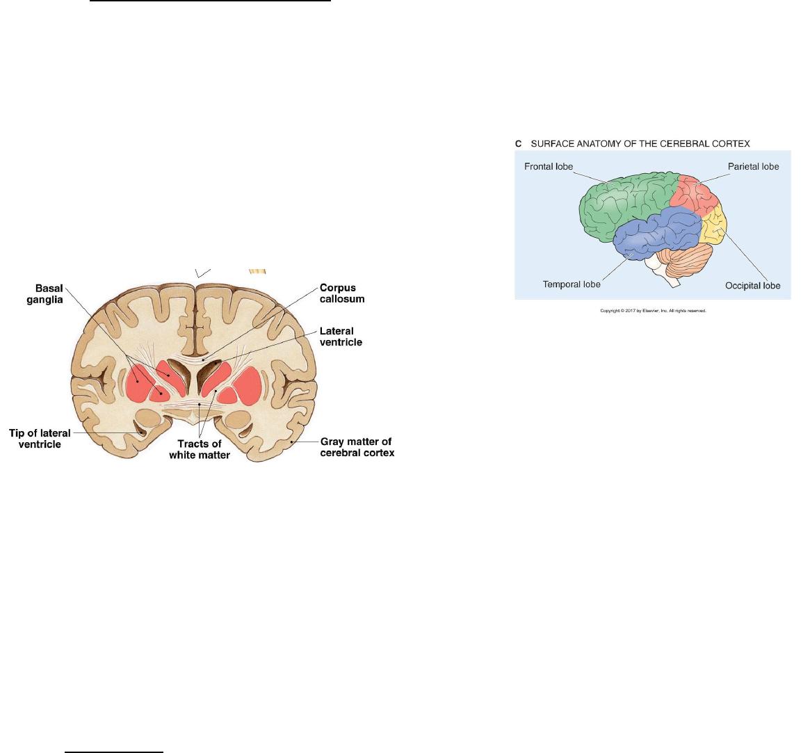
• Two hemispheres separated by corpus callosum
o Millions of neuronal axons that communicate with one another
• 4 major lobes
o Frontal– thoughts, voluntary motor
activity, speech, decision making
o Parietal- receiving & processing sensory
input, somatic sensing “touch”
o Temporal- auditory
o Occipital- visual
• White matter= signals
transmitted
o Information connected
o axon of neuron
o Myelinated nerve fibers,
fatty
• Gray matter= neural input & output, information processed
o Cell body of clustered neurons
o Neuronal cell bodies, dendrites, & glial cells
o 3 regions of gray matter
1) cerebral cortex- outer layer of cerebrum
2) basal ganglia- internally localized, involved in movement
control
3) limbic system- higher cognitive functions with emotional
response (cell bodies/nuclei bunch together to process)
a. amygdala- expression of emotions, fear
b. hippocampus- memory formation & learning
Cerebellum:
• not directly connected to muscle
o Information from cerebrum & muscle helps you with intent & pain

o “little brain” = processes information regarding movement
o Balance, movement, voluntary movement
o 3 distinct subconscious control
1) Vestibulocerebellum= maintains balance & eye movement
(equilibrium, info from spinal cord)
2) Spinocerebellum= enhance muscle tone & coordinates skilled,
voluntary movements; intent vs. response, corrects errors
(doing vs. intend on doing)
3) Cerebrocerebellum= provides input to cortical motor areas &
stores procedural memories (what muscles are doing)
Diencephalon:
• Brain stem & cerebellum
• Thalamus- right below brain
o Processes information
o Sensory input bound for cortex
o Arousal & memory function
o Voluntary motor control (moving your body)
▪ Basal nuclei, cerebellum
• Hypothalamus- below thalamus
o Connected to ANS nuclei in brainstem
o Endocrine system
▪ Releases hormones to anterior
▪ Projects neurons to posterior
o Homoestatic functions
• Epithalamus– next to/above brain
o Pineal gland/habenular nucleus (neurons process info)
o Connects limbic system to other parts of the brain
o Cardiac rhythm
Homeostatic role of hypothalamus:
• A lot of nuclei located here
• Autonomic control center
• Emotional response
• Regulation of body temperature, food intake, water balance, sleep/wake
cycle
• Control endocrine system
Brainstem:
• Underneath diencephalon, lots of nuclei
• Processing information, oldest & primitive part of brain
• Midbrain (top), pons (middle), medulla (bottom)
• 12 cranial nerves
• Ingoing & outgoing fibers pass through
Midbrain:
• Eye movement: CN III (oculomotor), CN IV (trochlear)
• Auditory and visual reflexes
Pons:
• Communication between cerebellum & cerebrum
• Mastication= CN V (trigeminal)
• Eye movement= CN VI (abducens)
• Facial muscles= CN VII (facial)
• Balance & hearing in ears= CN VIII (vestibulocochlear)
Medulla:
• Works with pons to regulate breathing, blood pressure, heart rate, &
digestion: CN IX (glossopharyngeal) & CN X (vagus)
• Neck muscles: CN XI (accessory)
• Tongue: CN XII (hypoglossal)
Reticular information:
• Extends through entire brainstem
• Baseline info for awareness
• Nuclei built on top of each other, information passed
Spinal cord:
• 31 segments form spinal nerve, grows slowly
• Ends L1–L2, nerves extend after= cauda equina (bunch of nerves)
Spinal nerves:
• Cord, rootlet, root, nerve
• Dorsal root= short axons from afferent fibers (dorsal front ganglion), info
comes in
• Ventral root= axons of efferent fibers, info goes out
Gray matter of spinal cord:
• Dorsal horn- interneurons, afferent neurons terminate
• Lateral horn- efferent nerve fibers, supplies cardiac & smooth muscle
• Ventral horn- efferent neurons, supplies skeletal muscle, motor
White matter of spinal cord:
• Ascending tracts- sensory inputs in dorsal & lateral
• Descending tracts- motor outputs in ventral & lateral
Reflex arcs:
• Natural instinct
• Segmental- same segment, very basic reflex
• Intersegmental- another segment, moves up or down
• Suprasegmental- travel to brainstem, moves up
Peripheral Nervous System: cranial & spinal nerve
• Functions: how body responds
o Carries messages to and from CNS
o Motor signals to chemical signals on target tissue
• Somatic= skin, joint, muscle, skeletal muscle
o External environment, afferent & efferent
o Voluntary control
• Autonomic= receiving & responding, smooth & cardiac muscle
o Internal environment
o Involuntary control
Nerves:
• Travelling to & from CNS
• Axon portion of many neurons; Schwann cells (myelination)
• Epineurium– encloses entire nerve, support & protection
• Perineurium- bundle of nerves (fascicles), supports blood vessels
• Endoneurium– separates individual neurons, capillaries
• Periphery- cell bodies of nerves in ganglia
Nerve classification:
• Where they originate from
• Sensory- to CNS
• Motor- from CNS
Nerve plexus: spinal cord to root to nerve to rami
• Originates from rami (nerve splits)
• Various pathways- anterior to plexus to main nerve
• 4 main plexus
Dermatomes of spinal nerves:
• Cutaneous innervation provided by single dorsal root & its ganglion
o Overlap caused by adjacent dorsal root
Lecture 8:
Cerebrospinal fluid:
• Found in choroid plexuses
o Pia mater and glial cells in ventricles
• Highly regulated
• Regulate BECF through mixing
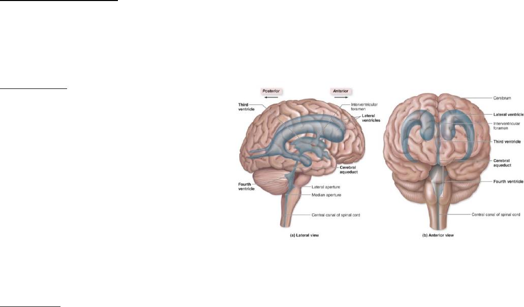
Ventricles: hallow compartments
• 4 ventricles interconnected
• 2 lateral ventricles- cerebrum
o Connected to each other
o Communicates with 3rd
via interventricular
foramen
• 3rd ventricle: diencephalon
o Cerebral aqueduct passes
through midbrain,
connects to 4th (water
passage)
• 4th ventricle: pons & cerebellum
o Joins with subarachnoid space at 2 lateral & median aperture
Meninges: brain tissue really soft
• Dura mater, outermost
o Tight, inelastic, thick
o Form dural or venous sinuses
o Blood leaving brain empties into sinuses
o CSF reentering at 1 sinus
• Arachnoid mater, innermost layer
o Tight junctions between cells
o Overlies subarachnoid space
o CSF blood barriers
o Arachnoid villi and granulations- dura to dural sinuses
• Pia mater, thinnest layer
o Neural tissues via glia limitans
• Leptomeninges, arachnoid & pia mater
o Sandwiches subarachnoid space
• Glia limitans
o Underlies pia
o Nearly complete layer of astrocyte endfeet
o Tightly to pia mater
• Physical barrier between environment
CSF Flow:
• 4 ventricles to subarachnoid space to villi(micro) or granulations(macro)
• Made from plasma
• Ependymal cells– ECF from CSF
• Choroid plexus- ependymal surrounding pia mater & blood vessel
o Blood flow 10x greater
o Capillaries- sealed, but leaky
BECF: dense, ecf through nervous system
• 20% brain volume
• Increases with sleep, cell shrinks
• Decreases when active, cell swells
• Changes nutrient routes
• Cell communicated with CSF
Blood brain barrier:
• Blood vessels & ECF
• Limits access of blood borne materials to interstitial fluid
o Exchange of material in blood in capillaries
• Brain tissue doesn’t get affected by this
• Anatomic restrictions- easy exchanges
o Tight junctions, thick basal lamina
o Astrocytes cling to blood vessels
• Physiological restrictions- water soluble substances transported
o Lipid soluble can penetrate through capillaries, incorporate easily
Circumventricular organs: around ventricles
• Lack BBB
o Hypothalamus- control output of hormones that enter blood
o Pineal & pituitary glands- output hormones into blood
o Vomiting center- notifies brain stem of poisonous substances
o Choroid plexus- permeable to produce CSF
Autonomic Nervous System: involuntary
• Regulating smooth & cardiac muscle, glands, secretory epithelia activity
• Sympathetic & Parasympathetic- opposed to one another
o CNS & PNS
o Innervates visceral organs
o 2 neuron chains
• Enteric– independent system, PNS
o Form plexuses
o Afferent, interneurons, & motor neurons surrounding GI (detect
chemicals)
• Preganglionic neuron (CNS) & postganglionic neuron (ganglion)
Sympathetic anatomy:
• Preganglionic- T1 & L3, exit through ventral roots, going to ganglia
• Sympathetic preganglionic neurons leave spinal nerve, enter sympathetic
trunk via rami communications
o Paravertebral ganglia, interconnected by bundles of axons to form
trunk
Rami communications:
• Spinal nerves to paraventricular ganglia
o White– preganglionic from T1-L3 to trunk, moving in
o Grey- postganglionic from sympathetic to spinal nerve, moving out
Paravertebral ganglia:
• Superior cervical ganglion- C1-C4, neck & head
• Middle cervical ganglion- C5-C6, thoracic viscera
• Inferior cervical ganglion- C7-C8, thoracic viscera
• Upper thoracic ganglia- T1-T5, thoracic viscera
• Remaining– abdominal and pelvic, prevertebral ganglia
Prevertebral ganglia: pass straight through
• Gets lower, abdominopelvic cavity
• Celiac ganglion