MODULE #3
THE FEMALE GAMETOGENESIS
LEARNING
OUTCOMES
After reading this module, the learner should be able to:
1. Define oogenesis and folliculogenesis
2. Discuss the processes of oogenesis and folliculogenesis
3. Describe the histological characteristics of follicles at different stages of
development.
INTRODUCTION
Oogenesis and folliculogenesis in females are very important reproductive
processes for the perpetuation of species. These two reproductive events are
composed of complex physiological processes that will lead to the release of the
egg known as ovulation. In this module, oogenesis and folliculogenesis will be
discussed with emphasis on the characteristics of ovarian follicles at different
stages of development.
This learning module is a compilation of notes and information directly obtained from print and online
sources and is only intended for use by MSU–General Santos students taking ANS150 (Physiology of
Reproduction in Farm Animals).
ABSTRACTION
o The ovary performs two major functions. One is the cyclic production of
fertilizable ova. The second is the production of a balanced ratio of steroid
hormones that maintain the development of the genital tract, facilitate
the migration of the early embryo, and secure its successful implantation
and development in the uterus.
o The follicle is the ovarian compartment that enables the ovary to fulfill its
dual function of gametogenesis and steroidogenesis. These functions
depend on the activity of ovarian follicles that consist of follicular cells and
oocytes.
o The activity and development of ovarian follicles are regulated by
gonadotropins secreted from the hypothalamus–pituitary axis.
o During follicular development, follicular cells such as granulosa and theca
cells differentiate into endocrine cells and secrete estrogens and
progestins.
o Follicle–stimulating hormone (FSH) from the pituitary stimulates ovarian
follicular development and promotes estradiol production by granulosa
cells in coordination with luteinizing hormone (LH).
o During the estrous cycle, the ovary contains primordial, primary,
secondary, and tertiary follicles, but follicle development that is dependent
on gonadotropins occurs from the secondary follicle stage to the ovulatory
phase.
o Some secondary follicles enter to gonadotropin–dependent development
upon FSH stimulation. This process is called follicular recruitment.
2
o The recruited secondary follicles develop into tertiary follicles that have
the follicular antrum. Of these tertiary follicles, one follicle develops to
reach the ovulatory phase and the others undergo atresia.
o In addition to gonadotropins, growth factors and cytokines are also
associated with such follicular development.
o In cattle, follicular development begins with the primary follicles that have
a layer of 11–20 cuboidal granulosa cells around the oocyte.
o At the secondary follicle stage, the follicles gain a second layer of granulosa
cells, and become responsive to gonadotropins. In fact, mRNA of
the FSH receptor mRNA is expressed in the granulosa cells of the
secondary follicles in cattle.
o At the tertiary follicle stage, the follicular cells such as the granulosa and
theca cells proliferate and differentiate into endocrine cells. These follicles
form an antral cavity that is filled with the fluid and the LH receptor gene
is expressed in the theca cells.
Oogenesis
o In contrast to males, most mammalian females are born with their lifelong
complement of gametes (and their ovaries remain housed within the
abdominal cavity).
o During fetal and early postnatal life immature germ cells (oogonia)
undergo mitosis, enter prophase of meiosis I, and become arrested
(dictyate phase).
o Oogonia in meiotic arrest grow in size (accumulate cytoplasm and begin
to form a zona pellucida) to yield primary oocytes.
o Early germ cells in the ovary, called oogonia, increase in number by
ordinary mitosis.
o Each oogonium contains the diploid number of chromosomes.
o After the oogonia cease to increase in number, they grow in size and
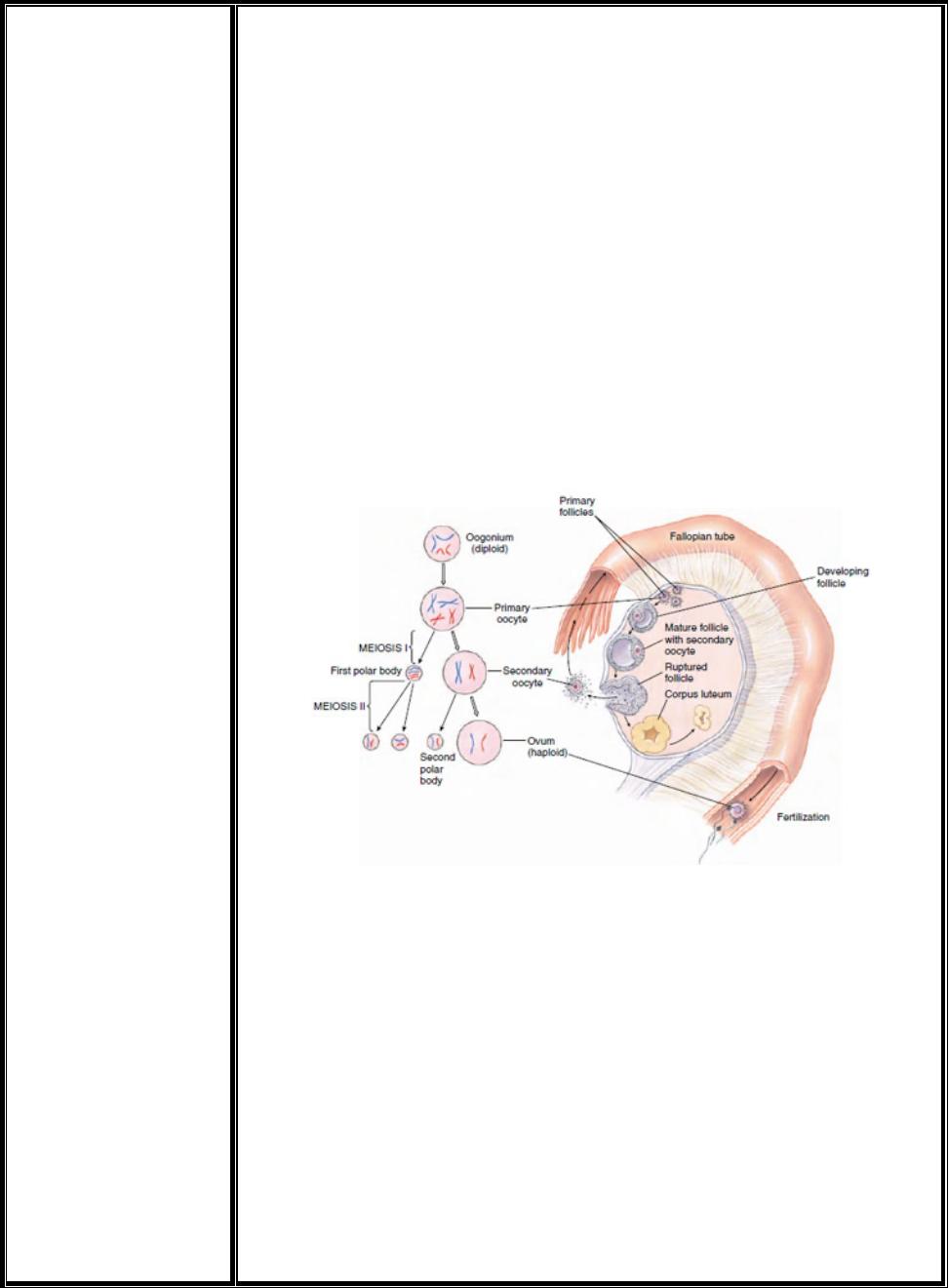
become primary oocytes (Figure 1).
o Before the first meiotic division, the chromosomes in each primary oocyte
meet in pairs, paternal and maternal homologues, just as in
spermatogenesis.
o When the first maturation (reduction) division occurs, the cytoplasm is
divided unequally. One of the two daughter cells, the secondary oocyte,
is large and receives most of the cytoplasm; the other is very small and is
called the first polar body (Figure 1).
o Each of these daughter cells, however, has received half of the
chromosomes.
o In the second meiotic division, the secondary oocyte divides into a large
ootid and a small polar body.
o If the first polar body also divides in this division, which sometimes
happens, there are three polar bodies and one ootid (Figure 1).
o The ootid develops into a functional ovum. The polar bodies are
nonfunctional, and they disintegrate.
o Formation of nonfunctional polar bodies is necessary to enable the egg to
dispose of excess chromosomes. Thus a mature ovum has the N (haploid)
number of chromosomes, the same as a sperm.
3
o However, each primary oocyte gives rise to only one functional gamete
instead of four as in spermatogenesis.
o In most vertebrates and many invertebrates, the egg does not actually
complete all the meiotic divisions before fertilization occurs.
o The general rule is that development is arrested during prophase I of the
first meiotic division.
o Meiosis resumes and is completed either at the time of ovulation (birds
and most mammals) or shortly after fertilization (many invertebrates,
teleost fishes, amphibians, and reptiles).
o In humans, the ova begin the first meiotic division at about the thirteenth
week of fetal development, then their development arrests in prophase I
as the primary oocyte until puberty, at which time one of these primary
oocytes typically develops each menstrual month into a functional egg.
o Meiosis II is completed only when the ovum is penetrated by a
spermatozoon
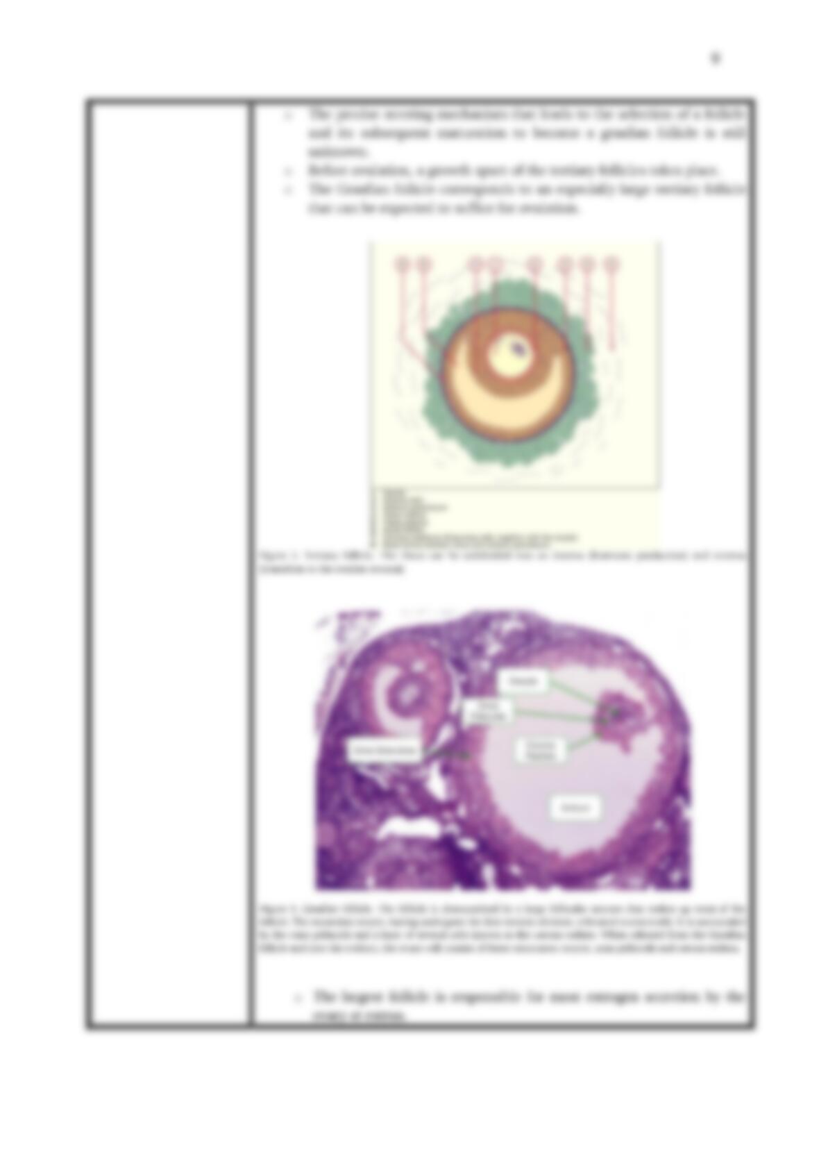
Figure 1. Oogenesis. Early germ cells (oogonia) increase by mitosis
during embryonic development to form diploid primary oocytes. After
puberty, each menstrual month a diploid primary oocyte is divided in the
first meiotic division into a haploid secondary oocyte and a haploid polar
body. If the secondary oocyte is fertilized, it enters the second meiotic
division. The double–stranded chromosomes separate into a large ootid
and small second polar body. Both ootid and second polar body now
contain the N amount of DNA. Fusion of the haploid egg nucleus with a
haploid sperm nucleus produces a diploid (2N) zygote.
Folliculogenesis
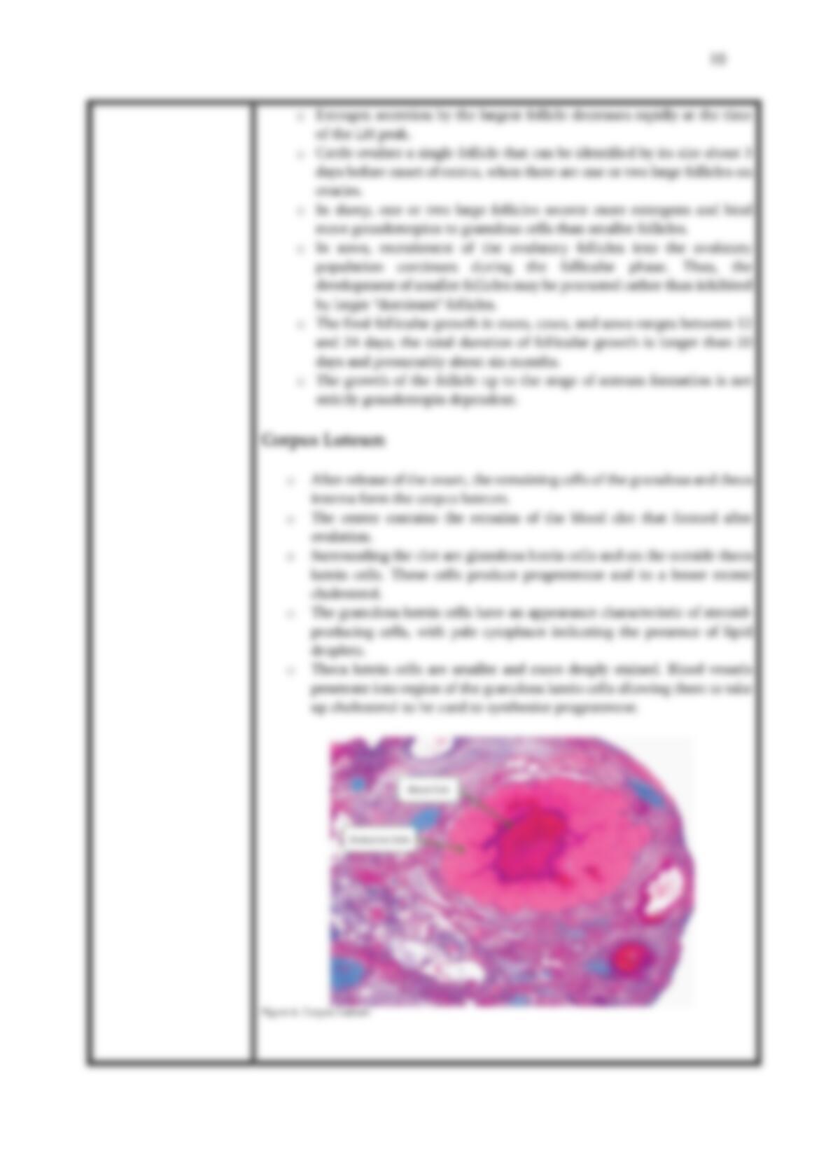
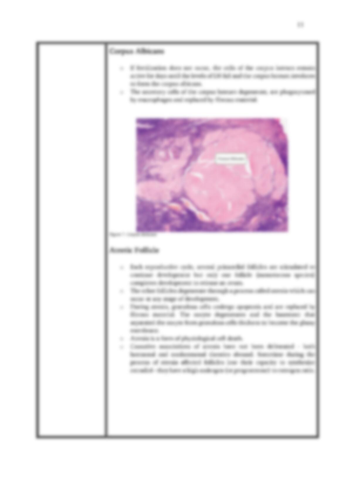
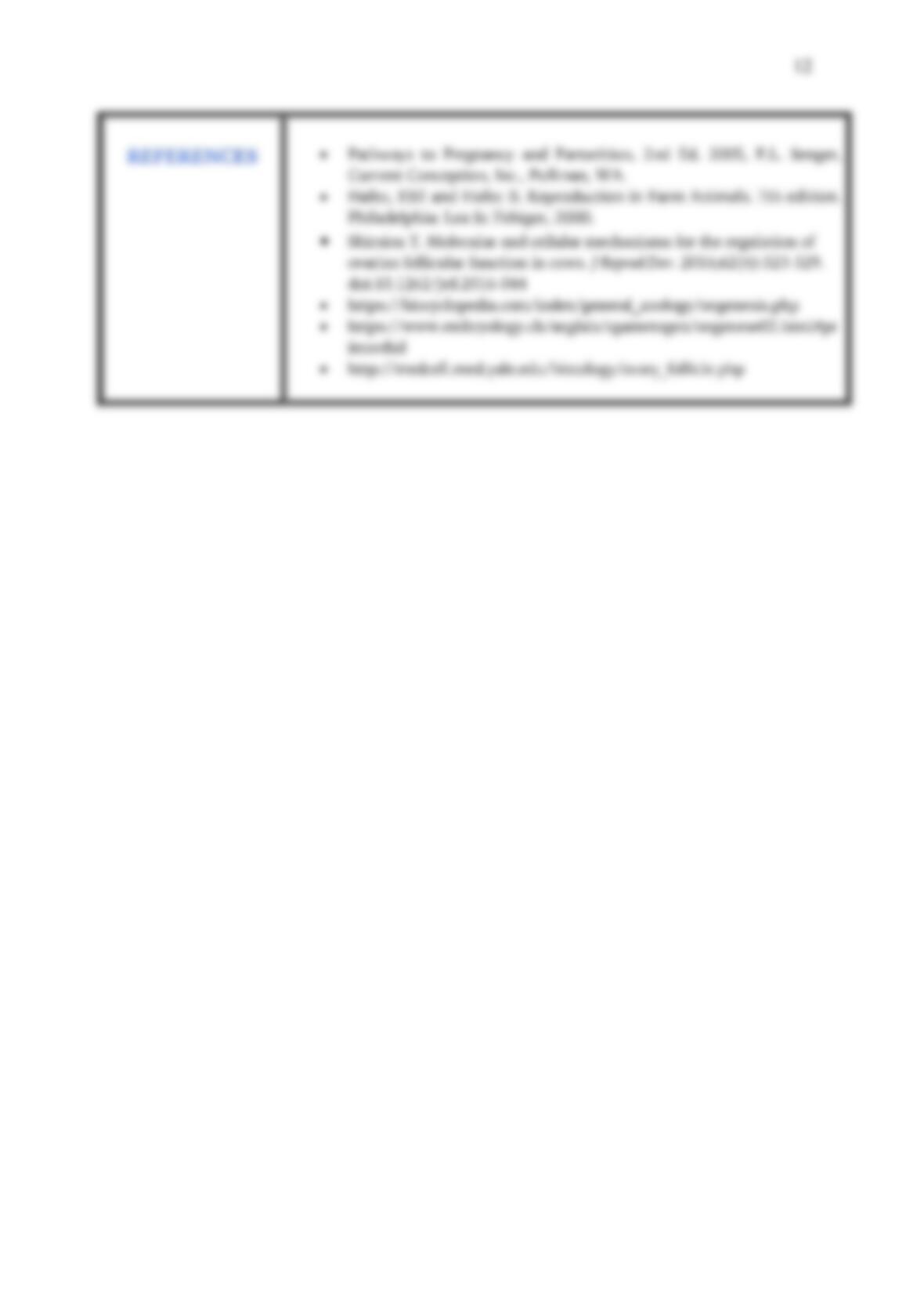
o Folliculogenesis is the maturation of the ovarian follicle, a densely
packed shell of somatic cells that contains an immature oocyte.
o Synchronously with ootidogenesis, the ovarian follicle surrounding the
ootid has developed from a primordial follicle to a preovulatory one.
4
o Folliculogenesis describes the progression of a number of
small primordial follicles into large preovulatory follicles that occurs in part
during the reproductive cycle.
o The primary role of the follicle is oocyte support.
o From the pool of follicles a female animal is born with, only very few of
them will be ovulated, as mostly will break down (in a process
called follicular atresia).
o From birth, the ovaries contain a number of immature, primordial
follicles. These follicles each contain a similarly immature primary
oocyte.
o In the primordial follicle reserve, formed during fetal life or soon after
birth, some primordial follicles begin to grow continuously throughout
life or at least until the reserve is exhausted.
o When any follicle is released from this reserve, it continues to grow
until ovulation or until the follicle degenerates, which is the case with
the majority of follicles
o At puberty, clutches of follicles begin folliculogenesis, entering a
growth pattern that ends in ovulation (the process where the oocyte
leaves the follicle) or in atresia (death of the follicle’s granulosa cells).
o During follicular development, primordial follicles undergo a series of
critical changes in character, both histologically and hormonally.
o First, they change into primary follicles and later into secondary