MODULE #1
THE MALE REPRODUCTIVE SYSTEM
LEARNING
OUTCOMES
After reading this module, the learner should be able to:
1. Identify the organs that comprise the male reproductive system in farm
animals
2. Discuss the functions of the reproductive structures in the male
reproductive system
3. Enumerate the parts of the testes and penis and explain their functions
INTRODUCTION
In order to ensure the survival of the species, animals must reproduce. For this
to be possible, the reproductive system of the male must complement the
reproductive tract of its female counterpart. The male reproductive system is
composed of several reproductive structures and organs that perform specific
physiological functions. This module will discuss the organs and functions of
that make up the male reproductive system as well as the important differences
between the male reproductive system of different farm animals.
This learning module is a compilation of notes and information directly obtained from print and online
sources and is only intended for use by MSU–General Santos students taking ANS150 (Physiology of
Reproduction in Farm Animals).
ABSTRACTION
The Male Reproductive Organs
o The male reproductive or genital organs consist in all species of the
testicle, epididymis, ductus deferens, spermatic cord, accompanying tunics
of spermatic cord and testicle (paired structures), accessory genital glands
(paired or single structures), penis, prepuce, and the male urethra.
The Testicle and Scrotum
o The testicle is the male essential reproductive gland that produces
spermatozoa through the process of spermatogenesis, and also
produces testosterone, the male hormone.
o The testicle has an ovoid shape, a head, a tail, a lateral surface, a medial
surface, a free border, and an epididymal border where the epididymis
is attached.
o The testicle is covered by a white fibrous capsule called tunica albuginea,
which sends, inside of the testicle, interlobular connective tissue walls
called septa, or septula when they are very small.
o The albuginea consists of collagen fibers and a few elastic fibers. The
branches of the testicular artery and vein travel within the albuginea.
This tunic also covers the ductus epididymidis.
2
o The septa/septula consist of collagen fibers, vessels, and nerves; divide
the parenchyma of the testicle in lobules; and build up a loose
connective tissue structure called mediastinum testis, which also contains
vessels and nerves.
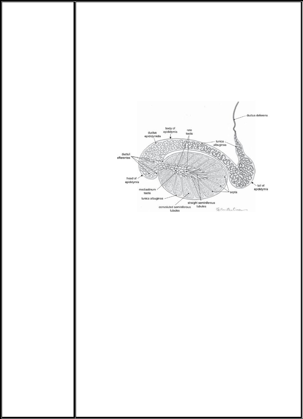
o The lobules contain the seminiferous tubules. The peripheral segment of
the seminiferous tubules is convoluted; the tubules end as straight
seminiferous tubules, which interconnect with each other inside of the
mediastinum testis in a network of ducts called rete testis.
o The rete testis is lined by a non–secretory cuboidal epithelium and
continues as ductuli efferentes, which are coiled (Fig 1).
Fig 1. Internal organization of testicle and epididymis
o The position in the scrotum and the orientation of the long axis of the
testis differ with the species (Fig 2).
o Testicular size varies throughout the year in seasonal breeders (ram,
stallion, camel).
o Removal of one testis results in considerable enlargement of the
remaining gonad (up to 80% increase in weight).
o In the unilateral cryptorchid, removal of the descended testis may be
followed by descent of the abdominal testis as it enlarges.
o The interstitial (Leydig) cells, which lie between the seminiferous
tubules, secrete male hormones into the testicular veins and lymphatic
vessels. The spermatogenic cells of the tubule divide and differentiate to
form spermatozoa.
o Just before puberty, the sustentacular (Sertoli) cells of the tubule form
a barrier, which isolates the differentiating germ cells from the general
circulation.
o Sertoli cells are the somatic cells of the testis that are essential for testis
formation and spermatogenesis.
o Sertoli cells facilitate the progression of germ cells to spermatozoa via
direct contact and by controlling the environment milieu within the
seminiferous tubules.
o The Sertoli cells do not increase in numbers after puberty is attained.
This may limit spermiogenesis.
3
o Sperm production increases with age in the postpubertal period and is
subject to seasonal changes in many species.
o Castration of prepubertal males suppresses sexual development.
Regressive changes in behavior and structure take place following
castration of adult males. Castration is a standard procedure in animal
husbandry to modify aggressive male behavior and to eliminate
undesirable carcass qualities, e.g., boar taint.
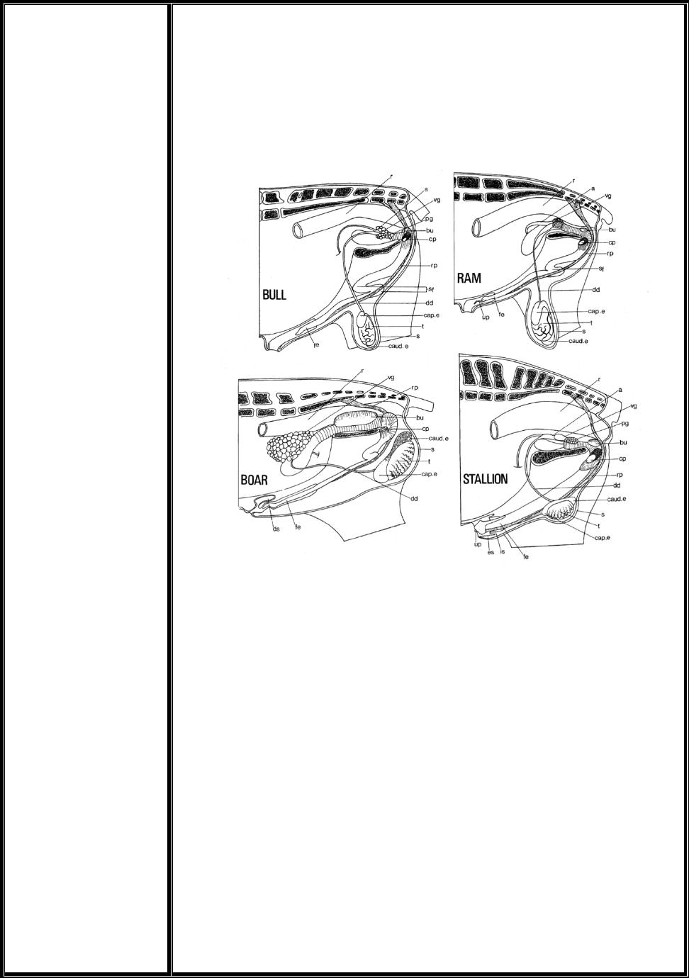
Fig 2. Diagram of the male reproductive tracts as seen in left lateral dissections. a, Ampulla; bu,
bulbourethral gland; cap. e, caput epididymidis, caud.· e, cauda epididymidis, cp, left crus of penis,
severed from the left ischium; dd, ductus deferens; ds, dorsal diverticulum of prepuce; es, prepefiile
prepuce; fe, free part of the penis; is, preputial fold;pg, prostate gland; r, rectum; rp, retractor penis
muscle; s, scrotum; sf, sigmoid flexure; t, testis; up, urethral process; vg, vesicular gland. (Adapted
from Popesko, Atlas der topographischen anatomie der Haustiere. Vol. 3, Jena: Fischer, 1968.)
The Epididymis and Ductus Deferens
o The epididymis is the first excretory organ of the male genital system.
o The epididymis is attached to the testicle and consists of the ductuli
efferentes and the ductus epididymidis, surrounded by the testicular
albuginea.
o The ductus epididymidis is very tortuous and extremely long in all
species. On the lateral aspect of the testicle, between it and the
epididymis, a large space is outlined under the name of testicular bursa.
o The epididymis has a head, a body, and a tail.
o The caput epididymidis (head), in which a variable number of efferent
ductules join the duct of the epididymis. It forms a flattened structure
applied to one pole of the testis.
o The narrow corpus epididymidis (body) terminates at the opposite pole
in the expanded cauda epididymidis (tail).
4
o The middle region of each efferent duct shows marked secretory
activity.
o The convoluted duct of the epididymis is very long (bull, 36 m; boar,
54 m). The wall of the duct of the epididymis has a prominent layer of
circular muscle fibers and a pseudostratified epithelium of columnar
cells.
o There is a progressive decrease in the height of the epithelium and
stereocilia and a widening of the lumen throughout the three segments.
The first two segments are concerned with sperm maturation, whereas
the terminal segment is for sperm storage.
o The ductus epididymidis continues inside of the tail of the epididymis
with the ductus deferens.
o The ductus deferens (vas deferens) is the continuation of, and extends from
the ductus epididymidis to the urethra. The ductus deferens is held in
place by the mesoductus deferens while running within the spermatic cord.
o The vas deferens transports mature sperm to the urethra in preparation
for ejaculation.
o The ductus deferens then enters the abdominal cavity and, switching
the direction, passes within the pelvic cavity over the dorsal aspect of
the urinary bladder. It crosses the ipsilateral ureter and opens on the
roof of the pelvic urethra, lateral to the colliculus seminalis (elevation on
the roof of the prostatic segment of the male urethra at the end of the