Advanced Survival in Patients with Multiple Irradiations for Brain
Melanoma Metastases and Associated Abscopal Effect
Sundip Patel1, Doon Gibbs, Onare Rice, Andrew Gifford
Brookhaven National Laboratory ,Upton New York 11953,USA
Department of Advance Radiation, Upton New York
Abstract
Brain metastases are often detected in melanoma patients. Central nervous system (CNS)
involvement is usually associated with poor prognosis. We describe a case of a female patient
with multiple melanoma brain metastases who survived for 49 months after detection of CNS
involvement and the first radiosurgery. She underwent several courses of irradiation (five
radiosurgeries, stereotactic hypofractionated radiotherapy, and whole brain radiotherapy),
chemotherapy (started in 12 months after the first radiosurgery), and targeted therapy (started
30 months after the first radiosurgery). The clear abscopal effect in the form of disappearance
of intradermal lesions was observed after the second radiosurgery without any systemic
treatment. We believe that the application of combination of different treatment modalities can
significantly prolong survival in certain patients with melanoma brain metastases, and can be
recommended even in case of poor prognosis.
Introduction
Melanoma is characterized by high probability of brain involvement. From the stage of
primary cutaneous melanoma, progression develops in about one-third of cases [1].
According to clinical and imaging data, central nervous system (CNS) metastases were
found in 2-20% of patients and in 36-54% of cases according to autopsy studies [1]. The
median overall survival for patients with melanoma brain metastases is 9.8 months [2].
Several treatments have been shown to significantly improve the life expectancy in these
patients. They are immunotherapy and targeted BRAF– and MEK-inhibitors [3, 4]. Data about
the effectiveness of conventional chemotherapy, whole brain radiotherapy, radiosurgery, and
surgery are more controversial [3, 5]. Much attention is paid to the investigation of the
abscopal effect which is often observed after a combination of immunotherapy with
irradiation, and is associated with a higher survival in patients with melanoma metastases
[6]. The irradiation alone induces the effect very rarely. We present a case of a female
patient with melanoma who was treated with several courses of irradiation (with five courses
of radiosurgery (single fraction) among them, whole brain radiotherapy and stereotactic
hypofractionated radiotherapy (three fractions)), which provided prolonged survival of 49
months from brain metastases detection. The interesting fact is that the radiosurgical
treatment was followed by the development of abscopal effect in the form of disappearance of
intradermal lesions without any systemic therapy.
Case Presentation
In Jan 2020, a 37-year-old woman underwent surgical resection of the melanoma in the
sacrum area. Further investigation performed at the end of 2015 (when the analysis had
become available) revealed malignant melanoma with V600M mutation in the BRAF gene. In
May 2012, due to progression, the inguinal–femoral lymphadenectomy was performed. The
patient was not recommended any systemic treatment. In June 2013, screening magnetic
resonance imaging (MRI) found multiple relatively small brain metastases without edema
and mass effect. The Karnofsky performance status (KPS) was 90 at that time. In the same
month (25 months from the disease onset), the patient was treated with Gamma– Knife
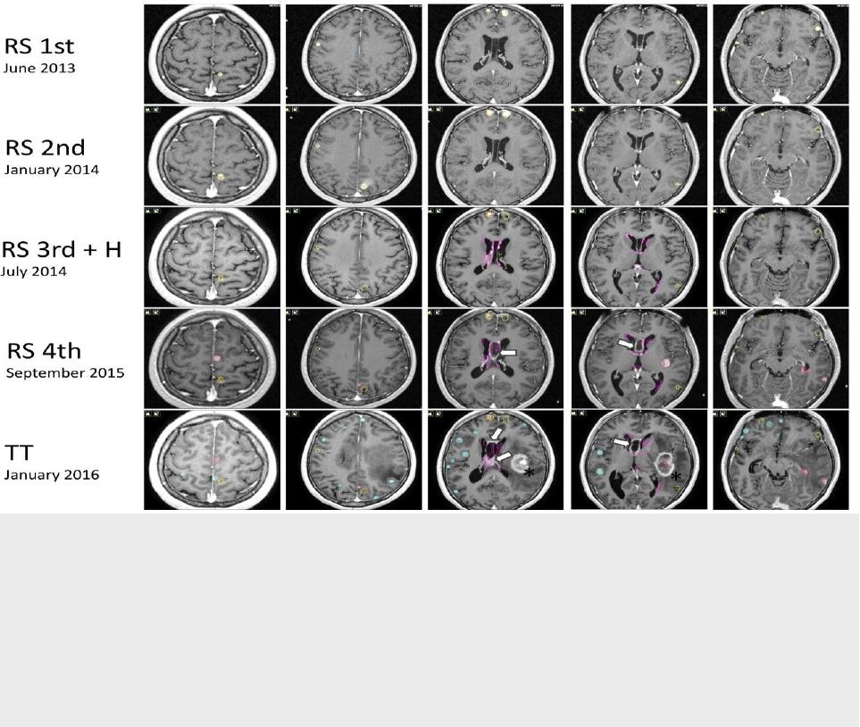
radiosurgery (Figure 1). During the session, overall 19 targets were irradiated with a
marginal dose of 24 Gy prescribed for 52-91% isodose line. The mean volume of treated
metastases composed 0.107 ccm (0.005-0.727 ccm). Despite the recommendation, the
systemic treatment was postponed. Local oncologists evaluated her prognosis as very poor
and refused to perform any systemic treatment. In six months, brain MRI demonstrated
growth in four of 19 previously irradiated metastases and emergence of five new lesions. At
the same period, the patient noticed the appearance of intradermal nodes on her arms and
chest.
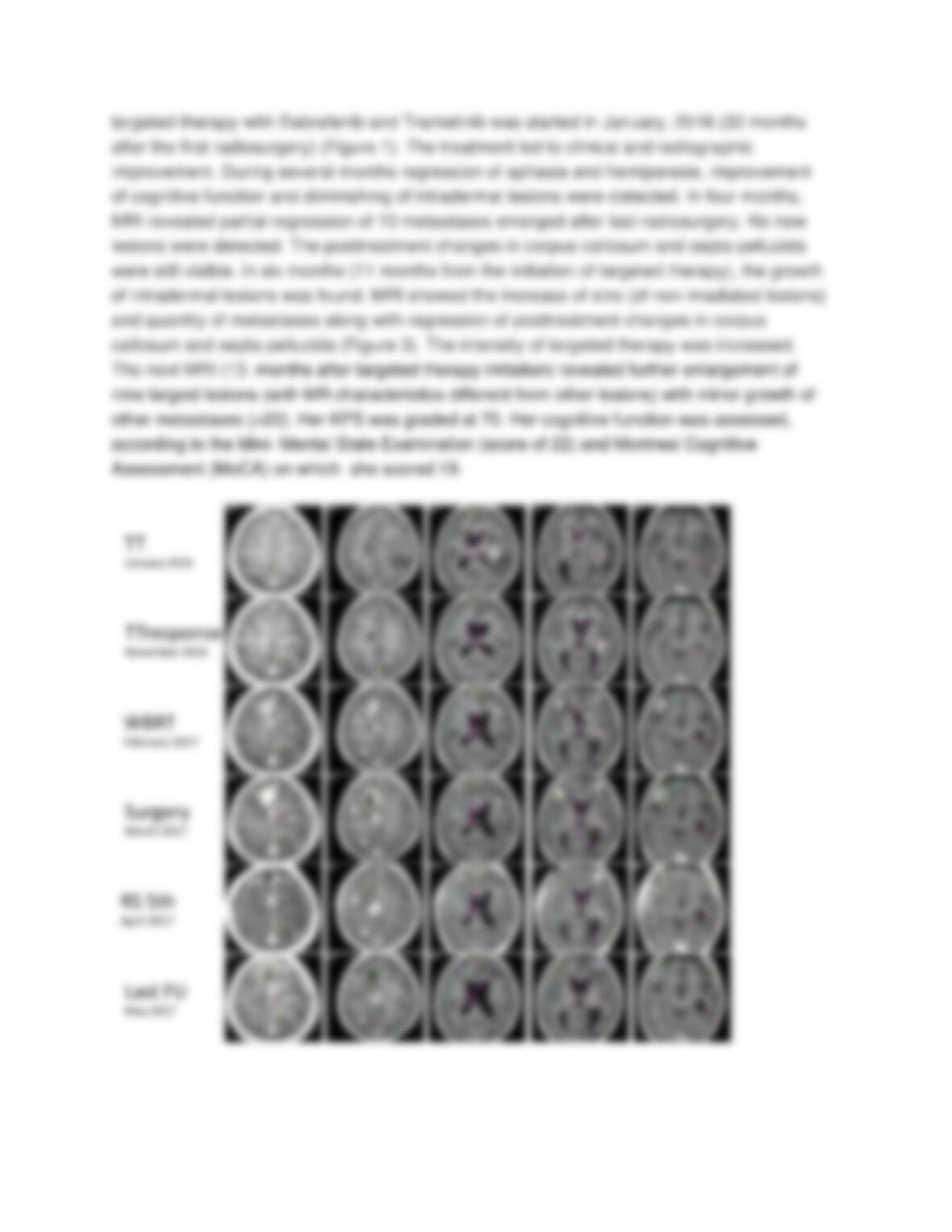
The color of the contour indicates the stage when the lesion was treated: yellow – the 1st
radiosurgery, orange – the 2nd radiosurgery, pink – the 3rd radiosurgery and

hypofractionation, red – the 4th radiosurgery, blue – lesions before the initiation of targeted
therapy. Arrows point on posttreatment changes after the 3rd irradiation in the area of septa
pellucida; asterisk indicates the hemorrhage which emerged in one of the lesion treated with
the 4th radiosurgery
The second course of radiosurgical irradiation of all new and recurring lesions was
performed with Gamma-Knife in January, 2014 (Figure 1). Nine lesions with the mean
volume of 0.197 ccm (0.01-0.389 ccm) were irradiated with the marginal dose of 20 and 24
Gy prescribed to the 50- 82% isodose line. The systemic treatment was not started. During
three months after radiosurgery, the patient noticed complete regression of intradermal
metastases without any systemic treatment. In six months, the new brain MRI showed
dissemination of the disease along the ventricular system with the emergence of multiple
small lesions, mainly, in the bodies of both lateral ventricles, and larger lesions in the
hypothalamic area, pineal region and the lateral parts of the fourth ventricle. No new
metastases in the brain parenchyma were revealed. The cerebrospinal fluid (CSF) cytology
was not performed. At this moment (37 months after the disease onset and 12 months after
the first radiosurgery), the systemic treatment with Cisplatin and Temozolomide was started.
In July 2014, the patient was treated with CyberKnife (Figure 1). The choice of the